Abstract
Bisphenol A (BPA) is a monomer use in manufacturing a wide range of chemical products which include epoxy resins and polycarbonate. It has been reported that BPA increases the cell proliferation activity of human breast cancer MCF-7 cells as well as 17-β estradiol (E2) and diethylstilbestrol (DES). However, BPA induces target genes through ER-dependent and ER-independent manners which are different from the actions induced by E2. Therefore, BPA may be unique in estrogen-dependent cell proliferation compared to other endocrine disrupting chemicals (EDCs). In the present study, to test whether ERα is essential to the BPA-induced proliferation on MCF-7 cells, we suppressed the ERα expression of MCF-7 cells by RNA interference (RNAi). Proliferation effects in the presence of E2, DES and BPA were not observed in ERα-knockdown MCF-7 cells in comparison with control MCF-7. In addition, a marker of proliferative potential, MIB-1 labeling index (LI), showed no change in BPA-treated groups compared with vehicle-treated groups on ERα-knockdown MCF-7 cells. In conclusion, we demonstrated that ERα has a role in BPA-induced cell proliferation as well as E2 and DES. Moreover, this study indicated that the direct knockdown of ERα using RNAi serves as an additional tool to evaluate, in parallel with MCF-7 cell proliferation assay, for potential EDCs.
Keywords: Bisphenol A (BPA), estrogen receptor α (ERα), RNA interference (RNAi), MCF-7, cell proliferation
I. Introduction
Bisphenol A (BPA) is a known endocrine disrupting chemical (EDC) that is a major component of epoxy and polycarbonate resin, which is widely used as an ingredient in the protective coatings on food containers and as adhesives used in packaging products [1, 8, 16]. It has been reported that BPA has weak estrogen activity approximately 1/1000- to 1/10,000-fold of 17-β estradiol (E2) [29], and interacts with estrogen receptors alpha (ERα) and beta (ERβ) [7, 10, 12]. Exposure of BPA induces the expression of progesterone receptor, and promoted MCF-7 breast cancer cell proliferation [9, 16, 21, 23, 25]. In experiments with rodents, exposure of high doses of BPA has been reported to induce reproductive toxicity and abnormal cellular development [15].
Between the 1940s to the 1970s, diethylstilbestrol (DES), a potent synthetic estrogen, was prescribed to prevent complications of pregnancy [28]. Prenatal DES exposure in women is known to cause genital abnormalities and carcinomas [2]. Perinatal exposure of laboratory mice to DES also elicits a spectrum of reproductive tract lesions [13].
Estrogens regulate many physiological processes, including normal cell proliferation, development, and tissue-specific gene regulation in the reproductive tract and in the central nervous and skeletal systems. Estrogens also influence the pathological processes of hormone-dependent diseases, such as breast, endometrial, and ovarian cancers, as well as osteoporosis. The biological actions of estrogens are mediated by binding to one of two specific estrogen receptors (ERs), ERα or ERβ, which belong to the nuclear receptor superfamily [11, 31]. The biological significance of the existence of two ERα and ERβ subtypes is at this moment unclear [17, 18].
Studies on estrogenic effects of EDCs have been mainly performed by MCF-7 cell proliferation assay (E-screen assay), ER reporter gene assay or yeast assays [3, 6, 25]. Interestingly, BPA exhibits characteristics of a distinct molecular mechanism of action at ERα, with BPA interacting differently within the ligand-binding domain compared with E2 [5]. In addition, BPA induces target genes through ER-dependent and ER-independent manners which are different from the actions induced by E2 [23, 24]. Therefore, BPA may be unique in estrogen-dependent cell proliferation compared to other EDCs. In the present study, to investigate whether ERα participate in the BPA-induced cell proliferation as well as E2 and DES, we performed the suppression of ERα expression in MCF-7 cells by RNAi.
II. Materials and Methods
Chemicals
17β-estradiol and diethylstilbestrol were purchased from Wako Pure Chemicals Industries, Ltd. (Wako, Osaka, Japan). Bisphenol A was purchased from Sigma-Aldrich (Sigma, St Louis, MO, USA). The chemicals were solubilized in dimethyl sulfoxide (DMSO; Sigma) and the control culture was exposed to a culture medium containing 0.1% DMSO.
Construction of shRNA expression vectors
Human ERα gene sequence-specific shRNA expression vector, piERα was constructed by iGENE (Tokyo, Japan) using piGENE-PUR-hU6 with resistant characteristics of ampicillin and puromycin. The piERα shRNA sequence was as follows: 5'-TTGTGTTTCAACATTCTCC-3' (the target sequence is located in the exon 4 of ERα cDNA).
Cell proliferation assays
In this study, we employed ER-positive human breast cancer cell lines MCF-7 and BT474 which have been shown to transiently induce the expression of EGFR mRNA and protein because they were estrogen responsive [30]. BT474 cells were chosen as the less responsive ones to estrogen stimulation. MCF-7 was kindly provided by Dr. Ichirou Mori (Wakayama Medical University, Wakayama, Japan), and BT474 was purchased from the American Type Culture Collection (ATCC, Rockville, MD, USA). They were grown in Dulbecco’s modified Eagle’s medium (GIBCO, Carlsbad, CA, USA) containing 10% fetal bovine serum. Twenty-four hr before treatment, cell culture medium was replaced with phenol red-free Dulbecco’s modified Eagle’s medium (GIBCO) containing 10% dextran-coated charcoal treated fetal bovine serum. MCF-7 and BT474 cells were seeded into 24-well plates at a density of 2×10⁴ cells/well and cultured further with or without 10−10~10−6 M of E2, 10−10~10−6 M of DES, or 10−8~10−4 M of BPA for 96 hr, and then the cell number was measured with the cell counting kit-8 (Dojindo, Kumamoto, Japan).
ERα gene silencing in MCF-7 cells using RNAi
MCF-7 cells were transfected with piERα vector or empty vector. Twenty-four hr before transfection, cell culture medium was replaced with phenol red-free Dulbecco’s modified Eagle’s medium (GIBCO) containing 10% dextran-coated charcoal treated fetal bovine serum. The cells were transfected with 2 µg of indicated plasmids using 5 µl of FuGENE HD transfection regent (Roche, Indianapolis, IN, USA). After 24 hr of transfection, the cells were cultured with 2 µg/ml puromycin for 48 hr. At the end of incubation, cells were washed with phosphate-buffered saline (PBS), dead cells were removed from culture flask. Living cells were seeded in 24-well plates at density of 5×10⁴ and cultured further with or without 10−10~10−6 M of E2, 10−10~10−6 M of DES, or 10−8~10−4 M of BPA for 96 hr.
Real-time RT-PCR
Total cellular RNA was isolated by the SV Total RNA Isolation System (Promega, Madison, WI, USA) according to the manufacturer’s recommended protocol. One µg of total RNA was converted to double-stranded cDNA using QuantiTect Reverse Transcription Kit (Qiagen, Valencia, CA, USA). Real-time RT-PCR analysis was done using a DNA Opticon system (MJ Research, Waltham, MA, USA). The level of ERα expression was normalized relative to that of the internal control, the β-actin gene. The reaction protocol consisted of the following cycles: 95°C for 10 min, 40 cycles of 94°C for 10 sec, 60°C for 15 sec, and 72°C for 15 sec, followed by 72°C for 5 min. Primers for PCR amplification were as follows: ERα forward, 5'-TGGAGATCTTCGACATGCTG-3'/reverse 5'-TCCAGAGACTTCAGGGTGCT-3', β-actin, 5'-CCCAGCACAATGAAGATCAA-3'/reverse, 5'-ACATCTGCTGGAAGGTGGAC-3'.
Immunocytochemistry
For immunostaining of ERα, the cells were cultured in two-well permanox slides, and were washed with PBS and fixed by 10% buffered formalin for 30 min. Cells were pretreated by boiling in Target Retrieval solution High pH (DAKO, Glostrup, Denmark) for 40 min. After peroxidase activity was blocked with 0.03% H2O2 for 15 min, cells were treated consecutively at room temperature with mouse anti-ERα (1D5; DAKO) diluted at 1:50 for 1 hr, and the signal was amplified with DAKO ENVISION+ kit (DAKO) according to the manufacturer’s recommendations. Immunoreactivity was visualized by incubation with 3,3'-diaminobenzidine tetrahydrochloride. Finally, cells were counterstained with hematoxylin.
MIB-1 labeling index assay
Intact MCF-7 cells, empty vector transfected-MCF-7 cells and ERα-knockdown MCF-7 cells were incubated with either BPA 10−5 M or vehicle control (0.1% DMSO) in cell-chamber for 96 hr. At the end of incubation, the cells were washed with PBS and fixed by 10% buffered formalin for 30 min. Endogenous peroxidase activity was blocked with 0.03% H2O2 for 15 min, and cells were permeabilized (0.4% Tween20, 1% normal horse serum in PBS for 1 hr) and then incubated for 1 hr at room temperature with mouse anti-Ki-67 (MIB-1; DAKO) diluted at 1:50. The signal was amplified with DAKO ENVISION+ kit (DAKO) according to the manufacturer’s recommendations. Immunoreactivity was visualized by incubation with 3,3'-diaminobenzidine tetrahydrochloride. Finally, cells were counterstained with hematoxylin. The MIB-1 LI was defined as the percentage of immunostained cells divided by the total number of cells in the evaluated area. All counts were performed at a magnification of ×400 using an ocular graticule consisting of 10×10=100 fields covering an area of 0.0625 mm2. Ten viable fields from the area of maximal labeling were chosen for counting. A thousand to 5000 cells were counted from each specimen.
Statistical analysis
Data are presented as means±standard deviation (SD) of more or three independent experiments. The differences between each treated value were evaluated by analysis of variance and Student’s t-test. P<0.05 was considered statistically significant.
III. Results
Cell proliferation analysis of intact MCF-7 cells and BT474 cells
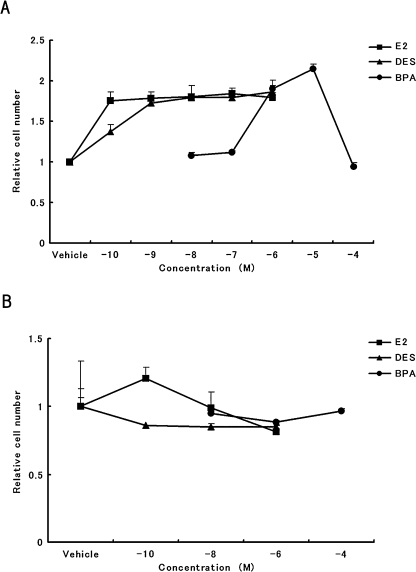
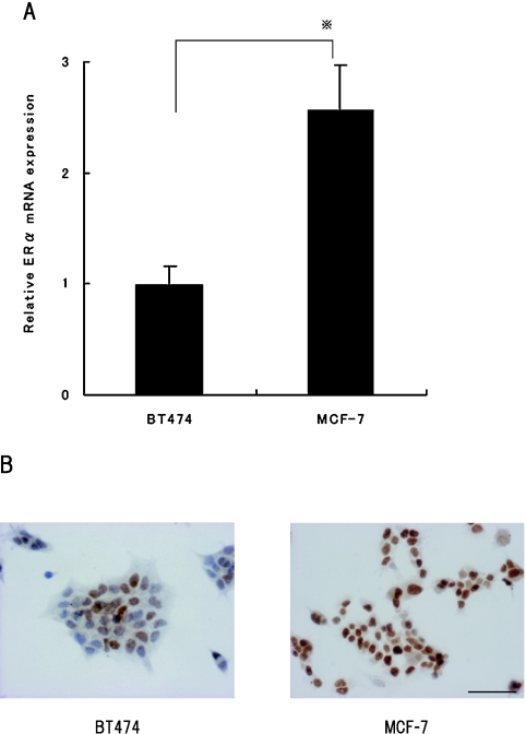
The human breast cancer MCF-7 and BT474 cells were treated for 96 hr with a concentration of 10−10 M~10−6 M of E2, 10−10 M~10−6 M of DES or 10−8 M~10−4 M of BPA. In the cell proliferation of MCF-7, a significant increase was detectable at concentration of 10−10 M~10−6 M of E2, 10−10 M~10−6 M of DES and 10−6 M and 10−5 M of BPA as compared with vehicle groups (0.1% DMSO) (Fig. 1). In the cell proliferation of BT474, a significant increase was not detected in any dose of E2-, DES- and BPA-treated groups compared with vehicle groups (0.1% DMSO) (Fig. 1). The expression level of ERα of MCF-7 and BT474 were examined by real-time RT-PCR and immunocytochemistry analysis (Fig. 2), the MCF-7 ERα mRNA was highly expressed approximately 2.6-fold compared with BT474; these results were further supported by immunocytochemistry analysis.
Fig. 1.
Effects of E2, DES and BPA on the cell proliferation in (A) MCF-7 cells and (B) BT474 cells. Cells were treated with the indicated concentrations of (closed square) E2, (closed triangle) DES or (closed circle) BPA for 96 hr, and then the cell number was counted. Bars indicate mean±SD values (n=3). Data are expressed as fold change from vehicle control.
Fig. 2.
The difference of ERα mRNA expression levels between MCF-7 cells and BT474 cells by (A) real-time RT-PCR and (B) immunocytochemistry. Real-time RT-PCR analysis was normalized by those of β-actin as an internal standard. Data are expressed as fold change from BT474 cells. * P<0.01. Original magnification: ×50. Bar=100 µm.
Effects of E2, DES and BPA on cell proliferation in ERα-knockdown MCF-7 cells using RNAi
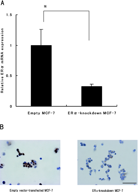
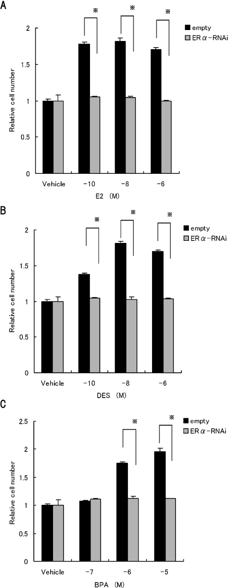
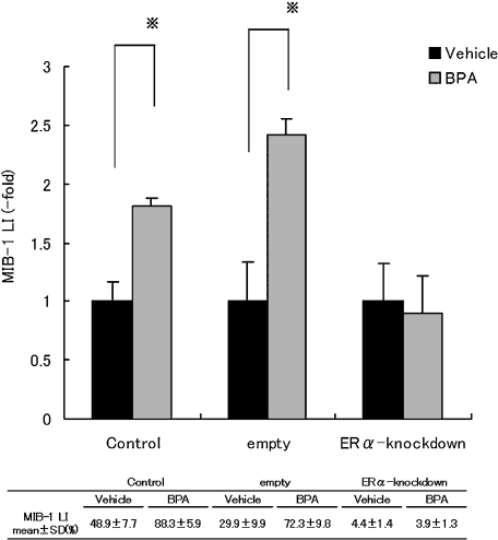
First, we evaluated the effect of RNAi-mediated knockdown of ERα; the degree of knockdown was assessed by real-time RT-PCR and immunocytochemistry analysis. Transfection of the ERα shRNA expression vector was observed to be significantly decreased by 3.1-fold compared with empty vector-transfected MCF-7; these results were further supported by immunocytochemistry analysis (Fig. 3). The cell proliferation of ERα-knockdown MCF-7 was not detected in any dose of E2, DES and BPA (Fig. 4). As another index for the cell proliferation activity, MIB-1 LI was analyzed by Ki-67 immunohistochemistry. The MIB-1 LI of intact MCF-7 (1.8±0.07-fold) and empty vector-transfected MCF-7 (2.4±0.1-fold) with 10−5 M of BPA were significantly increased as compared with vehicle-treated groups. In contrast, the MIB-1 LI of ERα-knockdown MCF-7 showed no change in BPA-treated groups (0.9±0.3-fold) (Fig. 5).
Fig. 3.
The efficacy of ERα-knockdown by RNAi was confirmed according to (A) real-time RT-PCR and (B) immunocytochemistry analysis. Real-time RT-PCR normalized by those of β-actin as an internal standard. Data are expressed as fold change from empty vector-transfected MCF-7. * P<0.05. Original magnification: ×50. Bar=100 µm.
Fig. 4.
Effects of E2, DES and BPA on the cell proliferation in ERα-knockdown MCF-7 cells. The ERα-knockdown MCF-7 and empty vector-transfected MCF-7 were treated with the indicated concentration of (A) E2, (B) DES or (C) BPA for 96 h, and then the cell number was counted. Data are expressed as fold change from vehicle control. * P<0.05.
Fig. 5.
Quantification of cell proliferation by MIB-1. Intact MCF-7, ERα-knockdown MCF-7 and empty vector-transfected MCF-7 were treated with concentration of 10−5 M of BPA or vehicle (0.1% DMSO). Data are expressed as fold change from vehicle control. Table presented the proportion of MIB-1-positive cells in each group. * P<0.01.
IV. Discussion
The biological effects of BPA have been widely studied over the past several decades. It has been reported that BPA can exert some of its effects by binding at the ERα or ERβ to induce estrogenic signals that modify estrogen-responsive gene expression [7, 10, 12]. However, in vitro effects of BPA might be different in individual cell types, including possible ER-independent actions [23, 27]. Although a major mechanism of BPA action is ER-regulated gene expression through the estrogen responsive element (ERE), high concentrations of BPA could regulate expression of growth- and development-related genes different from E2 in MCF-7 cells stably expressing HA-tagged ERα [24], indicating that BPA is unique in estrogen-dependent cell proliferation compared to other EDCs. In the present study, we demonstrated that the proliferation effects were induced in exposure of MCF-7 to BPA, which were dependent on the presence of functional ERα as well as E2- and DES-action.
The MCF-7 cell line is a human breast cancer cell line which possesses ERs and has been widely used as valuable materials in environmental toxicology. As shown in Figure 1, BPA-treated MCF-7 cells were shown to have lower cell proliferation in comparison to E2 and DES, but cell proliferation was not observed in BT474 cells with these stimuli. The effective doses of these EDCs were consistent with previous evidence [8, 20, 22]. To examine the reason for differential effects of cell proliferation between MCF-7 cells and BT474 cells, the ERα expression level was examined by real-time RT-PCR and immunocytochemical analysis. It was shown that the proliferative effects to these EDCs were correlated with ERα content between MCF-7 cells and BT474 cells, suggesting that regulation of ERα concentration may be a key component in estrogen responsiveness of target cells. Therefore, in this study, to investigate whether ERα is essential to the BPA-induced cell proliferation as well as E2 and DES, we carried out specific inhibition of the ERα gene using RNAi mediated by ERα-shRNA vector in MCF-7. As shown in Figure 4, we found that the cell proliferation of MCF-7 with BPA, E2 and DES was obviously inhibited by knockdown of ERα. As another index for the cell proliferation activity, the Ki-67 antigen is widely known as a cell growth marker [4, 26]. MIB-1 LI of ERα-knockdown MCF-7 cells showed no change by incubation with BPA. It is possible that ERα-mediated gene expression results in the BPA-induced cell proliferation of MCF-7. On the other hand, BPA was shown to exert both agonistic and antagonistic effects through ERα while behaving exclusively as an agonist through ERβ [7]. Although intact MCF-7 expressed both ERα and ERβ in our studies, the BPA-induced cell proliferations were inhibited only by ERα-knockdown, suggesting that ERβ plays no important role in BPA-induced cell proliferation, but this remains to be further investigated.
In this study, we carried out specific inhibition of the ERα gene using RNAi mediated by ERα-shRNA vector in MCF-7 for the following reasons. It has been reported that specific gene silencing using shRNA-expressed vector can provided stable suppression of a target gene by antibiotic selection in mammalian cells [14, 19]. Indeed, immunocytochemical analysis in our study showed that a reduction of ERα was observed in ERα-shRNA vector, but not in control vector. It was shown that gene suppression by shRNA vector was a useful method for long-term estrogenicity tests. However, the ERα expressions were not completely inhibited in ERα-knockdown MCF-7 cells, and few ERα expressions were observed in the cytoplasm of these cells (as shown in Fig. 3B). It may be due to low transfection efficiency of piERα shRNA vector in MCF-7 cells.
In conclusion, we demonstrated that ERα has an important role in BPA-induced cell proliferation, as well as E2 and DES. Moreover, this study indicated that the knockdown of ERα using RNAi serves as additional tool for evaluating, in parallel with E-screen assay, for potential EDCs.
V. Acknowledgments
The authors wish to thank Hideo Tsukamoto from the Teaching and Research Support Center, Tokai University School of Medicine for his technical support.
VI. References
- 1.Biedermann M., Grob K. Food contamination from epoxy resins and organosols used as can coatings: Analysis by gradient NPLC. Food Addit. Contam. 1998;15:609–618. doi: 10.1080/02652039809374688. [DOI] [PubMed] [Google Scholar]
- 2.Bornstein J., Adam E., Adler-Storthz K., Kaufman R. H. Development of cervical and vaginal squamous cell neoplasia as a late consequence of in utero exposure to diethylstilbestrol. Obstet. Gynecol. Surv. 1988;43:15–21. [PubMed] [Google Scholar]
- 3.Brotons J. A., Olea-Serrano M. F., Villalobos M., Pedraza V., Olea N. Xenoestrogens released from lacquer coatings in food cans. Environ. Health Perspect. 1995;103:608–612. doi: 10.1289/ehp.95103608. [DOI] [PMC free article] [PubMed] [Google Scholar]
- 4.Gentil Perret A., Mosnier J. F., Buono J. P., Berthelot P., Chipponi J., Balique J. G., Cuilleret J., Dechelotte P., Boucheron S. The relationship between MIB-1 proliferation index and outcome in pancreatic neuroendocrine tumors. Am. J. Clin. Pathol. 1998;109:286–293. doi: 10.1093/ajcp/109.3.286. [DOI] [PubMed] [Google Scholar]
- 5.Gould J. C., Leonard L. S., Maness S. C., Wagner B. L., Conner K., Zacharewski T., Safe S., McDonnell D. P., Gaido K. W. Bisphenol A interacts with the estrogen receptor alpha in a distinct manner from estradiol. Mol. Cell Endocrinol. 1998;142:203–214. doi: 10.1016/s0303-7207(98)00084-7. [DOI] [PubMed] [Google Scholar]
- 6.Harris C. A., Henttu P., Parker M. G., Sumpter J. P. The estrogenic activity of phthalate esters in vitro. Environ. Health Perspect. 1997;105:802–811. doi: 10.1289/ehp.97105802. [DOI] [PMC free article] [PubMed] [Google Scholar]
- 7.Hiroi H., Tsutsumi O., Momoeda M., Takai Y., Osuga Y., Taketani Y. Differential interactions of bisphenol A and 17 beta-estradiol with estrogen receptor alpha (ERalpha) and ERbeta. Endocrine J. 1999;46:773–778. doi: 10.1507/endocrj.46.773. [DOI] [PubMed] [Google Scholar]
- 8.Kim H. S., Han S. Y., Yoo S. D., Lee B. M., Park K. L. Potential estrogenic effects of bisphenol A estimated by in vitro and in vivo combination assays. J. Toxicol. Sci. 2001;26:111–118. doi: 10.2131/jts.26.111. [DOI] [PubMed] [Google Scholar]
- 9.Krishnan A. V., Stathis P., Permuth S. F., Tokes L., Feldman D. Bisphenol-A: an estrogenic substance is released from polycarbonate flasks during autoclaving. Endocrinology. 1993;132:2279–2286. doi: 10.1210/endo.132.6.8504731. [DOI] [PubMed] [Google Scholar]
- 10.Kurosawa T., Hiroi H., Tsutsumi O., Ishikawa T., Osuga Y., Fujiwara T., Inoue S., Muramatsu M., Momoeda M., Taketani Y. The activity of bisphenol A depends on both the estrogen receptor subtype and the cell type. Endocrine J. 2002;49:465–471. doi: 10.1507/endocrj.49.465. [DOI] [PubMed] [Google Scholar]
- 11.Matthews J., Gustafsson J. A. Estrogen signaling: a subtle balance between ER alpha and ER beta. Mol. Interv. 2003;3:281–292. doi: 10.1124/mi.3.5.281. [DOI] [PubMed] [Google Scholar]
- 12.Matthews J. B., Twomey K., Zacharewski T. R. In vitro and in vivo interactions of bisphenol A and its metabolite, bisphenol A glucuronide, with estrogen receptors alpha and beta. Chem. Res. Toxicol. 2001;14:149–157. doi: 10.1021/tx0001833. [DOI] [PubMed] [Google Scholar]
- 13.McLachlan J. A., Newbold R. R., Bullock B. C. Long-term effects on the female mouse genital tract associated with prenatal exposure to diethylstilbestrol. Cancer Res. 1980;40:3988–3999. [PubMed] [Google Scholar]
- 14.Miyai S., Itoh J., Kajiya H., Takekoshi S., Osamura R. Y. Pit-1 gene inhibition using small interfering RNAs in rat pituitary GH secreting cell line. Acta Histochem. Cytochem. 2005;38:107–114. [Google Scholar]
- 15.Morrissey R. E., George J. D., Price C. J., Tyl R. W., Marr M. C., Kimmel C. A. The developmental toxicity of bisphenol A in rats and mice. Fundam. Appl. Toxicol. 1987;8:571–582. doi: 10.1016/0272-0590(87)90142-4. [DOI] [PubMed] [Google Scholar]
- 16.Nagel S. C., vom Saal F. S., Thayer K. A., Dhar M. G., Boechler M., Welshons W. V. Relative binding affinity-serum modified access (RBA-SMA) assay predicts the relative in vivo bioactivity of the xenoestrogens bisphenol A and octylphenol. Environ. Health Perspect. 1997;105:70–76. doi: 10.1289/ehp.9710570. [DOI] [PMC free article] [PubMed] [Google Scholar]
- 17.Nilsson S., Mäkelä S., Treuter E., Tujague M., Thomsen J., Andersson G., Enmark E., Pettersson K., Warner M., Gustafsson J. A. Mechanisms of estrogen action. Physiol. Rev. 2001;81:1535–1565. doi: 10.1152/physrev.2001.81.4.1535. [DOI] [PubMed] [Google Scholar]
- 18.Oshima Y., Matsuda K., Yoshida A., Watanabe N., Kawata M., Kubo T. Localization of estrogen receptors alpha and beta in the articular surface of the rat femur. Acta Histochem. Cytochem. 2007;40:27–34. doi: 10.1267/ahc.06015. [DOI] [PMC free article] [PubMed] [Google Scholar]
- 19.Paddison P. J., Caudy A. A., Bernstein E., Hannon G. J., Conklin D. S. Short hairpin RNAs (shRNAs) induce sequence-specific silencing in mammalian cells. Genes Dev. 2002;16:948–958. doi: 10.1101/gad.981002. [DOI] [PMC free article] [PubMed] [Google Scholar]
- 20.Perez P., Pulgar R., Olea-Serrano F., Villalobos M., Rivas A., Metzler M., Pedraza V., Olea N. The estrogenicity of bisphenol A-related diphenylalkanes with various substituents at the central carbon and the hydroxy groups. Environ. Health Perspect. 1998;106:167–174. doi: 10.1289/ehp.98106167. [DOI] [PMC free article] [PubMed] [Google Scholar]
- 21.Ramos J. G., Varayoud J., Sonnenschein C., Soto A. M., Muñoz De Toro M., Luque E. H. Prenatal exposure to low doses of bisphenol A alters the periductal stroma and glandular cell function in the rat ventral prostate. Biol. Reprod. 2001;65:1271–1277. doi: 10.1095/biolreprod65.4.1271. [DOI] [PubMed] [Google Scholar]
- 22.Schafer T. E., Lapp C. A., Hanes C. M., Lewis J. B., Wataha J. C., Schuster G. S. Estrogenicity of bisphenol A and bisphenol A dimethacrylate in vitro. J. Biomed. Mater. Res. 1999;45:192–197. doi: 10.1002/(sici)1097-4636(19990605)45:3<192::aid-jbm5>3.0.co;2-a. [DOI] [PubMed] [Google Scholar]
- 23.Singleton D. W., Feng Y., Chen Y., Busch S. J., Lee A. V., Puga A., Khan S. A. Bisphenol-A and estradiol exert novel gene regulation in human MCF-7 derived breast cancer cells. Mol. Cell Endocrinol. 2004;221:47–55. doi: 10.1016/j.mce.2004.04.010. [DOI] [PubMed] [Google Scholar]
- 24.Singleton D. W., Feng Y., Yang J., Puga A., Lee A. V., Khan S. A. Gene expression profiling reveals novel regulation by bisphenol-A in estrogen receptor-alpha-positive human cells. Environ. Res. 2006;100:86–92. doi: 10.1016/j.envres.2005.05.004. [DOI] [PubMed] [Google Scholar]
- 25.Soto A. M., Sonnenschein C., Chung K. L., Fernandez M. F., Olea N., Serrano F. O. The E-SCREEN assay as a tool to identify estrogens: an update on estrogenic environmental pollutants. Environ. Health Perspect. 1995;103:113–122. doi: 10.1289/ehp.95103s7113. [DOI] [PMC free article] [PubMed] [Google Scholar]
- 26.Takeshita K., Murata S., Mitsufuji S., Wakabayashi N., Kataoka K., Tsuchihashi Y., Okanoue T. Clinicopathological characteristics of esophageal squamous papillomas in Japanese patients—with comparison of findings from western countries. Acta Histochem. Cytochem. 2006;39:23–30. doi: 10.1267/ahc.05052. [DOI] [PMC free article] [PubMed] [Google Scholar]
- 27.Wetherill Y. B., Akingbemi B. T., Kanno J., McLachlan J. A., Nadal A., Sonnenschein C., Watson C. S., Zoeller R. T., Belcher S. M. In vitro molecular mechanisms of bisphenol A action. Reprod. Toxicol. 2007;24:178–198. doi: 10.1016/j.reprotox.2007.05.010. [DOI] [PubMed] [Google Scholar]
- 28.Wilcox A. J., Baird D. D., Weinberg C. R., Hornsby P. P., Herbst A. L. Fertility in men exposed prenatally to diethylstilbestrol. N. Engl. J. Med. 1995;332:1411–1416. doi: 10.1056/NEJM199505253322104. [DOI] [PubMed] [Google Scholar]
- 29.Witorsch R. J. Low-dose in utero effects of xenoestrogens in mice and their relevance to humans; an analytical review of the literature. Food Chem. Toxicol. 2002;40:905–912. doi: 10.1016/s0278-6915(02)00069-8. [DOI] [PubMed] [Google Scholar]
- 30.Yarden R. I., Lauber A. H., El-Ashry D., Chrysogelos S. A. Bimodal regulation of epidermal growth factor receptor by estrogen in breast cancer cells. Endocrinology. 1996;137:2739–2747. doi: 10.1210/endo.137.7.8770893. [DOI] [PubMed] [Google Scholar]
- 31.Yoshida M., Nishi M., Kizaki Z., Sawada T., Kawata M. . Subcellular and subnuclear distributions of estrogen receptor α in living cells using green fluorescent protein and immunohistochemistry. Acta Histochem. Cytochem. 2002;34:413–422. [Google Scholar]


