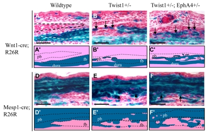
Fig. 7.
Cooperative control of neural crest-mesoderm boundary at coronal suture by Twist1 and EphA4. Either the neural crest marker Wnt1-Cre; R26R or the mesoderm marker Mesp1-Cre; R26R was crossed into mice with the indicated genotypes. Heads of E16.5 embryos were sectioned as in Fig. 2 and stained for lacZ. Schematics depicting key results are shown below each image. Note sharp mesoderm-neural crest boundary in wild-type embryo (A,A′,D,D′). Note both neural crest-derived cells and mesoderm-derived cells crossing boundary in mutant embryos. Also note increased severity of boundary defect in Twist1+/-; EphA4+/- embryo (C,C′,F,F′) compared with Twist1+/- embryo (B,B′,E,E′).

