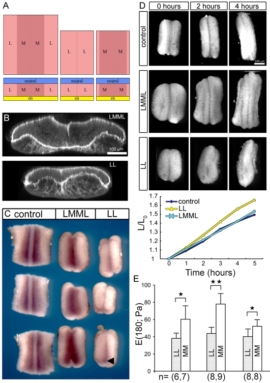
Fig. 3.
Mechanics of paraxial-medial and paraxial-lateral tissues.(A) Schematic diagram of explants carrying complete sets of paraxial tissues (lateral-medial-medial-lateral or LMML) and those carrying only paraxial lateral tissues (lateral-lateral or LL) of the dorsal axis. (B) Fibronectin fibril localization in transverse sections of LMML and LL explants shows the location of paraxial mesoderm outlined by fibronectin. (C) A view of XmyoD expression through the endodermal face of explants reveals a distinct boundary between paraxial-medial (XmyoD expressing) and paraxial-lateral (non-expressing) tissues of control dorsal isolates and LMML explants. XmyoD is absent from most of the length of LL explants that can occasionally express a small `wedge' of XmyoD in their most posterior third (arrowhead). All explants are positioned so that anterior is at the top of the panel. (D) Frames and explant lengths over time from a single representative time-lapse (n=6) demonstrate that LMML and LL explants elongate to the same degree as control explants. (E) The stiffness of LL and MM isolates show that MM explants are significantly more stiff than LL explants. *P<0.05; **P<0.01.

