Abstract
Chlamydia trachomatis is a leading cause of sexually transmitted infection worldwide and responsible for myriad of immunopathological changes associated with reproductive health. Delayed secretion of proinflammatory chemokine interleukin (IL)-8 is a hallmark of chlamydial infection and is dependent on chlamydial growth. We examined the effect of iron chelators on IL-8 production in HeLa 229 (cervix epitheloid cell, CCL2) cells infected with C. trachomatis. IL-8 production was induced by Iron chelator DFO and Mimosine, however, synergy with chlamydial infection was obtained with DFO only. Temporal expression of proinflammatory secreted cytokines IL-1beta, TNF-alpha, and IL-8 did not show synchrony in Chlamydia trachomatis infected cells. Secretion of IL-8 from Hela cells infected with C. trachomatis was not dependent on IL-1 beta and TNF- alpha induction. These results indicate towards involvement of iron in chlamydia induced IL-8 production.
1. Introduction
Chlamydia trachomatis is the most common sexually transmitted pathogen which causes severe sequelae in women including pelvic inflammatory disease, ectopic pregnancy, and infertility [1–4]. C. trachomatis is able to infect and multiply within a broad range of eukaryotic cells, including macrophages, smooth muscle, epithelial, and endothelial cells [5]. Chlamydial infection of epithelial cells at mucosal surface produces proinflammatory factors such as Interleukin (IL)-1α, IL-6, IL-8, IL-11, GRO-α, and granulocyte-macrophage colony-stimulating factor [6–8], which can lead to an acute inflammatory response characterized by neutrophil infiltration to the primary sites of infection, followed by a subepithelial accumulation of mononuclear leukocytes during the chronic phase of infection [9–11]. These cellular responses promote cellular proliferation and tissue damage of affected organs [12]. Most of the invasive bacterial pathogens often induce rapid but transient responses [13]. In contrast C. trachomatis infection induces delayed proinflammatory responses especially IL-8 production in epithelial cells and is dependent on bacterial replication [7, 14, 15]. In addition Tumor necrosis factor-alpha (TNF-α) and IL-1beta (IL-1β) have been reported to be the most powerful inducers of IL-8 in a multitude of cell lines and a major contributor in clearance process for intracellular pathogens [16, 17].
Bacterial iron chelator (desferal, deferoxamine mesylate) triggers inflammatory signals, including the production of CXC chemokine IL-8 in human intestinal epithelial cells (IECs) by activating ERK1/2 and p38 kinase pathways [18]. Chlamydia enters into persistence stage in the presence of iron-chelating drug (Desferal), thereby showing its dependence on iron for completion of developmental cycle [19]. Persistence can also be induced by antibiosis and tryptophan starvation induced by penicillin G and IFN-γ respectively [20–22]. Earlier model of C. pneumoniae persistence showed that after IFN-γ and penicillin treatment chlamydia-induced IL-8 expression was inhibited, while it stayed up regulated in iron-depletion [23]. In order to develop appropriate therapeutic regimen, it is essential to define the activation pathways where iron chelation controls IL-8 induction in chlamydial infection.
Thus, this study aimed to reveal (i) involvement of intracellular iron in chlamydia induced production of proinflammatory cytokine IL-8 from HeLa 229 cells and (ii) autocrine effect of TNF-α and IL-1β in production of IL-8 from C. trachomatis infected cells under iron deprived condition.
2. Materials and Methods
2.1. Materials
All the biochemicals were purchased from Sigma-Aldrich (Saint Louis, USA) unless otherwise mentioned: (i) Deferoxamine mesylate (DFO)—bacterial iron chelator; (ii) Mimosine—a plant based iron chelator; (iii) Ferric ammonium citrate (FAC)—iron source; (iv) Glucose oxidase (GO)—oxidant; (v) DETA-NANOate—nitric oxide producer; (vi) Pyrrolidin dithiocarbamate (PDTC)—nitric oxide inhibitor; (vii) Ebselen—antioxidant.
2.2. Propagation of Chlamydia Trachomatis
HeLa 229 cervix epitheloid cells (CCL2) were maintained in EMEM containing 10 μg/mL gentamicin and supplemented with 10% fetal bovine serum. Further HeLa cells maintained in 50 cm2 culture flask were infected with C. trachomatis serovar D for propagation. After 60 hours postinfection infected cells were scraped and sonicated (30 cycles of 0.6/0.4 seconds) to release elementary bodies (EBs) followed by centrifugation at 3000Xg to remove cellular debris. Supernatant containing EBs was purified on discontinuous gradients of Renograffin (Squibb, Montreal, Canada) as described previously [24]. Purified elementary bodies (EBs) were resuspended in isotonic sucrose-phosphate-glutamate (SPG) buffer and stored at −80°C. The infectivity of purified EBs was titrated by counting chlamydial inclusion forming units (IFUs) on the monolayer of HeLa 229 cells grown in a 96-well plate.
2.3. Infection
Subconfluent monolayer of HeLa cells in 12 well plates was inoculated with EBs at multiplicities of infection (moi) 2. The plates were then rocked for 2 hours at 37°C for homogenous infection after which the extracellular bacteria were removed by washing with phosphate buffered saline (PBS). Subsequently cells were cultured in Delbecco's Minimum Essential Media (Sigma, Saint Louis, USA) containing 10% Fetal bovine Serum (PAA Laboratories GmbH, Pasching, Austria), 10 μg/mL gentamicin (Sigma, Saint Louis, USA), 1 μg/mL Amphotericin B (Sigma, Saint Louis, USA) and incubated at 37°C in 5% CO2 environment. After 2 hours of infection, cells were treated with 0.5 mM deferoxamine (DFO), 1 mM mimosine, and 0.5 mM ferric ammonium citrate (FAC) at final concentration in different wells with their respective control. Further DETA-NANOate (1 mM) and glucose oxidase (10 U/mL) was added in combination with DFO, Mimosine and FAC. Antioxidant ebselen (2.5 mM) and PDTC (1 mM) added respective wells along with their controls and analysis was performed with 48 hpi supernatant. Supernatant of mock and CT infected cells were collected at 6, 12, 24, 30, 48, 72, and 96 hpi, centrifuged (Rota 4R-B/F, Plastocraft, Mumbai, India) at 12000Xg (10 min, 4°C), and stored at −80°C until cytokine assay.
2.4. Cytokine Assays Using ELISA
Levels of secreted cytokines (IL-8, TNF-α and IL-1β) in culture supernatants were determined using ELISA kit (Pierce Biotechnology Inc, Rockford, USA and (eBiosciences, San Diego, USA)) having detection limit of 2 pg/mL, 4 pg/mL, and 8 pg/mL. All the assays were performed in duplicate according to manufacturer's instructions.
2.5. Statistical Analysis
A statistical analysis was performed using GraphPad Prism software (version 5.0). Differences were tested for statistical significance by 2-way repeated measurements analysis of variance (ANOVA) followed by Bonferroni posttest. Every experiment was done twice in triplicate.
3. Results
3.1. Elevated Level of Exogenous IL-8 in C. trachomatis Infected Hela 229 Cells
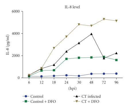
Level of IL-8 was significantly increased (P < 0.001) in culture supernatant of C. trachomatis infected cells compared to mock infected cells. Further significant increase in IL-8 level was observed in C. trachomatis infected cells in addition to iron chelator deferoxamine (DFO). In time dependent study significantly elevated level of IL-8 secretion was detected at 12 hpi, (P < 0.001) in C. trachomatis infected cells compared to control (mock infected Hela cells). Further in control cells two peaks were seen at 24 and 72 hpi through at 30 hpi representing a biphasic curve, whereas in C. trachomatis infected cells secreted IL-8 level represented a monophasic curve which showed consistently significantly increased levels from 12 hpi till 72 hpi (P < 0.001) (Figure 1).
Figure 1.
C. trachomatis infected HeLa 229 cells showing significantly (P < 0.001) increased levels of IL-8 as detected by ELISA in comparison to mock infected cells.
3.2. Secretion of IL-8 is Independent of Exogenous IL-1β and TNF-α in Chlamydia Infected Cells
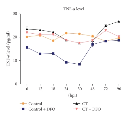
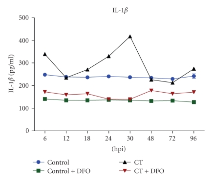
Temporal expression of IL-1β and TNF-α was assessed to ascertain the involvement of these cytokines in IL-8 induction. There was significant increase (P < 0.001) in level of secreted TNFα in culture supernatants during early (6, 12, and 18 hpi) and late phases (72 and 96 hpi) (Figure 2) whereas IL-1β levels were elevated at 30 hpi (Figure 3). However, at the midphase of C. trachomatis growth, level of secreted TNF-α declined, whereas IL-1β level did not show any deviation from basal level (Figures 2 and 3) and secretions of TNF-α, IL-1β, and IL-8 did not show synergy at different time points.
Figure 2.
TNF-α concentration as determined by ELISA in C. trachomatis infected culture supernatant was significantly (P < 0.001) increased at early time points (6, 12, and 18 hpi) and late time points (72 and 96 hpi), whereas there was decrease at midtime points (24, 30 and 48 hpi) in comparison to mock infected.
Figure 3.
Levels of IL-1β in supernatants of C. trachomatis infected culture were significantly decreased (P < 0.005) at early (6, 12, and 18 hpi) and late (72 and 96 hpi) time points in comparison to mock infected culture, whereas at midtime point no significant (P < 0.001) change was observed.
3.3. IL-8 Expression with Iron Chelator DFO and Mimosine in C. trachomatis Infected Hela Cells
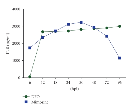
Mimosine, a plant based iron chelator, showed earlier induction of IL-8 at 6 hpi in C. trachomatis infected HeLa cells whereas in presence of DFO there was delayed (12 hpi) induction and was consistent at all time intervals (Figure 4). Gradual increase in IL-8 expression was detected, being highest at 30 hpi which subsequently declined at 48 hpi (Figure 4).
Figure 4.
Gradual increase was detected in levels of IL-8 in culture supernatants, highest at 30 hpi; thereafter, subsequent decrease was observed from 48 hpi onwords in C. trachomatis infected cells treated with Mimosine. DFO treated cells showed delayed secretion (12 hpi) but consistently elevated levels were detected at all the time point taken.
3.4. Chlamydia Induced IL-8 Production is Mediated by Reactive Nitrogen Species in Iron Restricted Condition (Evaluated at 48 hpi)
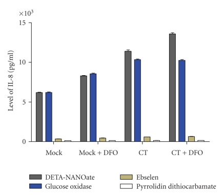
IL-8 production was significantly increased (P < 0.001) in C. trachomatis infected HeLa cells after stimulation with DETA-NANOate (Figure 5). Further significant increase (P < 0.001, Figure 5) was observed in IL-8 production in mock and CT infected cells upon co-stimulation with DFO and NO. However additive effect of DFO and NANOate was significantly higher (P < 0.005) in CT infected cells (Figure 5). Moreover glucose oxidase induced IL-8 production irrespective of CT infected or Mock (P < 0.005, Figure 5). Ebselen and PDTC decreased the production of IL-8 in all the conditions and significantly higher inhibition (P < 0.05, Figure 5) was observed with PDTC (Figure 5).
Figure 5.
IL-8 induction was significantly (P < 0.001) increased in C. trachomatis infected HeLa cells after stimulation with DETA-NANOate and there was synergic effect with DFO. Glucose oxidase treatment induced IL-8 production at same degree in both the C. trachomatis infected and Mock conditions (P < 0.005). Higher inhibition (P < 0.05) of IL-8 induction was observed after addition of nitric oxide specific inhibitor PDTC than ebselen.
4. Discussion
Chlamydial infections such as sexually transmitted or respiratory diseases occur frequently; however, course of these illnesses tends to be mild. Hence, they often pass unrecognized and untreated. These predominantly mild acute infections are considered as possible starting points of other chronic diseases, such as tubal inflammation with infertility, or reactive arthritis for C. trachomatis [25–27]. Disease outcome of Chlamydial infection is predominantly governed by mediators of inflammation and IL-8 leading the belligerence. Elevated level of IL-8 is dependent on chlamydial growth [7] and it is reported that in case of deferoxamine induced persistence, level of IL-8 remains higher [23]. These observations raise a question if chlamydia is directly involved in IL-8 induction or else intracellular level of iron is also a contributor?
In our study it was observed that in C. trachomatis infected HeLa 229 cells, level of secretory IL-8 increased significantly (P < 0.001) at 12 hpi till 72 hpi in comparison to mock infected cells showing thereby the probable involvement of early phase of chlamydial differentiation and metabolic activities responsible for induction and secretion of IL-8. This is in accordance with earlier study [28] wherein it has been shown that C. trachomatis and C. psittaci up regulate mRNA expression and secretion of the proinflammatory cytokines like IL-8, GROα, GM-CSF, and IL-6. Further IL-8 production in HeLa 229 cells infected with C. trachomatis was significantly delayed (from 24 hpi), unlike most invasive microorganisms that promote rapid but transient inflammatory response [7]. Elevated level of IL-8 might help epithelial cells at the mucosal surface to communicate with professional immune cells in response to pathogenic insult.
Further it was observed that IL-8 induction is independent of TNF-α and IL-1β in C. trachomatis infected HeLa 229 cells. Earlier studies had also suggested that TNF-α and IL-1β act as costimulatory signal for IL-8 induction in NF-κB dependent pathway [29]. Chlamydiae contains a tail serine protease (Tsp) that selectively cleaves the p65/RelA subunit of NF-κB to potentially interfere with host inflammatory response [30]. In a recent study lower level of TNF receptor 1 was observed on cell surface of chlamydia infected cells, inhibiting TNF mediated signaling [31] thereby enticing the possibility of TNF-α and IL1β independent expression of IL-8 in C. trachomatis infected HeLa 229 cells.
We also demonstrated that chlamydial infection acts synergistically with DFO to activate IL-8 secretion. Intracellular level of available iron has been shown to modulate inflammatory mediators and to regulate inflammatory process in several cell types. Two important signaling pathways represented by p38 and ERK1/2 are required for DFO-induced IL-8 secretion [18, 32–34]. IL-8 induced by C. trachomatis has been shown to be dependent on ERK and independent of p38 and Jun N-terminal MAPK [35] showing thereby that chlamydial infection and DFO follow the same course for IL-8 activation. However, further study of posttranscriptional mechanisms for DFO induced IL-8 involving p38 or ERK is warranted.
In our study DFO-induced IL-8 was increased by DETA-NANOate and H2O2 although extent of stimulation is less than that of NO in HeLa cells infected with C. trachomatis. It has been reported that NO is the initial factor in the signaling cascade that mediates IL-8 gene transcriptional activation by DFO. NF-κB is also an important transcriptional factor for IL-8 production in cancer cells. PDTC, one of the most effective inhibitors of NF-κB, inhibits the NO-dependent induction of the IL-8 gene [36]. It may be elucidating that NO plays important role in IL-8 induction in C. trachomatis infected cells treated with DFO.
In C. trachomatis infected cells, Mimosine treatment showed gradual increase and subsequent decrease in IL-8 expression. Conversely, delayed induction and consistent expression of IL-8 expression were observed with DFO. Thus synergy was observed only with DFO having bacterial origin, indicating that level of intracellular iron plays an important role in induction of proinflammatory responses. Further it may indicate towards involvement of iron sequestration mechanism of chlamydial origin and acquisition of iron from labile iron pool. Results of this study indicate the involvement of intracellular iron in activation of IL-8 in chlamydia infected HeLa 229 cells.
Acknowledgments
The authors wish to thank Indian Council of Medical Research (ICMR) India, for financial assistance to Harsh Vardhan in the form of fellowship and University Grant commission (UGC) India, for providing fellowship to Rishein Gupta, Hemchandra Jha and Apurb Rashmi Bhengraj. The study was funded by Indian Council of Medical Research.
References
- 1.Byrne GI, Ojcius DM. Chlamydia apoptosis: life and death decisions of an intracellular pathogen. Nature Reviews Microbiology. 2004;2(10):802–808. doi: 10.1038/nrmicro1007. [DOI] [PubMed] [Google Scholar]
- 2.Dautry-Varsat A, Balañá ME, Wyplosz B. Chlamydia-host cell interactions: recent advances on bacterial entry and intracellular development. Traffic. 2004;5(8):561–570. doi: 10.1111/j.1398-9219.2004.00207.x. [DOI] [PubMed] [Google Scholar]
- 3.Vats V, Rastogi S, Kumar A, et al. Detection of Chlamydia trachomatis by polymerase chain reaction in male patients with non-gonococcal urethritis attending an STD clinic. Sexually Transmitted Infections. 2004;80(4):327–328. doi: 10.1136/sti.2003.008839. [DOI] [PMC free article] [PubMed] [Google Scholar]
- 4.Singh V, Rastogi S, Garg S, et al. Polymerase chain reaction for detection of endocervical Chlamydia trachomatis infection in women attending a gynecology outpatient department in India. Acta Cytologica. 2002;46(3):540–544. doi: 10.1159/000326874. [DOI] [PubMed] [Google Scholar]
- 5.Hackstadt T. Cell biology. In: Stephens RS, editor. Chlamydia: Intracellular Biology, Pathogenesis, and Immunity. Washington, DC, USA: ASM Press; 1999. pp. 101–138. [Google Scholar]
- 6.Dessus-Babus S, Knight ST, Wyrick PB. Chlamydial infection of polarized HeLa cells induces PMN chemotaxis but the cytokine profile varies between disseminating and non-disseminating strains. Cellular Microbiology. 2000;2(4):317–327. doi: 10.1046/j.1462-5822.2000.00058.x. [DOI] [PubMed] [Google Scholar]
- 7.Rasmussen SJ, Eckmann L, Quayle AJ, et al. Secretion of proinflammatory cytokines by epithelial cells in response to Chlamydia infection suggests a central role for epithelial cells in chlamydial pathogenesis. The Journal of Clinical Investigation. 1997;99(1):77–87. doi: 10.1172/JCI119136. [DOI] [PMC free article] [PubMed] [Google Scholar]
- 8.Stephens RS. The cellular paradigm of chlamydial pathogenesis. Trends in Microbiology. 2003;11(1):44–51. doi: 10.1016/s0966-842x(02)00011-2. [DOI] [PubMed] [Google Scholar]
- 9.Patton DL, Kuo C-C. Histopathology of Chlamydia trachomatis salpingitis after primary and repeated reinfections in the monkey subcutaneous pocket model. Journal of Reproduction and Fertility. 1989;85(2):647–656. doi: 10.1530/jrf.0.0850647. [DOI] [PubMed] [Google Scholar]
- 10.Ward ME. Mechanisms of Chlamydia-induced disease. In: Stephens RS, editor. Chlamydia: Intracellular Biology, Pathogenesis, and Immunity. Washington, DC, USA: ASM Press; 1999. pp. 171–210. [Google Scholar]
- 11.Moreillon P, Majcherczyk PA. Proinflammatory activity of cell-wall constituents from gram-positive bacteria. Scandinavian Journal of Infectious Diseases. 2003;35(9):632–641. doi: 10.1080/00365540310016259. [DOI] [PubMed] [Google Scholar]
- 12.Wylie JL, Hatch GM, McClarty G. Host cell phospholipids are trafficked to and then modified by Chlamydia trachomatis . Journal of Bacteriology. 1997;179(23):7233–7242. doi: 10.1128/jb.179.23.7233-7242.1997. [DOI] [PMC free article] [PubMed] [Google Scholar]
- 13.Ulevitch RJ, Tobias PS. Recognition of gram-negative bacteria and endotoxin by the innate immune system. Current Opinion in Immunology. 1999;11(1):19–22. doi: 10.1016/s0952-7915(99)80004-1. [DOI] [PubMed] [Google Scholar]
- 14.Heine H, Müller-Loennies S, Brade L, Lindner B, Brade H. Endotoxic activity and chemical structure of lipopolysaccharides from Chlamydia trachomatis serotypes E and L2 and Chlamydophila psittaci 6BC. European Journal of Biochemistry. 2003;270(3):440–450. doi: 10.1046/j.1432-1033.2003.03392.x. [DOI] [PubMed] [Google Scholar]
- 15.Ingalls RR, Rice PA, Qureshi N, Takayama K, Lin JS, Golenbock DT. The inflammatory cytokine response to Chlamydia trachomatis infection is endotoxin mediated. Infection and Immunity. 1995;63(8):3125–3130. doi: 10.1128/iai.63.8.3125-3130.1995. [DOI] [PMC free article] [PubMed] [Google Scholar]
- 16.Rupp J, Kothe H, Mueller A, Maass M, Dalhoff K. Imbalanced secretion of IL-1β and IL-1RA in Chlamydia pneumoniae-infected mononuclear cells from COPD patients. European Respiratory Journal. 2003;22(2):274–279. doi: 10.1183/09031936.03.00007303. [DOI] [PubMed] [Google Scholar]
- 17.Kasahara K, Strieter RM, Standiford TJ, Kunkel SL. Adherence in combination with lipopolysaccharide, tumor necrosis factor or interleukin-1β potentiates the induction of monocyte-derived interleukin-8. Pathobiology. 1993;61(2):57–66. doi: 10.1159/000163762. [DOI] [PubMed] [Google Scholar]
- 18.Choi E-Y, Kim E-C, Oh H-M, et al. Iron chelator triggers inflammatory signals in human intestinal epithelial cells: involvement of p38 and extracellular signal-regulated kinase signaling pathways. The Journal of Immunology. 2004;172(11):7069–7077. doi: 10.4049/jimmunol.172.11.7069. [DOI] [PubMed] [Google Scholar]
- 19.Raulston JE. Response of Chlamydia trachomatis serovar E to iron restriction vitro and evidence for iron-regulated chlamydial proteins. Infection and Immunity. 1997;65(11):4539–4547. doi: 10.1128/iai.65.11.4539-4547.1997. [DOI] [PMC free article] [PubMed] [Google Scholar]
- 20.Ramsey KH, Miranpuri GS, Sigar IM, Ouellette S, Byrne GI. Chlamydia trachomatis persistence in the female mouse genital tract: inducible nitric oxide synthase and infection outcome. Infection and Immunity. 2001;69(8):5131–5137. doi: 10.1128/IAI.69.8.5131-5137.2001. [DOI] [PMC free article] [PubMed] [Google Scholar]
- 21.Matsumoto A, Manire GP. Electron microscopic observations on the effects of penicillin on the morphology of Chlamydia psittaci . Journal of Bacteriology. 1970;101(1):278–285. doi: 10.1128/jb.101.1.278-285.1970. [DOI] [PMC free article] [PubMed] [Google Scholar]
- 22.Pantoja LG, Miller RD, Ramirez JA, Molestina RE, Summersgill JT. Characterization of Chlamydia pneumoniae persistence in HEp-2 cells treated with gamma interferon. Infection and Immunity. 2001;69(12):7927–7932. doi: 10.1128/IAI.69.12.7927-7932.2001. [DOI] [PMC free article] [PubMed] [Google Scholar]
- 23.Peters J, Hess S, Endlich K, et al. Silencing or permanent activation: host-cell responses in models of persistent Chlamydia pneumoniae infection. Cellular Microbiology. 2005;7(8):1099–1108. doi: 10.1111/j.1462-5822.2005.00534.x. [DOI] [PubMed] [Google Scholar]
- 24.Scidmore MA. Current Protocols in Microbiology. chapter 11, Unit 11A.1. New York, NY, USA: John Wiley & Sons; 2005. Cultivation and laboratory maintenance of Chlamydia trachomatis . [DOI] [PubMed] [Google Scholar]
- 25.Pal S, Hui W, Peterson EM, de la Maza LM. Factors influencing the induction of infertility in a mouse model of Chlamydia trachomatis ascending genital tract infection. Journal of Medical Microbiology. 1998;47(7):599–605. doi: 10.1099/00222615-47-7-599. [DOI] [PubMed] [Google Scholar]
- 26.Mårdh P-A. Tubal factor infertility, with special regard to chlamydial salpingitis. Current Opinion in Infectious Diseases. 2004;17(1):49–52. doi: 10.1097/00001432-200402000-00010. [DOI] [PubMed] [Google Scholar]
- 27.Zeidler H, Kuipers J, Köhler L. Chlamydia-induced arthritis. Current Opinion in Rheumatology. 2004;16(4):380–392. doi: 10.1097/01.bor.0000126150.04251.f9. [DOI] [PubMed] [Google Scholar]
- 28.Quayle AJ. The innate and early immune response to pathogen challenge in the female genital tract and the pivotal role of epithelial cells. Journal of Reproductive Immunology. 2002;57(1-2):61–79. doi: 10.1016/s0165-0378(02)00019-0. [DOI] [PubMed] [Google Scholar]
- 29.Chaly YV, Selvan RS, Fegeding KV, Kolesnikova TS, Voitenok NN. Expression of IL-8 gene in human monocytes and lymphocytes: differential regulation by TNF and IL-1. Cytokine. 2000;12(6):636–643. doi: 10.1006/cyto.1999.0664. [DOI] [PubMed] [Google Scholar]
- 30.Lad SP, Li J, da Silva Correia J, et al. Cleavage of p65/RelA of the NF-κB pathway by Chlamydia . Proceedings of the National Academy of Sciences of the United States of America. 2007;104(8):2933–2938. doi: 10.1073/pnas.0608393104. [DOI] [PMC free article] [PubMed] [Google Scholar]
- 31.Paland N, Böhme L, Gurumurthy RK, Mäurer A, Szczepek AJ, Rudel T. Reduced display of tumor necrosis factor receptor I at the host cell surface supports infection with Chlamydia trachomatis . The Journal of Biological Chemistry. 2008;283(10):6438–6448. doi: 10.1074/jbc.M708422200. [DOI] [PubMed] [Google Scholar]
- 32.Weiss G, Fuchs D, Hausen A, et al. Iron modulates interferon-gamma effects in the human myelomonocytic cell line THP-1. Experimental Hematology. 1992;20(5):605–610. [PubMed] [Google Scholar]
- 33.O'Brien-Ladner AR, Blumer BM, Wesselius LJ. Differential regulation of human alveolar macrophage-derived interleukin-1β and tumor necrosis factor-α by iron. Journal of Laboratory and Clinical Medicine. 1998;132(6):497–506. doi: 10.1016/s0022-2143(98)90128-7. [DOI] [PubMed] [Google Scholar]
- 34.Golding S, Young SP. Iron requirements of human lymphocytes: relative contributions of intra- and extra-cellular iron. Scandinavian Journal of Immunology. 1995;41(3):229–236. doi: 10.1111/j.1365-3083.1995.tb03558.x. [DOI] [PubMed] [Google Scholar]
- 35.Buchholz KR, Stephens RS. The extracellular signal-regulated kinase/mitogen-activated protein kinase pathway induces the inflammatory factor interleukin-8 following Chlamydia trachomatis infection. Infection and Immunity. 2007;75(12):5924–5929. doi: 10.1128/IAI.01029-07. [DOI] [PMC free article] [PubMed] [Google Scholar]
- 36.Turpaev K, Litvinov D, Justesen J. Redox modulation of NO-dependent induction of interleukin 8 gene in monocytic U937 cells. Cytokine. 2003;23(1-2):15–22. doi: 10.1016/s1043-4666(03)00114-5. [DOI] [PubMed] [Google Scholar]


