Abstract
Fractures of the coronoid process of the ulna generally occur in relatively high-energy injuries and are commonly associated with injuries to other structures around the elbow. Damage to the coronoid process in addition to other elbow structures may complicate treatment. Several approaches have been used in the management of coronoid process fractures. This paper reports a method of coronoid process fracture fixation using suture anchors.
Keywords: Coronoid fracture, Suture anchor
Introduction
Fractures of the coronoid process of the ulna generally occur in relatively high-energy injuries and are commonly associated with injuries to other structures around the elbow. The term “terrible triad” refers to a combination of an elbow dislocation with fractures of the radial head and the coronoid process, first coined by Hotchkiss [4]. Damage to the coronoid process in addition to other elbow structures may complicate treatment. One study found that coronoid process fractures were present in 80% of elbow dislocations with radial head fractures and that patients with such injuries had the most frequent injury referred for delayed management [24].
Recurrent instability is common in patients with a posterior dislocation of the elbow and concomitant radial head and coronoid fractures [5, 11, 18], as is radiographic evidence of osteoarthrosis [5]. Treatment of the coronoid process and the anterior elbow capsule may be important for postoperative elbow stability [1, 9–11, 13, 15, 16, 20, 22]. Several approaches have been used in the management of coronoid process fractures. This paper reports a method of coronoid process fracture fixation using suture anchors. While suture anchors have been proposed for supplemental fixation of comminuted coronoid fractures [10], the authors are not aware of any articles discussing the use of suture anchors as the primary means of fixation of coronoid fractures.
Materials and Methods
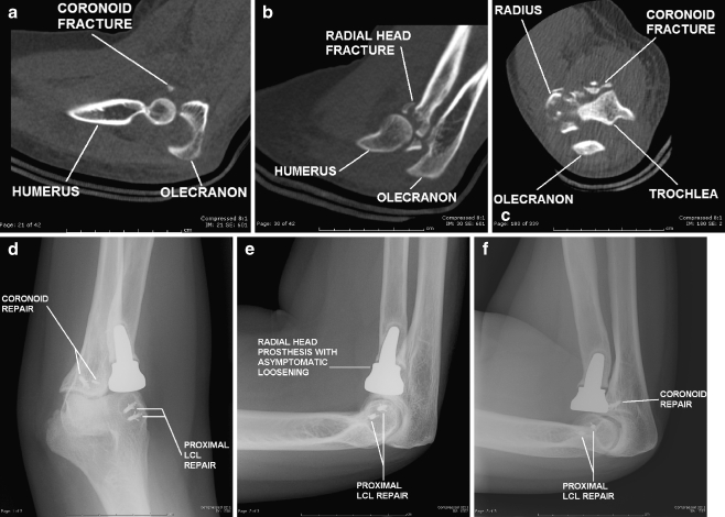
Three patients (aged 30 to 59 years) were referred for terrible triad injuries between August 2006 and September 2007. Two of the patients were female, while one was male. All of the patients were right-hand dominant, and injuries occurred in the left elbow in two patients and in the right elbow in one patient. The mechanisms of injury were a fall from a motorcycle, a fall down stairs, and a fall while walking, respectively. Initial radiographic imaging revealed posterior elbow dislocation with radial head and coronoid process fractures (Fig. 1) in two patients. Both of these patients were initially treated in the Emergency Department with closed reduction and splinting, and both demonstrated radiographic instability in the splint on initial presentation in the office. Initial radiographs of the third patient revealed fractures of the radial head, coronoid process, and lateral epicondyle.
Figure 1.
Imaging of a representative case: a 30-year-old right-hand dominant woman seen after a motorcycle accident. a Preoperative sagittal computed tomography (CT) scan demonstrating ulnohumeral subluxation with a coronoid fracture. b Preoperative sagittal CT scan demonstrating ulnohumeral subluxation with a comminuted radial head fracture. c Preoperative axial CT scan demonstrating a coronoid fracture with a comminuted radial head fracture and ulnohumeral subluxation. d Postoperative 22-month radiograph, anteroposterior view. e Postoperative 22-month radiograph, lateral view demonstrating asymptomatic loosening of the radial head implant. f Postoperative 22-month radiograph, radial head view demonstrating asymptomatic loosening of the radial head implant.
The patients underwent operative intervention between 5 and 16 days after the initial injury. All of the patients were noted to have comminuted radial head fractures that could not be reconstructed, and all therefore underwent radial head replacement. The coronoid injury was repaired using suture anchors in all three patients, with the sutures passing through the fracture fragment and the anterior elbow capsule in all cases. Injury to the lateral collateral ligament (LCL) was repaired using suture anchors in both patients with elbow dislocation as well as in the patient with the lateral epicondyle fracture.
Surgical Technique
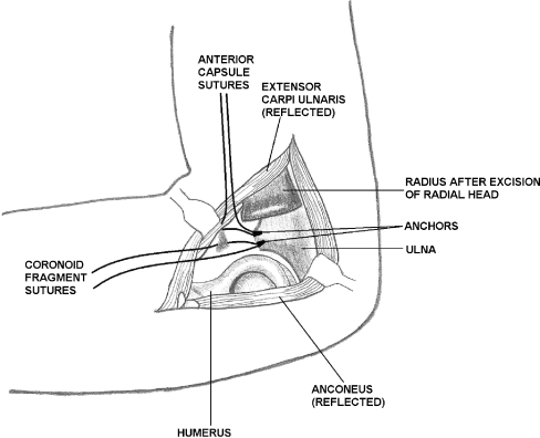
Surgical dissection is performed to allow for adequate exposure of the anterior aspect of the ulna, including the coronoid fracture fragment and the trochlear notch. In our cases, a posterolateral approach was used to dissect between the extensor carpi ulnaris and the anconeus (Fig. 2). Other approaches may, however, be used according to the nature of the injury. We found that the LCL was disrupted from the lateral epicondyle, and this injury facilitated access to the joint. Incision of the joint capsule revealed comminuted radial head fragments that were excised, allowing visualization of the coronoid fracture. Subperiosteal dissection along the anterior distal humerus may also allow further exposure of the ulnohumeral joint, facilitating debridement of loose bodies.
Figure 2.
Diagram of elbow joint from lateral side after excision of radial head showing placement of suture anchors for repair of the coronoid fracture and anterior elbow capsule. The injured lateral collateral ligament is not shown.
For larger fractures, an attempt at reduction of the fragment with a dental pick may be performed to assess the adequacy of bony stability as well as to plan the placement of the suture anchor(s). The drill guide is then placed in the fracture bed at the appropriately oriented angle, and the pilot hole for the initial suture anchor is made. The suture anchor is then placed into the hole in the standard fashion, and the fixation is tested to ensure no pull-out. The suture may then be passed through or around the fracture fragment if it is large enough or through the anterior capsule if the fracture is too small or comminuted. Two to three anchors are placed in this fashion along the breadth of the coronoid fracture base, with sutures incorporating the fracture fragment and anterior elbow capsule from medial to lateral. Additional suture anchors may be placed and the sutures passed, depending on the amount of fixation that is required. Once all of the suture anchors are placed, the sutures are tied sequentially from deep to superficial. Fixation may then be checked with gentle range of motion of the elbow. In our cases, immediate improvement of elbow alignment and stability was noted after repair of the coronoid injury.
Additional procedures, such as radial head management or collateral ligament repair, are then performed as necessary for elbow stability. Of note, if a radial head replacement is to be used, preparation of the radial shaft may follow placement of the suture anchors used for coronoid repair but should precede tying of the coronoid suture anchors. The wound is then irrigated and closed in layers and dressings applied. A posterior splint is then applied with the elbow in 90° of flexion.
Results
Follow-up in the three patients ranged from 7 to 22 months. One patient had obtained active range of motion from 25° to 135° of flexion, with full pronation and supination by the most recent 22-month postoperative visit. This patient developed postoperative recurrence of cubital tunnel syndrome for which she had previously been treated 5 years prior to her injury. She subsequently underwent left cubital tunnel release and ulnar nerve transposition and had an uncomplicated postoperative course. A second patient had obtained active range of motion of the elbow from 30° of flexion to 120° of flexion, with 70° of supination and 80° of pronation by the most recent 7-month postoperative visit. This patient had an uneventful postoperative course. The third patient had obtained active range of motion from 30° to 100° of flexion, with 80° of pronation and supination by the 8-month postoperative visit. This patient developed ulnar neuropathy postoperatively. She underwent left cubital tunnel release with elbow capsulectomy and release 9 months postoperatively and subsequently had improved active range of motion from 15° to 110° of flexion.
Radiographs revealed maintenance of concentric reduction (Fig. 1) in all three patients. Asymptomatic loosening of the radial head implant was noted in one patient, while mild heterotopic ossification was noted in another. Assessment of coronoid fracture healing was not possible due to overlap of the radial head implants on the lateral radiographs.
Discussion
This paper presents a novel approach to fixation of coronoid fractures with suture anchors. This approach has been used in three patients with terrible triad injuries to date. One patient had a small fracture of the coronoid tip, while the other two patients had larger fracture fragments. In all cases, the coronoid fragments were approached from the lateral aspect of the elbow after removal of a comminuted radial head fracture. None of the patients required a medial-sided repair or an external fixator, although LCL repair was necessary. We believe that this approach to fixation of coronoid fractures is simpler than more traditional approaches because less dissection is needed and because the required instrumentation is less complicated.
Repair of coronoid fractures has been described from lateral, medial [8, 10, 11, 15, 16, 22], posterior [7, 10–12, 15, 22], and anterior [21] approaches, as well as a combination of these [17]. Treatment of transverse fractures of the coronoid tip in the case of terrible triad injuries can be achieved through a lateral exposure by using a small extension of the existing capsuloligamentous and muscular damage and retracting the radial head out of the way [2, 7, 10–12, 14–16, 22]. If necessary for additional access to the coronoid, anterolateral dissection may be performed by elevating the wrist and common digital extensors as well as the supinator [16]. In our patients, we found that adequate access was achieved after removal of the radial head fragments, and additional anteromedial or anterolateral dissection was not required. Suture anchors may also be placed via isolated anterior or anteromedial approaches if necessary.
Multiple means of fixation of coronoid fractures have been proposed, depending on the nature of the fracture. Small coronoid tip fractures or anteromedial facet fractures may be repaired with suture fixation either alone or supplemented with one to two screws [2, 14–16, 22]. Various techniques have also been described for osteosynthesis of larger fractures. Acute treatment may consist of fixation with sutures through drill holes, threaded K-wires, cannulated screw fixation, plate and screw fixation, or buttress plating, all performed through a medial or posterior approach [2, 3, 7, 9, 10, 12, 14–18, 22]. In cases of significant comminution, supplemental fixation may be obtained using additional screws, a second plate, suture, threaded wires, or suture anchors [2, 9, 10, 14]. Comminuted Regan and Morrey type II fractures that are too fragmented to allow for stable internal fixation may also be fixed by lasso-type sutures in a similar manner [12]. Coronoid fractures that are not amenable to fixation (because of size, comminution, or delay in treatment) may be reconstructed or treated non-operatively [3, 6–8, 11, 12, 14, 16, 19, 23]. Seijas et al., however, reported a case of persistent ulnohumeral subluxation requiring joint transfixation after excision of coronoid fragments in a terrible triad injury [20]. Other authors recommend against excision of bone fragments as they may be useful for callus formation [9]. In the case of the small tip fracture of our patient, repair of the coronoid fragment was found to improve both the reduction and the stability of the elbow. Further improvement in the stability of the elbow was obtained by radial head replacement and LCL repair.
The technique of suture fixation of tip fractures involves passing sutures or wires through drill holes in the ulna to capture either the coronoid fragment itself or its anterior capsular attachment [10, 16, 18]. These sutures may be placed through drill holes in the fracture fragment when it is large enough or used to engage the capsular attachment otherwise [7, 10–12, 14, 15, 22]. The sutures are then secured to the proximal ulna through drill holes from the dorsal surface to the bed of the coronoid fracture near the joint using a Keith needle, suture passers, or some similar device [7, 11, 12, 14, 15, 22]. The repair of coronoid fractures with suture anchors as outlined above provides an additional technique in management of these demanding injuries. Suture anchors may be used, for example, to secure the anterior capsule to the remaining coronoid as an alternative to fragment excision. Some authors suggest that the sutures not be tied until the radial head is treated due to the likely need for subluxation of the elbow joint during radial head treatment and that repair of the coronoid should also precede repair of the LCL [7, 14]. We were able to tie the suture anchors after radial shaft preparation but before placement of the prosthesis in our patients as we felt that this repair would be stable to elbow manipulation during radial head placement. This allowed us to reconstruct the elbow progressively from deep to superficial without returning to address a component of the injury.
In summary, there are many approaches to management and fixation of coronoid fractures. This paper presents a method of primary fixation of coronoid process fractures and anterior joint capsule injuries by using suture anchors. This method may be particularly useful in the small coronoid process fractures, comminuted fractures, or osteoarticular fragments with little bone. The suture anchors may be placed through the exposure provided by a radial head fracture, obviating the need for additional exposure of the posterior aspect of the ulna for traditional suture fixation through drill holes and leading to less tissue trauma as well as better cosmesis and shorter operative time. The use of suture anchors also precludes the need for suture passers, Keith needles, or other such instrumentation that is required for traditional suture technique, thereby making it faster and easier. The technique of suture anchors may therefore be used to expand the repertoire of methods for fixation of coronoid fractures.
Contributor Information
Sylvan E. Clarke, Phone: +1-215-4567900, FAX: +1-215-3242426, Email: clarkes@einstein.edu
Sue Y. Lee, Phone: +1-215-4567903, FAX: +1-215-3242426, Email: LeeSu@einstein.edu
James R. Raphael, Phone: +1-215-4566759, FAX: +1-215-3242426, Email: RaphaelJ@einstein.edu
References
- 1.Cage DJ, Abrams RA, Callahan JJ, et al. Soft tissue attachments of the ulnar coronoid process. an anatomic study with radiographic correlation. Clin Orthop Relat Res. 1995;320:154–8. [PubMed]
- 2.Doornberg JN, Ring D. Coronoid fracture patterns. J Hand Surg [Am]. 2006;31:45–52. doi:10.1016/j.jhsa.2005.08.014. [DOI] [PubMed]
- 3.Doornberg JN, Ring DC. Fracture of the anteromedial facet of the coronoid process. J Bone Joint Surg Am. 2006;88:2216–24. doi:10.2106/JBJS.E.01127. [DOI] [PubMed]
- 4.Hotchkiss RN. Fractures and dislocations of the elbow. In: Rockwood CA Jr, Green DP, Bucholz RW, et al. editors. Rockwood and green’s fractures in adults. 4th ed. Philadelphia: Lippincott-Raven; 1996. p. 980–1.
- 5.Josefsson PO, Gentz CF, Johnell O, et al. Dislocations of the elbow and intraarticular fractures. Clin Orthop Relat Res. 1989;246:126–30. [PubMed]
- 6.Kohls-Gatzoulis J, Tsiridis E, Schizas C. Reconstruction of the coronoid process with iliac crest bone graft. J Shoulder Elbow Surg. 2004;13:217–20. doi:10.1016/j.jse.2003.12.003. [DOI] [PubMed]
- 7.McKee MD, Pugh DM, Wild LM, et al. Standard surgical protocol to treat elbow dislocations with radial head and coronoid fractures. surgical technique. J Bone Joint Surg Am. 2005;87(Suppl 1):22–32. doi:10.2106/JBJS.D.02933. [DOI] [PubMed]
- 8.Moritomo H, Tada K, Yoshida T, et al. Reconstruction of the coronoid for chronic dislocation of the elbow. use of a graft from the olecranon in two cases. J Bone Joint Surg Br. 1998;80:490–2. doi:10.1302/0301-620X.80B3.8328. [DOI] [PubMed]
- 9.Morrey BF. Complex instability of the elbow. Instr Course Lect. 1998;47:157–64. [PubMed]
- 10.O’Driscoll SW, Jupiter JB, Cohen MS, et al. Difficult elbow fractures: pearls and pitfalls. Instr Course Lect. 2003;52:113–34. [PubMed]
- 11.O’Driscoll SW, Jupiter JB, King GJ, et al. The unstable elbow. Instr Course Lect. 2001;50:89–102. [PubMed]
- 12.Pugh DM, Wild LM, Schemitsch EH, et al. Standard surgical protocol to treat elbow dislocations with radial head and coronoid fractures. J Bone Joint Surg Am. 2004;86-A:1122–30. [DOI] [PubMed]
- 13.Regan W, Morrey B. Fractures of the coronoid process of the ulna. J Bone Joint Surg Am. 1989;71:1348–54. [PubMed]
- 14.Ring D. Fractures of the coronoid process of the ulna. J Hand Surg [Am]. 2006;31:1679–89. doi:10.1016/j.jhsa.2006.08.020. [DOI] [PubMed]
- 15.Ring D, Jupiter JB. Fracture-dislocation of the elbow. J Bone Joint Surg Am. 1998;80:566–80. doi:10.1302/0301-620X.80B4.9165. [DOI] [PubMed]
- 16.Ring D, Jupiter JB. Reconstruction of posttraumatic elbow instability. Clin Orthop Relat Res. 2000;370:44–56. doi:10.1097/00003086-200001000-00006. [DOI] [PubMed]
- 17.Ring D, Jupiter JB, Sanders RW, et al. Transolecranon fracture-dislocation of the elbow. J Orthop Trauma. 1997;11:545–50. doi:10.1097/00005131-199711000-00001. [DOI] [PubMed]
- 18.Ring D, Jupiter JB, Zilberfarb J. Posterior dislocation of the elbow with fractures of the radial head and coronoid. J Bone Joint Surg Am. 2002;84-A:547–51. [DOI] [PubMed]
- 19.Schneeberger AG, Sadowski MM, Jacob HA. Coronoid process and radial head as posterolateral rotatory stabilizers of the elbow. J Bone Joint Surg Am. 2004;86-A:975–82. [DOI] [PubMed]
- 20.Seijas R, Joshi N, Hernandez A, et al. Terrible triad of the elbow—role of the coronoid process: a case report. J Orthop Surg (Hong Kong). 2005;13:296–9. [DOI] [PubMed]
- 21.Selesnick FH, Dolitsky B, Haskell SS. Fracture of the coronoid process requiring open reduction with internal fixation. a case report. J Bone Joint Surg Am. 1984;66:1304–6. [PubMed]
- 22.Tashjian RZ, Katarincic JA. Complex elbow instability. J Am Acad Orthop Surg. 2006;14:278–86. [DOI] [PubMed]
- 23.van Riet RP, Morrey BF, O’Driscoll SW. Use of osteochondral bone graft in coronoid fractures. J Shoulder Elbow Surg. 2005;14:519–23. doi:10.1016/j.jse.2004.11.007. [DOI] [PubMed]
- 24.van Riet RP, Morrey BF, O’Driscoll SW, et al. Associated injuries complicating radial head fractures: a demographic study. Clin Orthop Relat Res. 2005;441:351–5. doi:10.1097/01.blo.0000180606.30981.78. [DOI] [PubMed]


