Abstract
We report a case of Mycobacterium smegmatis granuloma in the soft tissues of the first web space of the left hand in a 67-year-old Caucasian woman. She was in good systematic health; there was no recollection of trauma to the hand. A combined regime of prolonged antibiotic therapy and surgical debridement was necessary to ultimately eradicate the infection. The natural history, microbiology, and treatment of this rare hand infection are discussed.
Keywords: Hand, Granuloma, Doxycyline, Mycobacterium smegmatis
Patient Report
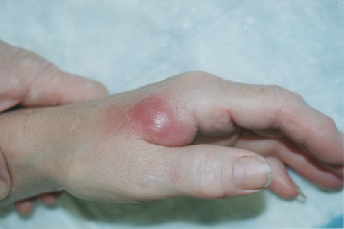
A 67-year-old Caucasian woman presented with a tender inflammatory mass measuring 2 cm in diameter located in the first web space of her non-dominant left hand (Fig. 1). She was in good systemic health, with no known immunocompromising disease. The patient had no recollection of any trauma to her hand, even minor. The only identified factor in her history that suggested a possible etiology was that she tended the gardens around her home. The infection did not respond to oral Cephalexin (Keflex—E. Lilly, Canada) therapy. She was taken to surgery; an elliptical skin excision was made to reveal a small amount of violaceous subcutaneous pus and an underlying mass of rubbery and spongy tissue, grayish brown in color. Frozen section analysis indicated a granulomatous reaction. The mass was resected in its entirety, and specimens were sent for culture and sensitivity analysis, including mycobacteria. Microscopic examination subsequently did not find any acid-fast bacilli.
Figure 1.
Clinical presentation of left hand.
Two weeks later, the surgical area exhibited low grade inflammation, with erythema, tenderness, and swelling. After discussing the case with an infectious disease specialist, the patient was placed empirically on 100 mg bid of doxycycline (Vibramycin—Pfizer, Canada). At 1 month, the Provincial Health Laboratory reported the growth of Mycobacterium smegmatis. The surgical area was improving, but due to financial reasons the patient was switched to oral ciprofloxacin (Cipro—Bayer, Canada) and trimethoprim-sulfamethoxazole (Bactrim—Roche, Canada).
Despite continuing on these two medications, the mass recurred over the next several weeks. Financial aid was sought and, at 9 months, was obtained to allow the patient to restart on doxycycline. However, the mass had progressed to 1 cm in diameter, and a decision was made to resect it a second time, which was completed in the tenth month. The second pathology report was identical to the first, i.e., granuloma. Again, acid-fast bacilli were not seen and, in this case, the culture was negative.
The surgical site healed uneventfully on the doxycycline and therapy was discontinued after 10 weeks. No further recurrence developed, and the patient had normal hand function.
Discussion
Mycobacterial diseases in humans are most commonly due to mycobacterium tuberculosis and mycobacterium leprae. Other mycobacterium infrequently cause human disease; although rare, they are nevertheless important pathogens. M. smegmatis was first recognized to be a human pathogen by Vonmoos et al. [2] in 1986. Since then, 25 additional cases of have been reported, 56–76% of which were skin or soft-tissue infections [2]. As a cause of human disease, M. smegmatis is second to Mycobacterium fortuitum, and can be found in normal human-genital secretions, as well as in lower animals, soil, dust, and water. Disseminated infections caused by M. smegmatis are commonly related to immunosuppression [4]. Clinical infections are often found to be a result of contaminated surgical or other wounds, caused by the use of contaminated solutions or lipid creams that act as carriers of the disease [4], as well as exit site infections, tunnel or pocket infections, and catheter-related infections [1].
In 1884, Lustgarten initially isolated M. smegmatis from syphilitic chancres. Subsequently, Alvarez and Travel gathered the fast-growing bacteria from normal human-genital secretions (smegma) one year later [2]. Classified together with other rapid growers M. fortuitum, M. abscessus, and M. chelonei, M. smegmatis falls into the Runyon group IV of medically important mycobacteria [4]. Isolates of this bacteria are similar to M. fortuitum, except for a negative 3-day arylsulfatase test; growth at 43°C–45°C; a low semiquantitative catalase test; and, in 50% of isolates, a late-developing, yellow-to-orange pigment. The bacteria are non-motile slender rods with branching or Y shapes, and are intrinsically resistant against many antibiotics due to their protective outer-lipid bilayer. While serving as an effective permeability barrier, the membrane also contains porins which mediate the diffusion of hydrophilic nutrients and serve functions analogous to those observed in Gram-negative bacteria [3].
The most effective treatment for M. smegmatis infections of skin, soft tissue, and bone is a combined regime of prolonged antibiotic therapy and surgical debridement. Although M. smegmatis is resistant to isoniazid and rifampin, it is susceptible to ethambutol. While cefoxitin and cefmetazole have modest antimicrobial activity, all the isolates reported to date have been susceptible or moderately susceptible to imipenem, as well as non-B-lactam drugs including doxycycline, sulfamthoxazole, amikacin, and ciprofloxacin. Of these, doxycycline has the lowest MIC measuring <0.25 at MIC50 and MIC90 [5]. Repeat surgical interventions in conjunction with the antibiotic treatment may be necessary for full eradication of infection.
Conclusions
Reviewing this case, the following observations are pertinent. Initial therapy with an oral B-lactam antibiotic (Cephalexin) proved unsuccessful; the high MIC’s reported for B-lactam antibiotics for M. smegmatis may explain this ineffectiveness. After the first surgery, the patient showed progressive improvement on doxycycline; however, financial constraints forced a change to oral ciprofloxacin and trimethoprim-sulfamethoxazole. This was followed by a gradual recurrence of the disease, inferring an ineffectiveness of this therapy. A second surgery combined with post-operative doxycycline for 10 weeks affected a cure, suggesting, at least in this particular case of M. smegmatis bacterial infection, that doxycycline was the drug of choice.
Acknowledgments
The authors wish to thank Dr. Rotstein-Coleman an infectious disease specialist at the Hamilton Health Sciences Center in Hamilton, Ontario for his clinical guidance in the management of this case.
References
- 1.Hong T, Butler WR, Hollis F, Floyd MM, Toney SR, Tang YW, et al. Characterization of a novel rapidly growing Mycobacterium species associated with sepsis. J Clin Microbiol. 2003;41(12):5650–3. [DOI] [PMC free article] [PubMed]
- 2.Newton JA Jr, Weiss PJ, Bowler WA, Oldfield EC 3rd. Soft-tissue infection due to Mycobacterium smegmatis: report of two cases. Clin Infect Dis. 1993;16(4):531–3. [DOI] [PubMed]
- 3.Niederweis M. Mycobacterial porins—new channel proteins in unique outer membranes. Molecular Microbiology. 2003;49(5):1167–1177(11). Available at: (http://www.ingentaconnect.com/content/bsc/mole/2003/00000049/00000005/art00003). [DOI] [PubMed]
- 4.Pennekamp A, Pfyffer GE, Wuest J, George CA, Ruef C. Mycobacterium smegmatis infection in a healthy woman following a facelift: case report and review of the literature. Ann Plast Surg. 1997;39(1):80–3. [DOI] [PubMed]
- 5.Wallace RJ Jr, Nash DR, Tsukamura M, Blacklock ZM, Silcox VA. Human disease due to Mycobacterium smegmatis. J Infect Dis. 1988;158(1):52–9. [DOI] [PubMed]


