Abstract
A case of osteoid osteoma of the scaphoid presenting as painful monoarticular arthritis is presented. Degenerative arthritis, associated with osteoid osteoma of the carpus, has not been described. The implications for treatment are discussed.
Keywords: Osteoid osteoma, Scaphoid, Arthritis
Introduction
Osteoid osteoma accounts for 10% of benign bone tumors. The vast majority of these occur in the appendicular skeleton with half occurring in the femur and tibia. There is a nearly 2:1 predominance in women, and the tumor is uncommon after the age of 30 [6]. Only 2% of all osteoid osteomas involve the carpus, and their rarity may make diagnosis challenging. Indeed, a review of the literature reveals scattered case reports of osteoid osteoma of the scaphoid [1–16]. We present a case report of a young woman with osteoid osteoma of her scaphoid who presented with marked radiographic and clinical evidence of radiocarpal degenerative arthritis.
Case Report
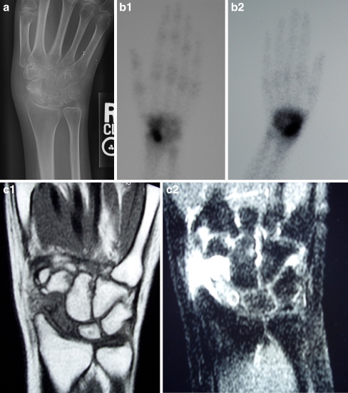
A 16-year-old woman presented with a 19-month history of worsening pain and swelling in her right hand and wrist. She denied prior trauma. She initially described very marked relief of symptoms with the use of aspirin and ibuprofen, although this efficacy waned over the last 6 months. Serologic workup performed by her primary care doctor highlighted a negative rheumatoid factor, negative lyme titers, a negative PPD, a white blood cell count of 6.2, a C-reactive protein of 0.6, and a sedimentation rate of 12. She was referred for orthopedic consultation and subsequent plain radiographs, magnetic resonance imaging (MRI), and triple-phase bone scan were obtained (Fig. 1). A presumptive diagnosis of indolent infection was made and she underwent open biopsy of her distal scaphoid pole via a volar approach. Specimens were taken of synovial fluid as well as bone and sent for bacterial, fungal, and mycobacterial cultures. At 8 weeks, these were negative.
Figure 1.
a Plain radiographs demonstrate a sclerotic lesion in the proximal scaphoid pole. Radiocarpal arthrosis with narrowing of the radiocarpal joint space and overgrowth of the radial styloid is noted. b Bone scan demonstrates intense uptake at the scaphoid with diffuse uptake across the carpus on delayed imaging. c MRI demonstrating homogeneous hypointense signal of the scaphoid on T1 sequences and hyperintense signal on T2 images with focal proximal hypointensity.
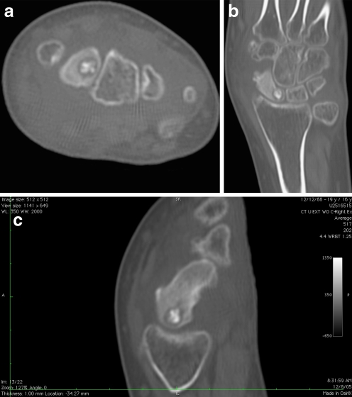
She was referred to this author for consultation at this point. Her review of systems revealed no constitutional signs or symptoms including fevers, chills, night sweats, nausea, vomiting, chest pain, shortness of breath, abdominal pain, other joint pain, low back pain or stiffness, or any unexpected weight loss or weight gain. Examination was notable for a dramatic subjective skin temperature difference between her left and right wrists. Her right wrist was markedly swollen dorsally and radially and held in 10° of ulnar deviation. Range of motion of her right wrist was painful through a limited arc of motion of 20° flexion (85° contralateral), 10° extension (80° contralateral), ulnar deviation of 10° (40° contralateral), and radial deviation of 0° (15° contralateral). Her right forearm was smaller in circumference compared to her left forearm. She was exquisitely tender over the proximal pole of her scaphoid. There was mild tenderness in her anatomic snuffbox and distal scaphoid pole. Watson’s maneuver was painful but did not reveal asymmetric instability. Her imaging studies were reviewed (Fig. 1). In addition, a computed tomography (CT) scan of her wrist was performed (Fig. 2).
Figure 2.
Axial, coronal, and scaphoid plane CT scans of the scaphoid demonstrating a well-circumscribed lytic lesion with central nidus of the proximal scaphoid. Note the marked soft tissue swelling.
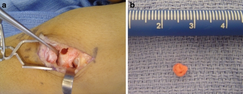
An open excisional biopsy was performed through a dorsal approach. Marked synovitis was encountered and a synovectomy was performed. The articular surface of the proximal scaphoid and lunate, as well as the corresponding articular facets on the distal radius, was found to be irregular with fibrillation and softening of the articular cartilage. A corticotomy was made in the proximal pole of her scaphoid under image intensification and a well-formed nidus was delivered into the field (Fig. 3). The lytic area was meticulously curretaged and packed with morselized cancellous allograft. Histologic examination of the lesion confirmed the diagnosis of osteoid osteoma.
Figure 3.
Intraoperative photographs of scaphoid corticotomy (a) and excised nidus (b).
At 2 weeks, the patient reported complete resolution of her pain. She was provided a removable splint for comfort and at 6 weeks postsurgery was begun on a program of active, active assisted, and passive range of motion, along with progressive strengthening. She was cleared for full return to activities as tolerated at 3 months postoperative. Her range of motion was unchanged from the 3 months preoperative measurements and at 18 months postoperatively, although she reported no discomfort or functional limitation in the use of her operative extremity. Her synovitic swelling was no longer evident. Plain radiographs at 18 months postoperative showed complete healing of her proximal pole without evidence of avascular necrosis. There was no progression in the radiographic appearance of degenerative arthritis.
Discussion
Osteoid osteoma of the carpus is rare. Murray et al. reported on 44 carpal bone tumors, 11 of which were osteoid osteoma and one was located in the scaphoid [14]. Lisanti and coworkers [9] identified the scaphoid as the most commonly affected carpal bone, followed by the capitate, hamate, and lunate. A total of 82 carpal osteoid osteomas were identified in their review between 1935 and 1996. Their review specifically identified a total of 30 cases of osteoid osteoma of the scaphoid. One case, involving the lunate, identified “malacia” of the lunate identified at the time of surgery. To our knowledge, this is the only mention of arthritic change of the wrist in association with this lesion.
The overwhelming symptom of osteoid osteoma is pain. This has been felt to be due to the presence of nerve fibers within the nidus and perinidal tissues and secondarily due to the stimulation of local nerve fibers by tissue edema [11]. Other authors have demonstrated increased local production of prostaglandin E2 in association with osteoid osteomas [10, 17].
It is well-established that prostaglandin E2 (PGE2) plays a key role in the pathogenesis of degenerative arthritis [8–12]. Both serum and synovial fluid from patients with osteoarthritis and rheumatoid arthritis contain increased levels of PGE2 compared to healthy controls. Prostaglandin E2 increases the production of proinflammatory cytokines, reactive oxygen species, and matrix metalloproteases which lead to increased pain and contribute to alteration of articular cartilage, synovium, and bone.
Secondary synovitis has been described in association with osteoid osteoma of the elbow and hip where a portion of the epiphysis is located within the joint capsule [2]. Additionally, synovitis of the first dorsal wrist compartment has been described secondary to cortical perforation of an osteoid osteoma of the radial styloid [2, 18]. Degenerative arthritis, associated with osteoid osteomas of the carpus, has not been described.
Our patient demonstrated marked radiographic and clinical evidence of radiocarpal degenerative arthritis in association with an osteoid osteoma. We speculate that this is an underreported entity as numerous authors cite resolution of pain following resection of osteoid osteoma from the wrist, but note no appreciable normalization of motion postoperatively [1, 3, 5, 11, 15, 16]. Furthermore, given the difficulty of establishing a radiographic diagnosis in these patients with radiographic changes noted often after delay of up to 25 months [9], prostaglandin-mediated alteration in synovium and articular cartilage proceed unchecked.
While symptomatic treatment with salicylates while awaiting spontaneous resolution of osteoid osteomas has been a long-accepted treatment, we posit that this may be contraindicated in the wrist due to the potential for irreversible prostaglandin-mediated articular changes. Carpal osteoid osteoma should remain in the differential diagnosis of the young patient presenting with painful monoarticular degenerative arthritis. Prompt diagnosis based on a high index of clinical suspicion and advanced radiographic imaging, including scintigraphy and MRI [6], followed by surgical excision could possibly lessen the possibility of degenerative arthritis in these patients and subsequently articular damage to the distal radius for osteoid osteomas of the carpus.
References
- 1.Cetti R, Christensen SE. Osteoid osteoma in the scaphoid bone. Case report. Scand J Plast Reconstr Surg. 1982;16(2):207–9. [DOI] [PubMed]
- 2.De Smet L. Synovitis of the wrist joint caused by an intra-articular perforation of an osteoid osteoma of the radial styloid. Clin Rheumatol. 2000;19(3):229–30. [DOI] [PubMed]
- 3.De Smet L, Fabry G. Osteoid osteoma of the hand and carpus: peculiar presentations and imaging. Acta Orthop Belg. 1995;61(2):113–6. [PubMed]
- 4.Fanning JW, Lucas GL. Osteoblastoma of the scaphoid: a case report. J Hand Surg Am. 1993;18(4):663–5. [DOI] [PubMed]
- 5.Garg V, Kapoor SK. Osteoid osteoma of scaphoid. J South Orthop Assoc. 2003;12(3):141–2. [PubMed]
- 6.Gitelis S, Schajowicz F. Osteoid osteoma and osteoblastoma. Orthop Clin North Am. 1989;20(3):313–25. [PubMed]
- 7.Joshi BH, Hogaboam C, Dover P, Husain SR, Puri RK. Role of interleukin-13 in cancer, pulmonary fibrosis, and other T(H)2-type diseases. Vitam Horm. 2006;74:479–504. doi:10.1016/S0083-6729(06)74019-5. [DOI] [PubMed]
- 8.Kojima F, Kato S, Kawai S. Prostaglandin E synthase in the pathophysiology of arthritis. Fundam Clin Pharmacol. 2005;19(3):255–61. [DOI] [PubMed]
- 9.Lisanti M, Rosati M, Spagnolli G, Luppichini G. Osteoid osteoma of the carpus. Case reports and a review of the literature. Acta Orthop Belg. 1996;62(4):195–9. [PubMed]
- 10.Makley JT, Dunn MJ. Prostaglandin synthesis by osteoid osteoma. Lancet 1982;2(8288):42. [DOI] [PubMed]
- 11.Marcuzzi A, Acciaro AL, Landi A. Osteoid osteoma of the hand and wrist. J Hand Surg Br. 2002;27(5):440–3. [DOI] [PubMed]
- 12.Martel-Pelletier J, Lajeunesse D, Fahmi H, Tardif G, Pelletier JP. New thoughts on the pathophysiology of osteoarthritis: one more step toward new therapeutic targets. Curr Rheumatol Rep. 2006;8(1):30–6. [DOI] [PubMed]
- 13.Muren C, Hoglund M, Engkvist O, Juhlin L. Osteoid osteomas of the hand. Report of three cases and review of the literature. Acta Radiol. 1991;32(1):62–6. [PubMed]
- 14.Murray PM, Berger RA, Inwards CY. Primary neoplasms of the carpal bones. J Hand Surg Am. 1999;24(5):1008–13. [DOI] [PubMed]
- 15.Nascimento RJ, Varney TE, Kasparyan NG, Warhold LG. Intracortical osteoid osteoma of the scaphoid. Orthopedics 2007;30(7):573–4. [DOI] [PubMed]
- 16.Themistocleous GS, Chloros GD, Mavrogenis AF, Khaldi L, Papagelopoulos PJ, Efstathopoulos DG. Unusual presentation of osteoid osteoma of the scaphoid. Arch Orthop Trauma Surg. 2005;125(7):482–5. doi:10.1007/s00402-005-0003-7. [DOI] [PubMed]
- 17.Wold LE, Pritchard DJ, Bergert J, Wilson DM. Prostaglandin synthesis by osteoid osteoma and osteoblastoma. Mod Pathol. 1988;1(2):129–31. [PubMed]
- 18.Volpin G, Shtarker H, Oliver S, Katznelson A, Stahl S. [Osteoid osteoma of the wrist joint resembling tenosynovitis]. Harefuah 2006;145(12):885–8. (942–3). [PubMed]


