Abstract
Purpose
We have reported the radiological and clinical outcome of scarf osteotomy in the treatment of moderate to severe hallux valgus among adolescent children.
Method
Data were collected retrospectively between April 2001 and June 2006. The pre- and post-operative intermetatarsal angle (IMA), hallux valgus angle (HVA) and distal metatarsal articular angle (DMAA) were determined. Patients were followed up for a mean of 37.6 months.
Results
Thirteen patients with 19 operated feet were available at the time of the latest follow-up. There was significant improvement in the mean post-operative IMA, which was maintained to the last follow-up. There was statistically significant improvement in the 6-week post-operative HVA and DMAA. However, this was lost at the final follow-up. The mean American Orthopaedic Foot and Ankle Society score for the whole group was 80 (54–100).
Conclusion
This study indicates that scarf osteotomy should be used with caution in symptomatic adolescent hallux valgus, as there is a high recurrence rate.
Keywords: Scarf osteotomy, Adolescent bunions, Hallux valgus
Introduction
There are over 130 described surgical procedures for the management of symptomatic hallux valgus in adults. No one procedure has been proven to be superior to the other [1]. Adolescent hallux valgus deformity differs from that of adults by the presence of a grossly increased distal metatarsal articular angle (DMAA) and the absence of medial subluxation [2]. Symptomatic deformities are best treated surgically as the conservative management including shoe wear modifications fails to halt the progression of the deformity [3]. The choice of the surgery depends on the degree of hallux valgus deformity. Severe deformities with a grossly abnormal distal metatarsal articular angle (DMAA) are best treated by a diaphyseal osteotomy that allows translation such as the scarf osteotomy [2, 4–6]. The addition of a closing wedge osteotomy of the proximal phalanx (Akin’s procedure) increases the corrective abilities of the procedure. In addition, the scarf osteotomy confers the advantage of inherent biomechanical stability. It also prevents shortening of the first ray and allows early mobilisation [7, 8]. There are no published reports in the English literature of scarf osteotomy in the management of adolescent children with symptomatic hallux valgus. The aim of this paper is to report the radiological and clinical outcome of scarf osteotomy in the treatment of moderate to severe hallux valgus among adolescent children.
Patients and methods
We retrospectively reviewed the clinical notes and radiographs of all adolescent patients who had surgery for symptomatic hallux valgus between April 2001 and July 2006. Fifteen patients (22 feet) underwent a scarf osteotomy of the first metatarsal. All procedures were carried out at a tertiary referral children’s hospital. Two patients (three feet) were lost to follow-up. There were 11 girls (16 feet) and 2 boys (3 feet) with data available for assessment during the latest follow-up. Six patients underwent bilateral procedures. The mean age at surgery was 14.3 years (12–18). Patients were followed up for a mean of 37.6 months (22.5–76.3).
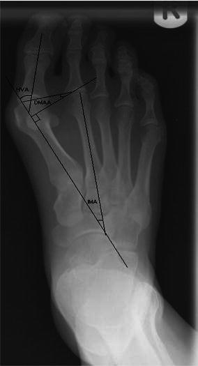
Pre-operative and 6-week post-operative (anteroposterior and lateral) radiographs were available for all patients. Nine patients (13 feet) had radiographs during further follow-ups for pain or recurrence of deformity. The intermetatarsal angle (IMA), hallux valgus angle (HVA) and distal metatarsal articular angle (DMAA) were measured pre- and post-surgery. Lines were constructed along the centre of the anatomical axes of the first metatarsal, second metatarsal and proximal phalanx of the hallux (Fig. 1). The angle formed by the intersection of the axes of the first and second metatarsals is the IMA. The value is normally 7°–9° [3]. The angle formed by the first metatarsal and the proximal phalanx of the hallux represents the HVA, normally 10°–15° [9]. A perpendicular to the anatomical axis of the first metatarsal was constructed. The angle was subtended by the inclination of the articular surface of the first metatarsal head, and this line represented the DMAA, normally <8° [9].
Fig. 1.

Pre-operative radiograph demonstrating the angles measured. HVA hallux valgus angle, IMA intermetatarsal angle, DMAA distal metatarsal articular angle
At the latest follow-up all patients were scored according to the American Orthopaedic Foot and Ankle Society Score (AOFAS) [10].
Two patients in our study also had an Akin’s procedure. This was necessary to improve the correction and probably influenced the clinical and radiological outcome in these two patients. One patient underwent a proximal interphalangeal (PIP) joint fusion for a flexion deformity of the PIP joint of the second toe.
Statistical analysis: Data were checked for normality using Statsdirect 2.7.2. The paired Student's ‘t’ test for normally distributed data and the Mann–Withney U test for non-normally distributed data and ANOVA were used to determine statistical significance with a P value of 0.05 or less considered to be significant.
Results
Table 1 shows the mean HVA, IMA and DMAA for the whole group. ANOVA was used to compare the pre-operative, 6 weeks post-operative and last measured IMA, DMAA and HVA and is shown in Table 2. There was no correlation between age at surgery and pre-operative IMA, HVA and DMAA, 6 weeks post-operative IMA, DMAA and HVA and final follow-up IMA, DMAA and HVA.
Table 1.
HVA, IMA and DMAA measurements
| All patients | Mean pre-op angle (°) | Mean 6 weeks post-op angle (°) | Mean last F/U angle (°) |
|---|---|---|---|
| HVA | 34 | 15 | 25 |
| IMA | 14 | 8.7 | 8.5 |
| DMAA | 17.4 | 8 | 15.6 |
Table 2.
ANOVA scores for comparison of IMA, HVA and DMAA preoperatively, at 6 weeks post-operatively and at the final follow-up
| All patients | Pre-op to 6 weeks post-op | Pre-op to last X-ray | 6 weeks post-op to last X-ray | |||
|---|---|---|---|---|---|---|
| Diff. in means (change) | P value | Diff. in means (change) | P value | Diff. in means (change) | P value | |
| 95% CI | 95% CI | 95% CI | ||||
| IMA (°) | 5.5 | <0.0001 | 5.3 | <0.0001 | −0.2 | 0.5 |
| 3.4 to 7.2 | ||||||
| 3.8 to 7.2 | −1.8 to 2.1 | |||||
| HVA (°) | 19.4 | <0.0001 | 9.2 | 0.0003 | −10.1 | <0.0001 |
| 15.2 to 23.6 | 4.4 to 14 | −5.3 to −14.1 | ||||
| DMAA (°) | 9.5 | <0.0001 | 1.9 | 0.32 | −7.6 | 0.0002 |
| 6.1 to 1286 | −1.9 to 5.7 | −3.8 to −11.5 | ||||
There was significant improvement in the mean IMA that was maintained at the final follow-up. There was also statistically significant improvement in the 6-week post-operative HVA, but this was lost at the last follow-up. The initial mean improvement of 19.4° of the HVA was reduced to 9.2° at the last follow-up. The DMAA showed a similar trend. The significant 6-week post-operative improvement of 9.5° was reduced to 1.9° at final follow-up.
The mean AOFAS score for the whole group was 80 (54–100). Six patients with seven operated feet were symptomatic (pain), and the deformity had recurred. Their mean AOFAS score was 68 (54–80). Seven other patients with ten operated feet were satisfied, and their mean AOFAS score was 94 (72–100). One patient was included in both groups as the patient was satisfied with one side, but not with the other. One patient had other significant disabilities that affected the AOFAS score obtained and was excluded from the analysis. There was no statistically significant correlation between age and AOFAS score. However, there was a statistically significant difference in the AOFAS score between the symptomatic and asymptomatic group, P < 0.001 (95% CI 14.9–33.7). There was no statistically significant difference between these two groups when their age, gender, pre-operative HVA, IMA and DMAA were compared.
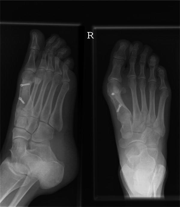
There were two (9%) cases of superficial wound infection. Methicillin-resistant Staphylococcus aureus (MRSA) was cultured from one patient, and the infection was treated successfully with 2 weeks of oral antibiotics. The other patient complained of prominent stitch. This was removed by surgical exploration and subsequently developed a superficial infection requiring 1 week of oral antibiotics. One patient developed complex regional pain syndrome 6 weeks post-operatively. This resolved spontaneously with physiotherapy and NSAIDs by 6 months. None of the patients in the study group reported symptoms of transfer metatarsalgia, restriction of movement or callus-related problems at the metatarsophalangeal (MTP) or IP joint. All patients achieved clinical and radiological bony union at the osteotomy site by the end of 3 months (Fig. 2).
Fig. 2.

Three months postoperative radiographs following scarf osteotomy
Discussion
A number of published studies have reported on the outcome of various procedures for hallux valgus in skeletally immature patients. No single technique has proved to be better than others, and all have a significant rate of recurrence in this age range. Scranton and Zuckerman [11] noticed a 36% failure rate with distal metatarsal osteotomy for adolescent hallux valgus. He proposed that elective bunion surgery in adolescents should only be performed in the face of progressive, painful deformity where both the patient and the patient’s parents fully understand the goals and risks of surgery.
Ball and Sullivan [12] recommended abandonment of the Mitchell procedure for juvenile bunions due to recurrence of hallux valgus and restricted motion of the first metatarsophalangeal joint. They found that only 61% (11 of 18) of the patients were satisfied. They also noted a significant incidence of metatarsalgia postoperatively. De's [13] experience of the procedure was similar. He found that excessive metatarsal shortening and dorsal tilting occurred, and this led to persistent metatarsalgia. In another study by Geissele et al. [14], only 59% of the patients had good results. He pointed out that the one factor most highly correlated with both decreased risk of recurrence of angular deformity and patient satisfaction was a reduction of the IMA. This was not borne out in our study.
Patients who had undergone double metatarsal osteotomy for the correction of moderate to severe hallux valgus deformity were reported to have a high complication rate of first MTP joint stiffness, making the procedure unsuitable for a high-functioning adolescent [15, 16].
We believed that the Z or “scarf” osteotomy offered potential advantages over some other methods for the treatment of adolescent hallux valgus. Z osteotomy of the first metatarsal was first described by Meyer [17] in 1926 and popularised by Weil and Bourouk [18, 19]. “Scarf” is a term used to describe a carpentry technique. The technique involves securing beams of timber longitudinally after notching or grooving the ends, thus allowing a degree of overlap. The same principle is applied surgically. The ‘Z’ osteotomy facilitates overlap of the bony edges of the metatarsal, which are then secured via headless screw fixation.
Cadaveric biomechanical studies have shown scarf osteotomy to be more stable under loaded conditions than the basal osteotomy [7, 20]. The absolute stability achieved by screw fixation results in early bony union [8]. In addition, the diaphyseal osteotomy results in a large surface area being available for bone-to-bone contact. This also contributes to the inherent stability of the osteotomy and encourages bony union [7, 20].
Furthermore, there are several studies that show good long-term results following scarf osteotomy in adults. In the study by Aminian et al. [21] involving 27 feet, the complication rate was 1.1%, including superficial infection and recurrence. He concluded that the scarf osteotomy provides a predictable and effective correction of moderate to severe hallux valgus deformities. In another study by Galli et al. [22] involving 25 patients, 19 patients were declared to be very satisfied, 4 satisfied and only 1 dissatisfied at 24-month follow-up. None of the patients had pain, and one patient was dissatisfied with the aesthetic result. Prospective studies by Deenik et al. [23] and Lorei et al. [24] have also showed good results for scarf osteotomy in adults.
In our study group, initially we were pleased with the short-term outcome. Early radiological evaluation revealed significant improvement in the mean HVA, IMA and DMAA, and patients were happy with the outcome. This finding is consistent with that reported in the literature for scarf osteotomy in the adult population [25–27].
None of our patients suffered from transfer metatarsalgia or stiffness of the first MTP joint. None of our patients were immobilised with plaster. They were allowed to mobilise fully weight bearing as tolerated using wood-soled shoes post-operatively for up to 6 weeks. With this regimen the bony union rate in our series of patients was 100% by the 3rd post-operative month, and there were no incidents of osteotomy displacement. This was confirmed both clinically and radiologically in all cases.
The medium term follow-up of our patients revealed that although there was a sustained correction of the IMA, the initial correction of the DMAA and HVA was short lived. This correlated with patient dissatisfaction at the outcome of the procedure. These findings are not dissimilar to those for other hallux valgus procedures. To our knowledge there is only one published study on scarf osteotomy for hallux valgus in children, which was done by Salmeron et al. [28]. The article is in the French literature, and the findings of the study are similar to ours in that 9 out of 19 French patients were reported to have had a poor outcome with residual pain and cosmetic problems.
The reasons for poor outcome following scarf osteotomy in children are not clear. Some authors have postulated a relationship between pes planus and hallux valgus in the adolescent age group [11, 29, 30]. Any factor that causes stretching of the tissues on the medial side of the hallux on weight bearing may play a role in recurrence of the deformity. The balance between abductor hallucis that inserts on the plantar and medial surface of the proximal phalanx and the adductor hallucis that inserts on the lateral surface is lost. The adductor hallucis overpowers the abductor hallucis, and this may favour the gradual recurrence of the deformity. Adaptive secondary bony changes can then occur that influence the alignment of the metatarsal articular surface leading to a change in the DMAA. The recurrence of adolescent hallux valgus following the scarf procedure is no different than that following other surgical techniques. It is reasonable to assume that deformity recurrence is a consequence of the patient group rather than the weakness of any specific technique.
A limitation of this study is the small sample size, even though it is similar to that found in other studies [31–33]. In addition, the retrospective nature of the study has its inherent deficiencies. However, we feel it is useful to report our medium-term results as the outcome had a clear trend.
We believe that the scarf osteotomy as a treatment option for symptomatic adolescent hallux valgus should be used sparingly. Patients should be warned and advised regarding the potential for recurrence. Where possible, the procedure should be deferred until skeletal maturity is attained and the outcome is more predictable.
Contributor Information
H. L. George, Phone: +44-151-2525553, FAX: +44-151-2525921
L. A. James, Email: drjames@doctors.org.uk, Email: mrleroyjames@aol.com
References
- 1.Kelikian H. Hallux valgus, allied deformities of the forefoot and metatarsalgia. Philadelphia: WB Saunders; 1965. pp. 1–5. [Google Scholar]
- 2.Robinson AHN, Limbers JP. Aspects of current management. Modern concepts in the treatment of hallux valgus. J Bone Joint Surg Br. 2005;87(8):1038–1045. doi: 10.1302/0301-620X.87B8.16467. [DOI] [PubMed] [Google Scholar]
- 3.Kilmartin TE, Barrington RL, Wallace WA. A controlled prospective trial of a foot orthosis for juvenile hallux valgus. J Bone Joint Surg (Br) 1994;76-B:210–214. [PubMed] [Google Scholar]
- 4.Barouk LS. Scarf osteotomy for hallux valgus correction: local anatomy, surgical technique, and combination with other forefoot procedures. Foot Ankle Clin. 2000;5:525–558. [PubMed] [Google Scholar]
- 5.Dereymaeker G. Scarf osteotomy for correction of hallux valgus: surgical technique and results as compared to distal chevron osteotomy. Foot Ankle Clin. 2000;5:513–524. [PubMed] [Google Scholar]
- 6.Smith AM, Alwan T, Davies MS. Perioperative complications of the scarf osteotomy. Foot Ankle Int. 2003;24:222–227. doi: 10.1177/107110070302400304. [DOI] [PubMed] [Google Scholar]
- 7.Newman AS, Negrine JP, Zecovic M, Stanford P, Walsh WR. A biomechanical comparison of the Z step-cut and basilar cresenteric osteotomies of the first metatarsal. Foot Ankle lnt. 2000;21:584–587. doi: 10.1177/107110070002100710. [DOI] [PubMed] [Google Scholar]
- 8.Zygmunt KH, Gudas CJ, Laros GS. Z-bunionectomy with internal screw fixation. J Am Podiatr Med Assoc. 1989;79:322–329. doi: 10.7547/87507315-79-7-322. [DOI] [PubMed] [Google Scholar]
- 9.Gudas CJ, Marcinko DE. The complex deformity known as hallux abductor valgus. In: Marcinko DE, editor. Comprehensive textbook of hallux valgus reconstruction. St Louis: Mosby; 1992. pp. 1–17. [Google Scholar]
- 10.Kitaoka HB, Alexander IJ, Adelaar RS, Nunley JA, Myerson MS, Sanders M. Clinical rating systems for the ankle-hindfoot, midfoot, hallux and lesser toes. Foot Ankle Int. 1994;15(7):349–353. doi: 10.1177/107110079401500701. [DOI] [PubMed] [Google Scholar]
- 11.Scranton PE, Zuckerman JD. Bunion surgery in adolescents: results of surgical treatment. J Pediatr Orthop. 1984;4:39–43. doi: 10.1097/01241398-198401000-00009. [DOI] [PubMed] [Google Scholar]
- 12.Ball J, Sullivan JA. Treatment of juvenile bunion by Mitchell osteotomy. Orthopedics. 1985;8(10):1249–1251. doi: 10.3928/0147-7447-19851001-09. [DOI] [PubMed] [Google Scholar]
- 13.De S. Distal metatarsal osteotomy for adolescent hallux valgus. J Pediatr Orthop. 1984;4:32–38. doi: 10.1097/01241398-198401000-00008. [DOI] [PubMed] [Google Scholar]
- 14.Geissele AE, Stanton RP. Surgical treatment of adolescent hallux valgus. J Paediatr Orthop. 1990;10(5):642–648. doi: 10.1097/01241398-199009000-00014. [DOI] [PubMed] [Google Scholar]
- 15.Johnson AE, Georgopoulous G, Erickson MA, Eilert R. Treatment of adolescent hallux valgus with the first metatarsal double osteotomy: The Denver experience. J Pediatr Orthop. 2004;24:358–362. doi: 10.1097/01241398-200407000-00003. [DOI] [PubMed] [Google Scholar]
- 16.Coughlin M. Juvenile bunions. In: Mann R, Coughlin M, editors. Surgery of the foot & ankle. St Louis: Mosby; 1993. pp. 297–339. [Google Scholar]
- 17.Meyer M. Eine neue modifikation der hallux-valgus-operation. Zentralbl Chirg. 1926;53B5:32–38. [Google Scholar]
- 18.Weil LS, Borelli AN. Modified scarf bunionectomy: our experience in more than 1000 cases. J Foot Surg. 1991;30:609–622. [Google Scholar]
- 19.Bourouk LS. Osteotomie Scarf du premier metatarsien. Med Chir Pied. 1994;10:111–120. [Google Scholar]
- 20.Tnka HJ, Parks B, Ivanic G, Chu IT, Easley ME, Schon LC, Myerson MS. Six-first metatarsal shaft osteotomies: mechanical and immobilisation comparisons. Clin Orthop Relat Res. 2000;381:256–265. doi: 10.1097/00003086-200012000-00030. [DOI] [PubMed] [Google Scholar]
- 21.Aminian A, Kelikian A, Moen T. Scarf osteotomy for hallux valgus deformity: an intermediate followup of clinical and radiographic outcomes. Foot Ankle Int. 2006;27(11):883–886. doi: 10.1177/107110070602701103. [DOI] [PubMed] [Google Scholar]
- 22.Galli M, Muratori F, Visci F, Pezzillo F, Aulisa AG. Middle term results of I metatarsal “scarf” osteotomy. Clin Ter. 2007;158(3):209–212. [PubMed] [Google Scholar]
- 23.Deenik AR, Pilot P, Brandt SE, van Mameren H, Geesink RG, Draijer WF. Scarf versus chevron osteotomy in hallux valgus: a randomized controlled trial in 96 patients. Foot Ankle Int. 2007;28(5):537–541. doi: 10.3113/FAI.2007.0537. [DOI] [PubMed] [Google Scholar]
- 24.Lorei TJ, Kinast C, Klärner H, Rosenbaum D. Pedographic, clinical, and functional outcome after scarf osteotomy. Clin Orthop Relat Res. 2006;451:161–166. doi: 10.1097/01.blo.0000229297.29345.09. [DOI] [PubMed] [Google Scholar]
- 25.Crevoisier X, Mouhsin E, Ortolano V, Udin B, Butoit M. The scarf osteotomy for the treatment of hallux valgus deformity: a review of 84 cases. Foot Ankle lnt. 2001;22:970–976. doi: 10.1177/107110070102201208. [DOI] [PubMed] [Google Scholar]
- 26.Kristen KH, Berger C, Stelzig S, Thalhammer E, Posch M. The scarf osteotomy for the correction of hallux valgus deformities. Foot Ankle lnt. 2002;23:221–229. doi: 10.1177/107110070202300306. [DOI] [PubMed] [Google Scholar]
- 27.Jone S, Al Hussainy HA, Ali F, Betts RP, Flowers MJ. Scarf osteotomy for hallux valgus. A prospective clinical and pedobarographic study. J Bone Joint Surg Br. 2004;86-B(6):830–836. doi: 10.1302/0301-620X.86B6.15000. [DOI] [PubMed] [Google Scholar]
- 28.Salmeron F, Sales De Gauzy J, Galy C, Darodes P, Cahuzac JP. Scarf osteotomy of hallux valgus in children and adolescents. Rev Chir Orthop Reparatrice Appar Mot. 2001;87(7):706–711. [PubMed] [Google Scholar]
- 29.Inman VT. Hallux valgus: A review of etiologic factors. Orthop Clin North Am. 1974;5:59. [PubMed] [Google Scholar]
- 30.Kalen V, Brecher A. Relationship between adolescent bunions and flatfeet. Foot Ankle. 1988;8:331. doi: 10.1177/107110078800800609. [DOI] [PubMed] [Google Scholar]
- 31.Aronson J, Nguyen L, Aronson E. Early results of the modified Peterson bunion procedure for adolescent hallux valgus. J Pediatr Orthop. 2001;21:65–69. doi: 10.1097/01241398-200101000-00014. [DOI] [PubMed] [Google Scholar]
- 32.Coughlin M. Juvenile hallux valgus. Foot Ankle Int. 1995;16:682–697. doi: 10.1177/107110079501601104. [DOI] [PubMed] [Google Scholar]
- 33.Peterson H, Newman S. Adolescent bunion deformity treated with double osteotomy and longitudinal pin fixation of the first ray. J Pediatr Orthop. 1993;13:80–84. doi: 10.1097/01241398-199301000-00016. [DOI] [PubMed] [Google Scholar]


