

Abstract
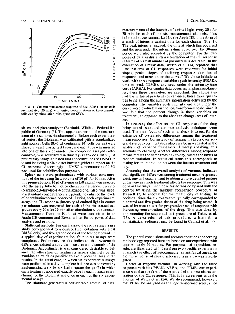
Chemiluminescence is the result of the respiratory burst generated by phagocytic cells after stimulation by antigen. The measurement of chemiluminescence represents a sensitive means for detecting antigenic stimulation and immune cell function. Although the kinetics of chemiluminescence reactions have been described, appropriate statistical methods for the evaluation of data from chemiluminescence assays have not been reported. Based on examination of data from several studies in which the chemiluminescence response of spleen cells was investigated after stimulation with the particulate antigen zymosan, recommendations are made for the design and statistical evaluation of such studies. Three parameters were used in assessing the chemiluminescence response; peak intensity of the emitted light, time to peak, and the area under the intensity-time curve. The data indicated that peak intensity alone provides an adequate characterization of the chemiluminescence response. Since percent change in response upon treatment is of interest, analysis on the log scale is appropriate, and the statistical procedure of choice in evaluating data of this type is a trend analysis. The need for a balanced allocation of treatments to avoid potential bias is demonstrated. The methods proposed are illustrated with data from two studies in which the effect of preincubation with low concentrations of ketoconazole, an antifungal agent, on the chemiluminescence response of BALB/cBY spleen cells was examined.
Full text
PDF

Selected References
These references are in PubMed. This may not be the complete list of references from this article.
- Allen R. C., Stjernholm R. L., Steele R. H. Evidence for the generation of an electronic excitation state(s) in human polymorphonuclear leukocytes and its participation in bactericidal activity. Biochem Biophys Res Commun. 1972 May 26;47(4):679–684. doi: 10.1016/0006-291x(72)90545-1. [DOI] [PubMed] [Google Scholar]
- Babior B. M., Kipnes R. S., Curnutte J. T. Biological defense mechanisms. The production by leukocytes of superoxide, a potential bactericidal agent. J Clin Invest. 1973 Mar;52(3):741–744. doi: 10.1172/JCI107236. [DOI] [PMC free article] [PubMed] [Google Scholar]
- DeChatelet L. R., Long G. D., Shirley P. S., Bass D. A., Thomas M. J., Henderson F. W., Cohen M. S. Mechanism of the luminol-dependent chemiluminescence of human neutrophils. J Immunol. 1982 Oct;129(4):1589–1593. [PubMed] [Google Scholar]
- Fromtling R. A., Fromtling A. M., Staib F., Müller S. Effect of uremia on lymphocyte transformation and chemiluminescence by spleen cells of normal and Cryptococcus neoformans-infected mice. Infect Immun. 1981 Jun;32(3):1073–1078. doi: 10.1128/iai.32.3.1073-1078.1981. [DOI] [PMC free article] [PubMed] [Google Scholar]
- Heberer M., Ernst M., Dürig M., Allgöwer M., Fischer H. Measurement of chemiluminescence in freshly drawn human blood. II. Clinical application of zymosan-induced chemiluminescence. Klin Wochenschr. 1982 Dec 1;60(23):1443–1448. doi: 10.1007/BF01720991. [DOI] [PubMed] [Google Scholar]
- Mantel N. Assessing laboratory evidence for neoplastic activity. Biometrics. 1980 Sep;36(3):381–399. [PubMed] [Google Scholar]
- Tukey J. W., Ciminera J. L., Heyse J. F. Testing the statistical certainty of a response to increasing doses of a drug. Biometrics. 1985 Mar;41(1):295–301. [PubMed] [Google Scholar]
- Welch W. D., Davis D., Thrupp L. D. Effect of antimicrobial agents on human polymorphonuclear leukocyte microbicidal function. Antimicrob Agents Chemother. 1981 Jul;20(1):15–20. doi: 10.1128/aac.20.1.15. [DOI] [PMC free article] [PubMed] [Google Scholar]