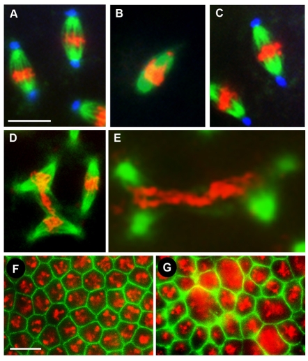
Fig. 3.
Suppression of nopo by mnk. (A-E) Representative mitotic spindles in syncytial embryos from wild-type (A), nopoZ1447 (B) and mnk nopoZ1447 females (C-E). (A-C) Microtubules are in green, DNA in red, and centrosomes in blue. nopo (B) is suppressed by mnk, as evidenced by the restoration of elongated spindles with attached centrosomes (C). (D,E) Microtubules are in green and DNA in red. Aberrant mitotic figures with DNA shared by two spindles are observed in mnk nopo-derived embryos. (F,G) Cellularized embryos (2-3 hours). Actin is in green and DNA in red. Developmental arrest of nopo is suppressed by mnk. Cellularized mnk nopoZ1447-derived embryos show large DNA masses (G) compared to wild type (F). Scale bars: 10 μm in A-E; 20 μm in F,G.

