

Abstract
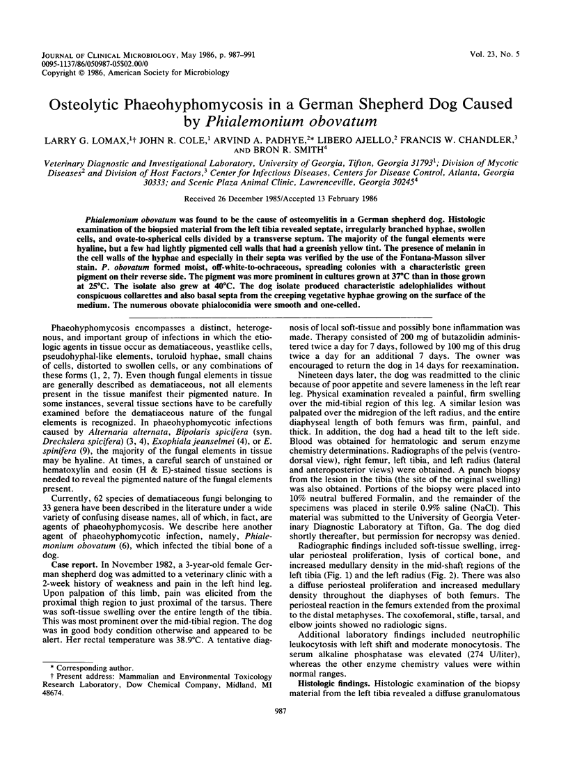
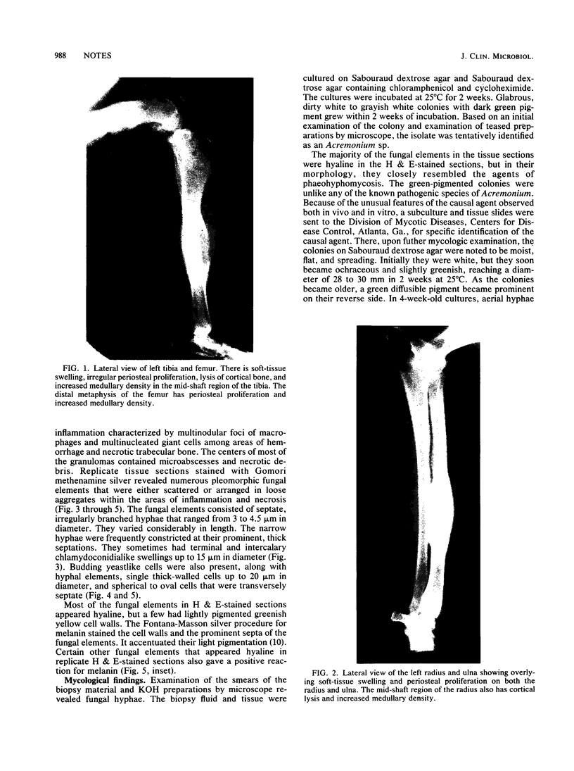
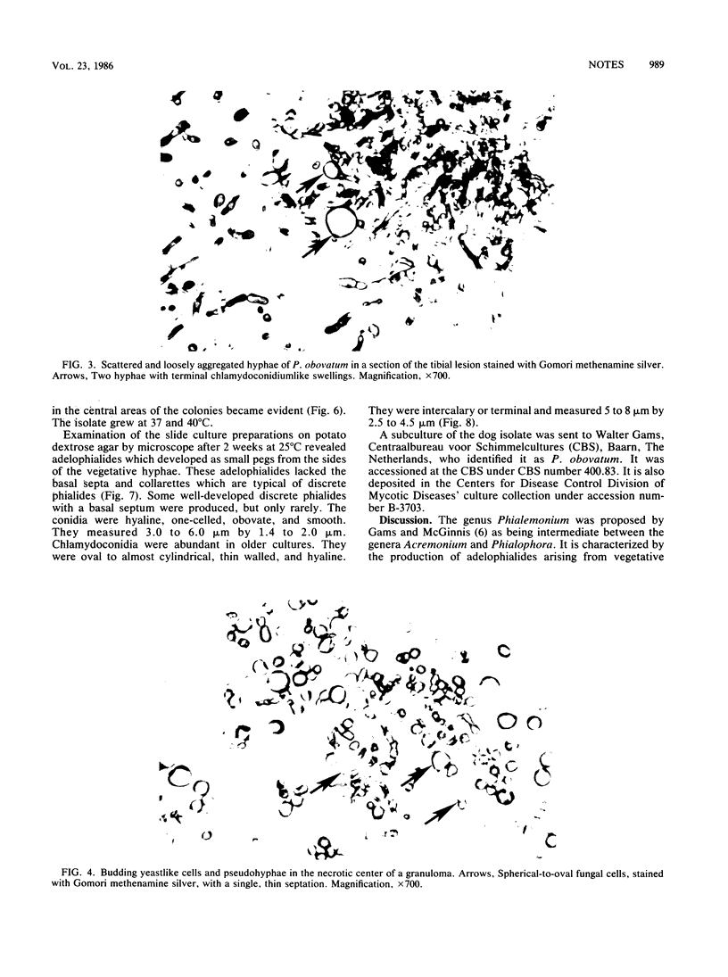
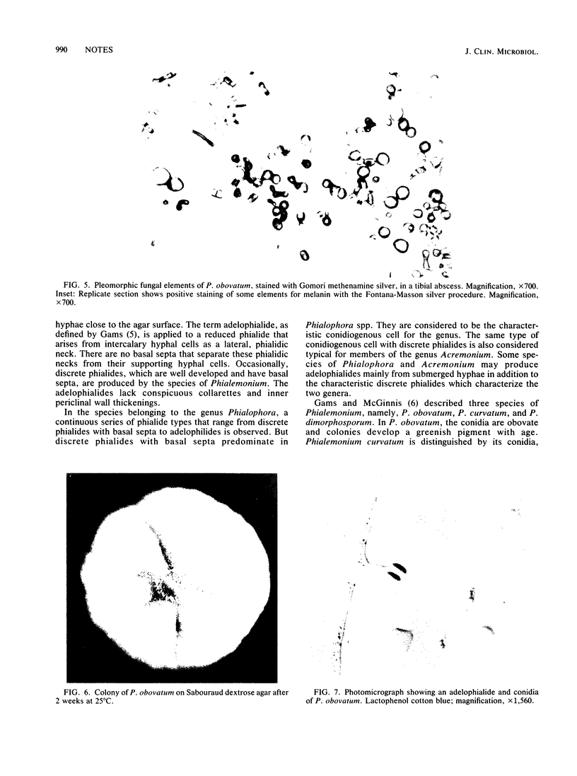
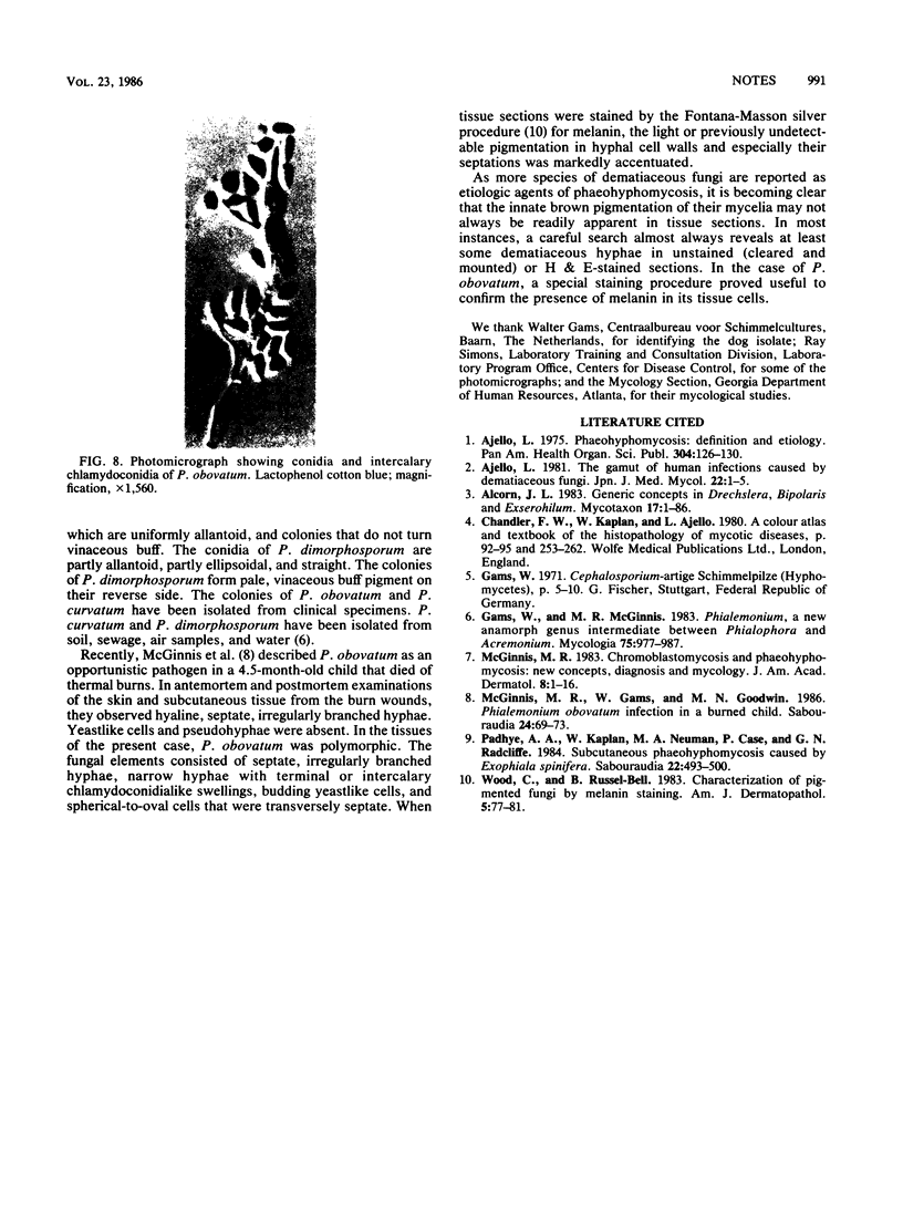
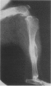
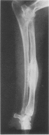
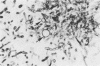
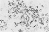
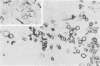
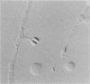
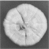
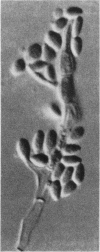
Phialemonium obovatum was found to be the cause of osteomyelitis in a German shepherd dog. Histologic examination of the biopsied material from the left tibia revealed septate, irregularly branched hyphae, swollen cells, and ovate-to-spherical cells divided by a transverse septum. The majority of the fungal elements were hyaline, but a few had lightly pigmented cell walls that had a greenish yellow tint. The presence of melanin in the cell walls of the hyphae and especially in their septa was verified by the use of the Fontana-Masson silver stain. P. obovatum formed moist, off-white-to-ochraceous, spreading colonies with a characteristic green pigment on their reverse side. The pigment was more prominent in cultures grown at 37 degrees C than in those grown at 25 degrees C. The isolate also grew at 40 degrees C. The dog isolate produced characteristic adelophialides without conspicuous collarettes and also basal septa from the creeping vegetative hyphae growing on the surface of the medium. The numerous obovate phialoconidia were smooth and one-celled.
Full text
PDF
Images in this article
Selected References
These references are in PubMed. This may not be the complete list of references from this article.
- McGinnis M. R. Chromoblastomycosis and phaeohyphomycosis: new concepts, diagnosis, and mycology. J Am Acad Dermatol. 1983 Jan;8(1):1–16. doi: 10.1016/s0190-9622(83)70001-0. [DOI] [PubMed] [Google Scholar]
- Padhye A. A., Kaplan W., Neuman M. A., Case P., Radcliffe G. N. Subcutaneous phaeohyphomycosis caused by Exophiala spinifera. Sabouraudia. 1984;22(6):493–500. [PubMed] [Google Scholar]
- Wood C., Russel-Bell B. Characterization of pigmented fungi by melanin staining. Am J Dermatopathol. 1983 Feb;5(1):77–81. doi: 10.1097/00000372-198302000-00015. [DOI] [PubMed] [Google Scholar]