

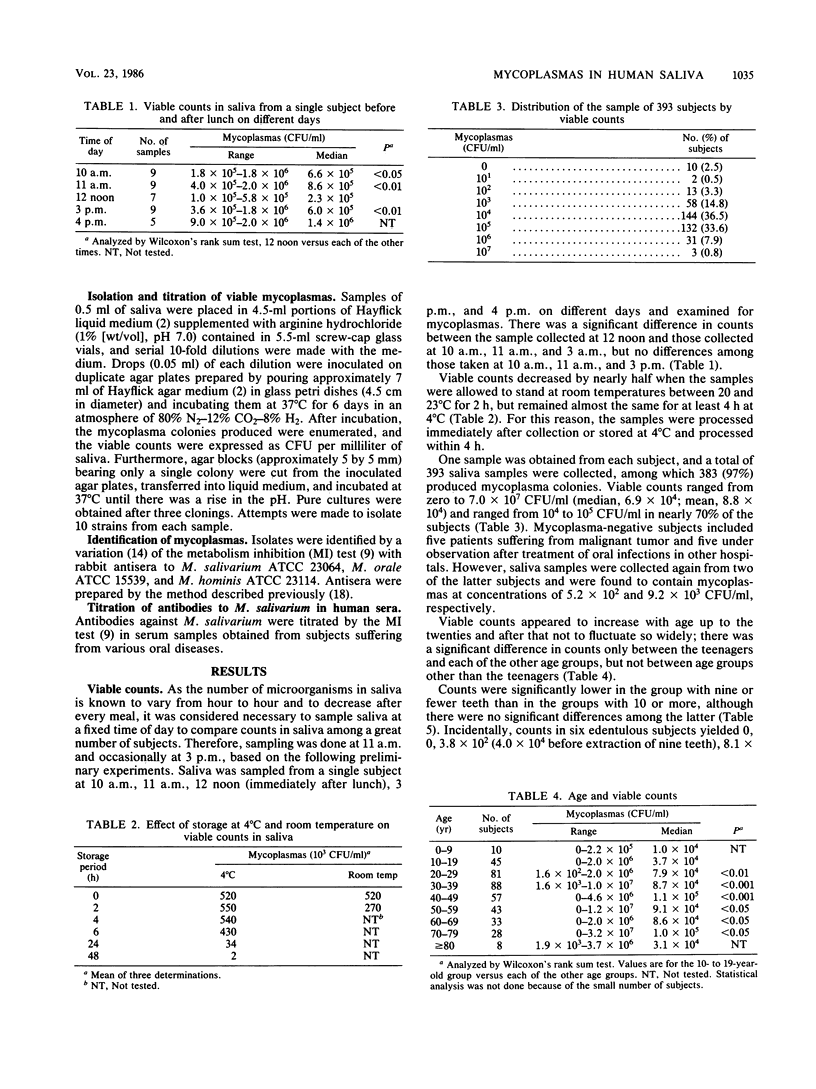
Abstract
Saliva samples collected from 393 subjects with and without oral diseases were examined for concentrations of mycoplasmas and Mycoplasma species. Mycoplasmas were isolated from 383 (97%) of the 393 subjects. Viable counts ranged from zero to 7.6 X 10(7) CFU/ml (median, 6.9 X 10(4)) and were significantly (P less than 0.01) higher in diseased subjects, except for those with arthrosis temporomandibularis, than in controls. Of 1,400 isolates, 897 (64%), 442 (30%), and 8 (1%) were identified as Mycoplasma salivarium, M. orale, and M. hominis, respectively, and the remaining 73 isolates (5%) were unidentifiable. More than two-thirds of the isolates from diseased subjects versus only half from controls were identified as M. salivarium. In diseased subjects other than those with ostitis (especially those with arthrosis temporomandibularis), the incidence of M. salivarium was higher than that of M. orale, whereas the former occurred about as frequently as the latter in the controls. Antibodies to M. salivarium were also measured in sera from some subjects by the metabolism inhibition test. Sera with metabolism inhibition titers of 16 or greater were rated positive. There was no significant difference in the prevalence of antibodies between diseased subjects (60%) and controls (40%), but the mean titers (97 to 220) of all positive sera from diseased subjects were two to four times those for sera from controls. In addition, a fourfold or greater rise or fall of antibody titers to the organism was shown in paired sera from some subjects. On the basis of these results, M. salivarium was strongly suggested to participate etiologically in some cases of oral infection.
Full text
PDF


Selected References
These references are in PubMed. This may not be the complete list of references from this article.
- Engel L. D., Kenny G. E. Mycoplasma salivarium in human gingival sulci. J Periodontal Res. 1970;5(3):163–171. doi: 10.1111/j.1600-0765.1970.tb00711.x. [DOI] [PubMed] [Google Scholar]
- Hayflick L. Tissue cultures and mycoplasmas. Tex Rep Biol Med. 1965 Jun;23(Suppl):285+–285+. [PubMed] [Google Scholar]
- Ichimaru H., Nakamura M. The components of Mycoplasma salivarium and its growth medium that are responsible for film formation. J Gen Microbiol. 1979 Jun;112(2):389–392. doi: 10.1099/00221287-112-2-389. [DOI] [PubMed] [Google Scholar]
- Kumagai K., Iwabuchi T., Hinuma Y., Yuri K., Ishida N. Incidence, species, and significance of Mycoplasma species in the mouth. J Infect Dis. 1971 Jan;123(1):16–21. doi: 10.1093/infdis/123.1.16. [DOI] [PubMed] [Google Scholar]
- Mishima K. [Hemolysis of oral mycoplasmas (author's transl)]. Kokubyo Gakkai Zasshi. 1973 Dec;40(4):389–403. doi: 10.5357/koubyou.40.389. [DOI] [PubMed] [Google Scholar]
- Parkinson C. F. Histamine release from human leukocytes when stimulated by Mycoplasma salivarium. Infect Immun. 1975 Mar;11(3):595–597. doi: 10.1128/iai.11.3.595-597.1975. [DOI] [PMC free article] [PubMed] [Google Scholar]
- Parkinson C. F. Reactivity of mast cells to Mycoplasma salivarium. Infect Immun. 1975 Mar;11(3):598–600. doi: 10.1128/iai.11.3.598-600.1975. [DOI] [PMC free article] [PubMed] [Google Scholar]
- Purcell R. H., Taylor-Robinson D., Wong D. C., Chanock R. M. A color test for the measurement of antibody to the non-acid-forming human Mycoplasma species. Am J Epidemiol. 1966 Jul;84(1):51–66. doi: 10.1093/oxfordjournals.aje.a120627. [DOI] [PubMed] [Google Scholar]
- SCHIMKE R. T., BARILE M. F. ARGININE METABOLISM IN PLEUROPNEUMONIA-LIKE ORGANISMS ISOLATED FROM MAMMALIAN CELL CULTURE. J Bacteriol. 1963 Aug;86:195–206. doi: 10.1128/jb.86.2.195-206.1963. [DOI] [PMC free article] [PubMed] [Google Scholar]
- Sobeslavsky O., Chanock R. M. Peroxide formation by mycoplasmas which infect man. Proc Soc Exp Biol Med. 1968 Nov;129(2):531–535. doi: 10.3181/00379727-129-33362. [DOI] [PubMed] [Google Scholar]
- Uchida A. Isolation and enumeration of mycoplasmas in dental plaques. Bull Tokyo Med Dent Univ. 1981 Dec;28(4):117–123. [PubMed] [Google Scholar]
- Watanabe T. Biological and serological studies on oral Mycoplasma. Bull Tokyo Med Dent Univ. 1967 Dec;14(4):447–460. [PubMed] [Google Scholar]
- Watanabe T., Matsuura M., Seto K. Proteolytic activity of mycoplasmas and ureaplasmas isolated freshly from human saliva. Med Microbiol Immunol. 1985;173(5):251–255. doi: 10.1007/BF02124942. [DOI] [PubMed] [Google Scholar]
- Watanabe T., Mishima K., Fujita O., Horikawa T., Noguchi T. Possible role of mycoplasmas in periodontal disease. Bull Tokyo Med Dent Univ. 1972 Mar;19(1):93–109. [PubMed] [Google Scholar]
- Watanabe T., Mishima K., Horikawa T. Proteolytic activities of human mycoplasmas. Jpn J Microbiol. 1973 Mar;17(2):151–153. doi: 10.1111/j.1348-0421.1973.tb00719.x. [DOI] [PubMed] [Google Scholar]
- Watanabe T., Taylor-Robinson D. Serological tests on L-phase variants of some bacteria. Med Microbiol Immunol. 1973;158(4):291–298. doi: 10.1007/BF02121415. [DOI] [PubMed] [Google Scholar]