Abstract
This study examined the association of cytokine gene polymorphisms with intrahepatic bile duct wall fibrosis in human clonorchiasis. A total of 240 residents in Heilongjiang, China underwent ultrasonography, blood sampling, and stool examination. Single nucleotide polymorphism (SNP) sites for IFN-γ (+874 T/A), IL-10 (-1,082 G/A, -819 C/T, -592 C/A), TNF-α (-308 G/A), and TGF-β1 (codon 10 T/C, codon 25 G/C) genes were observed with the TaqMan allelic discrimination assay. No significant correlation was observed between individual cytokine gene polymorphisms and intrahepatic duct dilatation (IHDD). Among individuals with clonorchiasis of moderate intensity, the incidence of IHDD was high in those with IFN-γ intermediate-producing genotype, +874AT (80.0%, P = 0.177), and in those with TNF-α low-producing genotype, -308GG (63.0%, P = 0.148). According to the combination of IFN-γ and TNF-α genotypes, the risks for IHDD could be stratified into high (intermediate-producing IFN-γ and low producing TNF-α), moderate, and low (low-producing IFN-γ and high producing TNF-α) risk groups. The incidence of IHDD was significantly different among these groups (P = 0.022): 88.9% (odds ratio, OR = 24.0) in high, 56.5% (OR = 3.9) in moderate, and 25.0% (OR = 1) in low risk groups. SNP of IFN-γ and TNF-α genes may contribute to the modulation of fibrosis in the intrahepatic bile duct wall in clonorchiasis patients.
Keywords: Clonorchis sinensis, IFN-γ, IL-10, TNF-α, TGF-β1, single nucleotide polymorphism, bile duct, fibrosis
INTRODUCTION
Clonorchis sinensis is endemic in most of East Asian countries, and its prevalence depends upon the distribution of intermediate hosts and fish-eating habits of residents. Clonorchiasis is a major health problem in those endemic areas because it results in serious pathological changes in the intrahepatic bile ducts and surrounding tissues. The pathological changes include peri-ductal inflammation, biliary mucosal hypertrophy, bile duct dilatation, peri-ductal fibrosis and wall thickening, metamorphosis of mucus-secreting cells, and neoplastic changes in the biliary epithelial cells [1]. The degree of pathological change varies according to intensity and chronicity of the infection [2].
Peri-ductal fibrosis and the wall thickening is a major pathological change in clonorchiasis and is generally induced by complicated interactions between infiltrating cells, cytokines, and the extracellular matrix. The over-production of the extracellular matrix during bile duct fibrosis in schistosomiasis is regulated by cytokines produced by activated Kupffer cells in the liver [3]. The activated Kupffer cells produce several kinds of cytokines such as TGF-α, TGF-β1, TNF-α, PDGF, IL-10, and IFN-γ. Of the cytokines, IFN-γ, IL-10, TNF-α, and TGF-β1 are known to modulate hepatic fibrosis in alcoholic liver disease and hepatitis C, and production of these cytokines is affected by gene polymorphisms [4]. Schistosoma japonicum egg deposition in the liver results in hepatic fibrosis, and the degree of fibrosis is modulated by MHC class II and IL-13 gene polymorphisms [5].
Peri-ductal hepatic fibrosis in clonorchiasis can be estimated by sonography of the liver [6], but its correlation with cytokine gene polymorphisms is still unknown. We postulate that the peri-ductal fibrosis in clonorchiasis is modulated by cytokines and also by their genetic polymorphisms. Our study was designed to demonstrate and elucidate any correlation of cytokine gene single nucleotide polymorphisms with liver pathology in patients with clonorchiasis.
MATERIALS AND METHODS
Subjects and examinations
A total of 531 residents, whole available residents of one village, were examined in a clonorchiasis endemic area in Heilongjiang, China, in 2004. Feces from the subjects were examined by the Kato-Katz method and the number of eggs per gram (EPG) was quantified [7]. The subjects were classified into 3 groups based on fecal examination results: 1) EPG = 0, negative infection, 2) EPG = 1-299, and 3) EPG ≥ 300. Genetic analysis of the subjects was performed through blood samples. Infected individuals were treated with praziquantel at a dose of 25 mg/kg and administered 3 times over a single day. The subjects agreed to participate in this study by oral communication, and 240 individuals of the 531 finished all examinations and their data are included in this study.
Ultrasonography
Two experienced radiologists with no clinical information about participating subjects performed ultrasonographic studies using a SonoAce 5500 ultrasound scanner (Medison, Seoul, Korea) equipped with a 3-6 convex-array transducer. Intrahepatic duct dilatation (IHDD) and periductal echogenicity (PDE) were ultrasonographically identified as previously described [6].
DNA extraction and analysis of cytokine gene polymorphisms
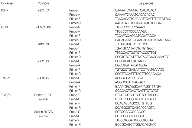
Fingertip blood from participating subjects was absorbed on filter papers and transported to the laboratory in Seoul, Korea. DNA was extracted from the filter papers using the Chelex-100 resin protocol (BioRad, Hercules, California, USA). Specific primers and probes were designed in order to analyze single nucleotide polymorphisms (SNP) of the IFN-γ, IL-10, TNF-α, and TGF-β1 genes (Table 1) [8-10]. Also the TaqMan (Applied Biosystems, Foster City, California, USA) real-time PCR allelic discrimination assay was applied [9]. TaqMan probes and PCR primers were used to analyze SNP genotypes. A pair of TaqMan probes contained each polymorphism site and complementary nucleotides for each genotype. A mixture of 10 ng of DNA, 900 nM of primers, 200 nM of probes, and 2X TaqMan Universal PCR mix (Perkin-Elmer, Applied Biosystems) underwent reaction with real-time PCR conditions of 50℃ for 2 min, 95℃ for 10 min and 92℃ for 30 sec 60℃ for 1 min for 50 cycles. The products were sequenced and sequences were grouped by SNP.
Table 1.
Probe and primer combinations for cytokine polymorphism analysis using TaqMan real-time PCR allelic discrimination assay
Statistical analysis
A total of 7 known SNP sites were analyzed for linkage of genotypes, clonorchiasis intensity, and sonographic data using the SPSS 12 software. The SNP sites which were examined were IFN-γ (+874 T/A), IL-10 (-1,082 G/A, -819 C/T, -592 C/A), TNF-α (-308 G/A), and TGF-β1 (codon 10 T/C, codon 25 G/C).
RESULTS
Demography of subjects and fecal examination
A total of 240 subjects (124 males and 116 females) were examined with a median age of 40 years (range, 9 to 80 years). Fecal examination detected eggs of C. sinensis in 171 subjects, with light infection in 65.5% and moderate infection in 35.5% of positives. Samples were negative for screening of other helminth eggs. Individuals under 10 years of age did not have positive fecal samples. Positive fecal samples were seen in subjects aged 10-20 years (31.3%), 21-30 years (66.7%), 31-40 years (76.4%), 41-50 years (82.7%), 51-60 years (80.3%), and 61 years or older (38.1%).
Ultrasonography
Ultrasonography revealed a diagnosis of IHDD in 106 (44.2%) and PDE in 101 (42.1%) of the 240 subjects.
Analysis of cytokine gene polymorphisms
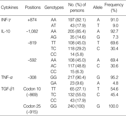
The intermediate-producing genotype, AT (17.9%) and low-producing genotype, AA (82.1%) were observed at +874 T/A for IFN-γ analysis [10]. The low-producing genotype, AA (85.4%) and the intermediate-producing genotype, AG (14.6%) at -1,082 G/A, TT (45.0%), TC (49.2%), and CC (5.8%) genotypes at -819 C/T, and AA (45.0%), AC (48.8%), and CC (6.3%) genotypes at -592 C/A were also observed for IL-10 analysis [11]. The high-producing genotype, GA (9.6%) and the low-producing genotype, GG (90.4%) were detected at -308 G/A for TNF-α analysis. The high-producing genotype, TC (55.0%) or TT (27.1%), and the low-producing genotype CC (17.9%) for TGF-β1 were found at codon 10 T/C and the genotype GG only at codon 25 G/C (Table 2) [12]. This cytokine gene polymorphism data from Chinese subjects were similar to those seen in Koreans [11,12].
Table 2.
Genotype and allele frequencies of IFN-γ, IL-10, TNF-α and TGF-β1 genes in subjected population (n = 240)
Relation between cytokine gene SNPs and pathology caused by clonorchiasis
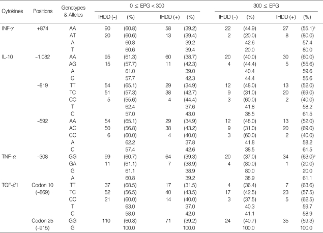
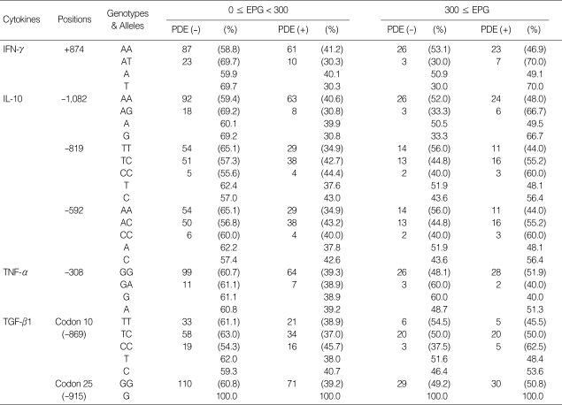
Participants were divided into 3 groups based on EPG counts: negative, EPG 1-299, and EPG ≥ 300, and were further divided into 6 groups based on degree of IHDD (Table 3) and PDE (Table 4). All groups were compared to assess genotype and allele frequencies, but none of the comparisons was statistically significant. Gene polymorphisms of IL-10 and TGF-β1 revealed no differences in the incidence of bile duct fibrosis among individuals with negative, light, and moderate infections. Among individuals with clonorchiasis of EPG 300 or more, the intermediate-producing IFN-γ genotype, +874AT, revealed a higher incidence of fibrosis than the low-producing genotype, +874AA (80.0% vs 55.1%; odds ratio, OR = 3.3; P = 0.177), and the low-producing TNF-α genotype, -308GG, revealed a higher incidence of fibrosis than the high-producing genotype, -308GA (63.8% vs 20.0%; OR = 6.8; P = 0.148). However, these values did not reach statistical significance.
Table 3.
Distribution of genotypes and alleles of 4 cytokines by EPG groups of Clonorchis sinensis in relation to intrahepatic duct dilatation (IHDD)
aP = 0.177 (OR = 3.3, 95% CI 0.6-17.0), AT vs AA; bP = 0.148 (OR = 6.8, 95% CI 0.7 - 65.2), GG vs GA.
Table 4.
Distribution of genotypes and alleles of 4 cytokines between 2 EPG groups of Clonorchis sinensis in relation to periductal echogenicity (PDE)
We analyzed the associations between cytokine genotypes and peri-ductal fibrosis in terms of IHDD. Combinations of IFN-γ and TNF-α genotypes were classified into 3 groups stratified for IHDD risks: high, moderate, and low risk groups. The incidence of IHDD was 25.0% in the low risk group (OR = 1), 56.5% in the moderate risk group (OR = 3.9), and 88.9% in the high risk group (OR = 24.0), and the differences were statistically significant by linear by linear association analysis (P = 0.022, Table 5).
Table 5.
Association of IFN-γ and TNF-α genotype combinations with intrahepatic duct dilatation (IHDD) in persons moderately infected with Clonorchis sinensis
aIFN-γ: intermediate producing genotype, +874AT; low producing, +874AA. TNF-α: high producing genotype -308GA; low producing, -308GG; bLinear by linear association analysis (P = 0.022).
OR, odds ratio; CI, confidence interval.
DISCUSSION
The present study first investigated relations between host SNPs and liver pathology in clonorchiasis. The SNP genotype frequencies of the 4 cytokine genes at the polymorphic sites revealed no differences when examined by EPG counts. Furthermore, the cytokine genotype frequencies had no significant correlation with the liver pathologies determined by ultrasonography. IHDD is the result of fibrosis of the bile duct wall, and PDE is the result of inflammation surrounding the bile duct [13]. Although IHDD was the primary parameter evaluated in this analysis, it had no significant correlation with genotype or allele frequencies of individual cytokines.
However, a significant correlation was observed when the cytokine genotypes of IFN-γ and TNF-α were grouped in combination according to the risks for fibrosis [11,12]. Among individuals with clonorchiasis of EPG ≥ 300, the high risk group, intermediate-producing IFN-γ (+874AT) and low-producing TNF-α (-308GG) genotypes, was significantly correlated with development of IHDD (Table 5), but not with that of PDE. When the low risk group, low-producing IFN-γ(+874AA) and high-producing TNF-α(-308GA), was used as a reference (OR = 1), the moderate (OR = 3.9) and high (OR = 24.0) risk groups had significantly increased frequencies of IHDD (P = 0.022). The incidence of IHDD frequency was significantly different between different genotype groups despite the inclusion of only 59 total subjects with clonorchiasis of EPG ≥ 300. In our study, 9 of 59 individuals (15.3%) belonged to the high risk group for fibrosis, and 8 of these individuals (88.9%) developed IHDD. Fibrosis in this group was severe and persisted after praziquantel treatment, and therefore IHDD cases by liver ultrasonography must be strongly considered as clonorchiasis even if they are cured in endemic areas.
A few individuals, 4 of 59 (6.8%) belonged to the low risk group for fibrosis, and the bile duct walls in these individuals had minimal fibrosis and only one of these individuals (25%) had IHDD. It is possible that this group of SNPs may contribute to the majority of negative hepatic ultrasonographic findings in clonorchiasis patients. Though the numbers of this low risk group were only 4 and looked insufficient, we may suggest the tendency of the tissue reaction compared with that of other groups.
Most subjects (46/59, 78.0%) belonged to the moderate risk group for fibrosis, having intermediate-producing IFN-γ (+874AT) and high-producing TNF-α (-308GA) genotype or low-producing IFN-γ (+874AA) and low-producing TNF-α (-308GG) genotype. More than half (26/46, 56.5%) of the individuals in the moderate risk group had IHDD, which was similar to the frequencies of clonorchiasis IHDD cases as previously reported in China [14]. The individuals in this moderate risk group may develop fibrosis secondary to C. sinensis infection and resolve after treatment, and this may be the most representative hepatic disease progression in clonorchiasis.
It is well-known that TNF-α, IFN-γ, IL-10, and TGF-β1 polymorphisms affect cytokine production in either suppression or stimulation. In several liver diseases, low-producing IL-10 and high-producing IFN-γ and TGF-β1 genotypes are known to promote hepatic fibrosis, but the action of TNF-α is unclear [10]. Similarly, rejection in kidney transplantation was reported to occur more frequently among individuals with high risk IFN-γ and TNF-α genotype combinations than those of moderate or low risk genotypes [12].
Fibrosis during clonorchiasis occurs as a consequence of periductal inflammation and surrounds the bile duct to induce the ductal wall thickening. Inflammation sites in the ductal wall contain numerous inflammatory or immune cells which secrete multiple cytokines, and the cytokines are mixed in the tissue and may modulate tissue reactions in complicated ways according to genotype polymorphisms. Complicated tissue reactions in the liver may explain the reason that liver ultrasonography in infected humans have displayed low sensitivity and specificity [6,14]. A small proportion of clonorchiasis patients will have negative liver scans on ultrasonography as false negatives. Conversely, some egg negative individuals from endemic areas will show IHDD on scans as false positives [6]. Most ultrasonographic false positives may have residual bile duct fibrosis after treatment although a portion may be actively infected but found egg negative. This suggests that the livers of infected individuals respond to the liver fluke in different ways depending on the genetic polymorphisms of hosts. The IFN-γ and TNF-α SNPs may contribute to the different tissue responses observed by ultrasonography.
IFN-γ stimulates cellular immunity by activation of Th1 lymphocytes, cytolytic T lymphocytes, and natural killer cells. A study on IFN-γ gene polymorphism associated with Schistosoma mansoni liver pathology revealed that, +2,109 A/G genotype promoted hepatic fibrosis and +3,810 G/A genotype inhibited it [15]. Our study suggests that +874AT genotype of IFN-γ may play a key role for bile duct fibrosis in clonorchiasis.
Henri et al. [3] suggested that bile duct fibrosis was additionally promoted by TNF-α, but another study indicated that TNF-α gene polymorphisms in Schistosoma mansoni infection did not directly affect liver fibrosis [16]. In our study on TNF-α the low-producing genotype produced more fibrosis than the high-producing genotype (63% vs 20%), and this indicates that TNF-α may suppress fibrosis in the liver.
The genotypes and allelic frequencies of IFN-γ, IL-10, TNF-α, and TGF-β1 genes in Chinese subjects were similar to those seen in Koreans [11,12], and based on these similarities the genotype polymorphisms associated with clonorchiasis were also expected to be similar to those in Koreans.
In conclusion, individuals with combination genotypes of the intermediate-producing IFN-γ (+874AT) and the low-producing TNF-α (-308GG) have a higher risk of developing liver fibrosis than other combinations of genotypes in clonorchiasis. Such SNP polymorphisms may contribute to the various sonographic findings in clonorchiasis.
ACKNOWLEDGEMENTS
We appreciate Ms. Zhuo Ji, Mr. Gui Yu, and other staff of Heilongjiang CDC for their kind technical and administrative cooperation during this study. This study was financially supported by the grant from the Seoul National University College of Medicine Research Fund, #800-20040066.
References
- 1.Hong ST. Clonorchis sinensis. In: Miliotis MD, Bier JW, editors. International Handbook of Foodborne Pathogens. New York, USA: Marcel Dekker, Inc; 2003. pp. 581–592. [Google Scholar]
- 2.Lee SH, Lee JI, Huh S, Yu JR, Chung SW, Chai JY, Hong ST. Secretions of the biliary mucosa in experimental clonorchiasis. Korean J Parasitol. 1993;31:13–20. doi: 10.3347/kjp.1993.31.1.13. [DOI] [PubMed] [Google Scholar]
- 3.Henri S, Chevillard C, Mergani A, Paris P, Gaudart J, Camilla C, Dessein H, Montero F, Elwali NE, Saeed OK, Magzoub M, Dessein AJ. Cytokine regulation of periportal fibrosis in humans infected with Schistosoma mansoni: IFN-gamma is associated with protection against fibrosis and TNF-alpha with aggravation of disease. J Immunol. 2002;169:929–936. doi: 10.4049/jimmunol.169.2.929. [DOI] [PubMed] [Google Scholar]
- 4.Powell EE, Edwards-Smith CJ, Hay JL, Clouston AD, Crawford DH, Shorthouse C, Purdie DM, Jonsson JR. Host genetic factors influence disease progression in chronic hepatitis C. Hepatology. 2000;31:828–833. doi: 10.1053/he.2000.6253. [DOI] [PubMed] [Google Scholar]
- 5.Hirayama K. Genetic factors associated with development of cerebral malaria and fibrotic schistosomiasis. Korean J Parasitol. 2002;40:165–172. doi: 10.3347/kjp.2002.40.4.165. [DOI] [PMC free article] [PubMed] [Google Scholar]
- 6.Choi D, Hong ST, Lim JH, Cho SY, Rim HJ, Ji Z, Yuan R, Wang S. Sonographic findings of active Clonorchis sinensis infection. J Clin Ultrasound. 2004;32:17–23. doi: 10.1002/jcu.10216. [DOI] [PubMed] [Google Scholar]
- 7.Hong ST, Choi MH, Kim CH, Chung BS, Ji Z. The Kato-Katz method is reliable for diagnosis of Clonorchis sinensis infection. Diagn Microbiol Infect Dis. 2003;47:345–347. doi: 10.1016/s0732-8893(03)00113-5. [DOI] [PubMed] [Google Scholar]
- 8.Dai CY, Chuang WL, Hsieh MY, Lee LP, Hou NJ, Chen SC, Lin ZY, Hsieh MY, Wang LY, Tsai JF, Chang WY, Yu ML. Polymorphism of interferon-gamma gene at position +874 and clinical characteristics of chronic hepatitis C. Transl Res. 2006;148:128–133. doi: 10.1016/j.trsl.2006.04.005. [DOI] [PubMed] [Google Scholar]
- 9.Goyal A, Kazim SN, Sakhuja P, Malhotra V, Arora N, Sarin SK. Association of TNF-beta polymorphism with disease severity among patients infected with hepatitis C virus. J Med Virol. 2004;72:60–65. doi: 10.1002/jmv.10533. [DOI] [PubMed] [Google Scholar]
- 10.Bataller R, North KE, Brenner DA. Genetic polymorphisms and the progression of liver fibrosis: a critical appraisal. Hepatology. 2003;37:493–503. doi: 10.1053/jhep.2003.50127. [DOI] [PubMed] [Google Scholar]
- 11.Roh EY, Park MH, Park HJ, Ha J, Kim S, Ahn C. Association of polymorphisms in the IL-10 and IFN-gamma genes with allograft dysfunction following kidney transplantation in Koreans. J Korean Soc Transplant. 2003;17:34–42. [Google Scholar]
- 12.Park JY, Park MH, Park H, Ha J, Kim SJ, Ahn C. TNF-alpha and TGF-beta1 gene polymorphisms and renal allograft rejection in Koreans. Tissue Antigens. 2004;64:660–666. doi: 10.1111/j.1399-0039.2004.00330.x. [DOI] [PubMed] [Google Scholar]
- 13.Choi D, Hong ST. Imaging diagnosis of clonorchiasis. Korean J Parasitol. 2007;45:77–85. doi: 10.3347/kjp.2007.45.2.77. [DOI] [PMC free article] [PubMed] [Google Scholar]
- 14.Choi MS, Choi D, Choi MH, Ji Z, Li Z, Cho SY, Hong KS, Rim HJ, Hong ST. Correlation between sonographic findings and infection intensity in clonorchiasis. Am J Trop Med Hyg. 2005;73:1139–1144. [PubMed] [Google Scholar]
- 15.Chevillard C, Moukoko CE, Elwali NE, Bream JH, Kouriba B, Argiro L, Rahoud S, Mergani A, Henri S, Gaudart J, Mohamed-Ali Q, Young HA, Dessein AJ. IFN-gamma polymorphisms (IFN-gamma +2109 and IFN-gamma +3810) are associated with severe hepatic fibrosis in human hepatic schistosomiasis (Schistosoma mansoni) J Immunol. 2003;171:5596–5601. doi: 10.4049/jimmunol.171.10.5596. [DOI] [PubMed] [Google Scholar]
- 16.Moukoko CE, El Wali N, Saeed OK, Mohamed-Ali Q, Gaudart J, Dessein AJ, Chevillard C. No evidence for a major effect of tumor necrosis factor alpha gene polymorphisms in periportal fibrosis caused by Schistosoma mansoni infection. Infect Immun. 2003;71:5456–5460. doi: 10.1128/IAI.71.10.5456-5460.2003. [DOI] [PMC free article] [PubMed] [Google Scholar]


