Abstract
The antiinflammatory action of aspirin generally has been attributed to direct inhibition of cyclooxygenases (COX-1 and COX-2), but additional mechanisms are likely at work. These include aspirin’s inhibition of NFκB translocation to the nucleus as well as the capacity of salicylates to uncouple oxidative phosphorylation (i.e., deplete ATP). At clinically relevant doses, salicylates cause cells to release micromolar concentrations of adenosine, which serves as an endogenous ligand for at least four different types of well-characterized receptors. Previously, we have shown that adenosine mediates the antiinflammatory effects of other potent and widely used antiinflammatory agents, methotrexate and sulfasalazine, both in vitro and in vivo. To determine in vivo whether clinically relevant levels of salicylate act via adenosine, via NFκB, or via the “inflammatory” cyclooxygenase COX-2, we studied acute inflammation in the generic murine air-pouch model by using wild-type mice and mice rendered deficient in either COX-2 or p105, the precursor of p50, one of the components of the multimeric transcription factor NFκB. Here, we show that the antiinflammatory effects of aspirin and sodium salicylate, but not glucocorticoids, are largely mediated by the antiinflammatory autacoid adenosine independently of inhibition of prostaglandin synthesis by COX-1 or COX-2 or of the presence of p105. Indeed, both inflammation and the antiinflammatory effects of aspirin and sodium salicylate were independent of the levels of prostaglandins at the inflammatory site. These experiments also provide in vivo confirmation that the antiinflammatory effects of glucocorticoids depend, in part, on the p105 component of NFκB.
Salicylates, which include aspirin, are probably the oldest class of antiinflammatory agents still in use. As discovered by Vane (1), many of aspirin’s therapeutic effects are clearly due to inhibition of prostaglandin synthesis. However, not all of aspirin’s antiinflammatory effects can be accounted for by simple inhibition of prostaglandin synthesis via inhibition of cyclooxygenases 1 and 2 (COX-1 and COX-2). For example, therapeutic serum concentrations of salicylate, the major metabolite of aspirin and a poor inhibitor of COX-1 and COX-2, correlate better with clinical antiinflammation than serum concentrations of aspirin (2). Indeed, we have found that aspirin and sodium salicylate inhibit neutrophil activation by interfering with signal transduction pathways independently of prostaglandin synthesis (3, 4); others have reported that aspirin and sodium salicylate, but not other inhibitors of COX-1 and COX-2, disrupt signal transduction between cell surface receptors and transcription of “inflammatory” cytokines by inhibiting the inhibitor of NFκB (IκB) kinase β, thereby preventing translocation of the transcriptional regulator NFκB to the nucleus (5–9). More recently, Serhan’s laboratory (10) has found that aspirin, but not sodium salicylate, promotes synthesis of such antiinflammatory eicosanoids as 15-epi-lipoxin A4, and Schwenger et al. (11) have observed that salicylates promote apoptosis via activation of the p38 mitogen-activated protein kinase. Finally, salicylates uncouple oxidative phosphorylation leading to ATP catabolism, and we have established that pharmacologically relevant concentrations of sodium salicylate diminish intracellular ATP concentrations in vitro, thereby releasing micromolar amounts of adenosine, an autacoid with potent antiinflammatory properties (12), into extracellular fluids (13). To analyze the antiinflammatory effects of aspirin and sodium salicylate in vivo, we compared their inhibition of carrageenan-induced inflammation with that of the equipotent COX-1 and COX-2 inhibitor indomethacin and with that of dexamethasone. We also compared their efficacy in wild-type mice with that in animals with targeted disruption of either COX-2 or NFκB (p105).
METHODS
Materials.
Acetylsalicylic acid, sodium salicylate, indomethacin, carrageenan (type I), adenosine deaminase (ADA; type IV; calf intestinal), and dexamethasone were obtained from Sigma, and 3,7-dimethyl-1-propargylxanthine (DMPX) was obtained from Research Biochemicals (Natick, MA). All other reagents were of the highest grade available.
Mice.
BALB/c mice were purchased from Taconic Farms. Animals with targeted mutation of COX-2 (female B6; 129–Ptgs2fm1Jed) were purchased from The Jackson Laboratory. Breeding pairs of NFκB knockout mice (p105; B6; 129–Nfkb1fm1Bal) and wild-type controls (C57BL/6; 129) were purchased from The Jackson Laboratory, and the mice were bred in the New York University School of Medicine animal facility.
Induction of Air Pouches and Carrageenan-Induced Inflammation.
To induce an air pouch, 10- to 12-week-old mice were injected subcutaneously on the back with 3 ml of air. Every other day, the pouches were reinflated with 1 ml of air for a total period of 6 days. Mice were treated for 3 consecutive days and 1 h before the induction of inflammation with 0.2 ml of saline (control), or 0.2 ml of saline containing aspirin (200 mg/kg), sodium salicylate (200 mg/kg), or indomethacin (10 mg/kg) by gastric gavage. Inflammation was induced by injection of 1 ml of a suspension of 2% (wt/vol) carrageenan in PBS into the air pouch, as we have previously described (14, 15). After 4 h, the mice were killed; the pouches were flushed with 2 ml of PBS; and exudates were harvested. Aliquots were diluted to 1:1 with 0.01% (wt/vol) methylene blue in PBS, and cells (>95% neutrophils) were counted in a standard hematocytometer chamber (American Optical). There was no difference between the number of leukocytes present in the inflammatory exudates of male or female BALB/c, C57BL/6, 129, or NFκB (p105) knockouts (data not shown). All studies were carried out by using mice matched for age and gender. These studies were reviewed and approved by the Institutional Animal Care and Use Committee of the New York University Medical Center.
Injection of ADA and Adenosine Receptor Antagonist.
In some experiments, ADA (1.5 units/200 μl) or the adenosine antagonist DMPX (200 μg/200 μl) was injected with the carrageenan suspension into the air pouch. Control groups received 200 μl of PBS to correct the volume. Before dilution and injection into the air pouch, 50 μl of ADA (4,000 units/ml) was dialyzed against PBS overnight (4°C).
Adenosine Determination.
Aliquots of pouch exudates were added to an equal volume of 10% (wt/vol) trichloroacetic acid and kept on ice, and the organic phase was extracted with Freon/trioctylamine (31:9, vol/vol). The adenosine concentration in the aqueous phase was determined by reverse-phase HPLC, as we have previously described (14). Samples were applied to a SUPELCOSIL LC-18-T column (Supelco) and eluted with a linear gradient of 0.01 M ammonium phosphate (pH 5.5) and methanol (0–40%) formed over 45 min with a 1.5 ml/min flow rate. Adenosine was identified by retention time and by the characteristic ultraviolet ratio of absorbance at 250 nm and 260 nm, and the concentration was calculated by comparison with standards.
Prostaglandin E2 and Leukotriene B4 Determinations.
The concentration of prostaglandin E2 and leukotriene B4 in inflammatory exudates of mice was determined by using commercial ELISA kits (Cayman Chemicals, Ann Arbor, MI), following the instructions of the manufacturer.
RESULTS
In our initial studies, we tested the effects of aspirin, its major metabolite, sodium salicylate, and the equally nonselective COX-1 and COX-2 inhibitor, indomethacin, on the development of inflammation in the murine air-pouch model of inflammation (14). Mice were treated with oral aspirin or sodium salicylate at doses (200 mg/kg) required to achieve the millimolar serum concentrations needed for treatment of chronic inflammatory diseases (2) for 3 days: carrageenan-induced leukocyte accumulation in the air pouch was significantly decreased (Fig. 1 and Table 1). In contrast, treatment with indomethacin (at 10 mg/kg, its COX-1 and COX-2 inhibitory dose) failed to affect leukocyte accumulation in the inflamed air pouch (Fig. 1). These data suggest that indomethacin, which inhibits carrageenan-induced paw swelling in a COX-1- and COX-2-dependent manner (16), affects neutrophil accumulation and edema differentially. The data also suggest that inflammation in the air-pouch model, which more closely mimics inflammatory joint disease than does simple paw swelling, is provoked by mediators other than stable prostaglandins. Further evidence for this hypothesis is provided by the finding that aspirin’s pharmacologic precursor and major metabolite, sodium salicylate, was equipotent to aspirin in the air-pouch model, despite its relative inadequacy as an in vitro inhibitor of COX-1 and COX-2 (Fig. 1).
Figure 1.
Aspirin and sodium salicylate, but not indomethacin, are antiinflammatory in the murine air-pouch model of inflammation. Air pouches were formed on mice treated with saline, aspirin (200 mg/kg), sodium salicylate (200 mg/kg), or indomethacin 10 mg/kg), as described in Methods. Inflammation was induced by injection of 1 ml of a suspension of 2% (wt/vol) carrageenan in PBS into the air pouch, as we have previously described (14, 15). After 4 h, the mice were killed; the pouches were flushed with 2 ml of PBS; and the exudates were harvested. In some animals, DMPX (10 μM) or ADA (0.125 units/ml) was injected into the air pouch with the inflammatory stimulus. ADA and DMPX significantly diminished the inhibition of leukocyte accumulation mediated by treatment with either aspirin or sodium salicylate (P < 0.008 for all; n = 7–10 mice per group, studied on two separate occasions). We have previously established that the effects of ADA and DMPX in the murine air-pouch model are specific for adenosine A2 receptors, because neither agent reverses the antiinflammatory effects of dexamethasone and because the A1 receptor antagonist 1,3-dipropyl-8-cyclopentylxanthine does not reverse the antiinflammatory effect (14). The data are expressed as the mean percentages ± SEM of inhibition of leukocyte accumulation. Mean leukocyte accumulation in animals treated with saline was (2.6 ± 0.2) × 106 cells per ml.
Table 1.
Inhibition of prostaglandin synthesis by salicylates does not correlate with inhibition of inflammation
| Test group | Air pouch prostaglandin E2, pmol/ml | Air pouch leukotriene B4 pg/ml | Air pouch white cell count, n × 106 cells per ml | No. of mice |
|---|---|---|---|---|
| Control | 1,969 ± 300 | 92 ± 23 | 4.6 ± 0.2 | 6 |
| ADA (0.125 units/ml) | 2,030 ± 446 | 101 ± 23 | 4.5 ± 0.4 | 6 |
| DMPX (10 μM) | 2,330 ± 374 | 162 ± 32 | 5.3 ± 0.6 | 6 |
| Aspirin | 482 ± 91* | 91 ± 23 | 2.7 ± 0.2* | 4 |
| Aspirin + ADA | 421 ± 45* | 197 ± 31 | 4.1 ± 0.3† | 4 |
| Aspirin + DMPX | 423 ± 181** | 146 ± 44 | 4.4 ± 0.2† | 4 |
| Salicylate | 586 ± 134** | 217 ± 63 | 2.6 ± 0.2* | 4 |
| Salicylate + ADA | 602 ± 133** | 261 ± 60 | 4.1 ± 0.3‡ | 4 |
| Salicylate + DMPX | 765 ± 191** | 224 ± 39 | 4.3 ± 0.2‡ | 4 |
Data are presented as means ± SEM. Mice (BALB/c) were treated with saline, aspirin (100 mg/kg) or sodium salicylate (100 mg/kg) for 3 days by oral gavage during the induction of the air pouch. The pouches were injected with 2% (wt/vol) carrageenan, 0.125 units/ml ADA, 10 μM DMPX, or buffer. After 4 h, the animals were killed, and the exudates were collected and kept frozen (−20°C) until the prostaglandin E2 and leukotriene B4 contents were assayed by using a commercial ELISA kit.
, P < 0.01 vs. control, ADA, or DMPX alone (Student’s t test).
, P < 0.03 vs. control, ADA, or DMPX alone (Student’s t test).
, P < 0.01 vs. aspirin alone (Student’s t test).
, P < 0.01 vs. sodium salicylate alone (Student’s t test).
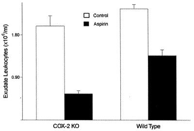
It is generally agreed that COX-2, the inducible form of cyclooxygenase, is responsible for the production of inflammatory eicosanoids at inflamed sites (17), although recent data suggest that COX-1 may be critical for inflammatory responses in mice (18, 19). We therefore determined whether aspirin retained its antiinflammatory properties in animals with targeted disruption of the gene for COX-2 (ref. 20; B6; 129–Ptgs2fm1Jed; The Jackson Laboratory). No difference was found in the response to aspirin between parental strain (wild-type; C57BL/6) animals and COX-2 knockouts (Fig. 2). The concentrations of prostaglandin E2 in the inflammatory exudates of COX-2 knockout mice were comparable with those found in controls, consistent with the observation that there is a compensatory increase of COX-1 activity in COX-2 knockout cells (21). As expected, aspirin treatment reduced the concentration of prostaglandin E2 in the inflammatory exudates of COX-2 knockout mice (Table 2).
Figure 2.
Aspirin is antiinflammatory in animals rendered deficient in COX-2 activity [COX-2 knockouts (KO)]. Air pouches were formed on female COX-2 knockout mice (C57BL/6; 129–Ptgs2fm1Jed; The Jackson Laboratory) or female wild-type mice (C57BL/6; 129), and the mice were treated with aspirin or saline, as described in Methods. There were significantly fewer leukocytes present in the air pouches of COX-2 knockout mice treated with aspirin (P < 0.01; n = 5) than in those of controls (n = 4). Similarly there were significantly fewer leukocytes present in the air pouches of wild-type mice treated with aspirin (P < 0.05; n = 5) than in those of controls (P < 0.05). The leukocyte counts are shown as means ± SEM.
Table 2.
High concentrations of prostaglandin E2 are present in the inflammatory exudates of COX-2 knockout mice
| Mouse type | Control
|
Aspirin
|
||
|---|---|---|---|---|
| Air pouch prostaglandin E2, pmol/ml | No. of mice | Air pouch prostaglandin E2, pmol/ml | No. of mice | |
| Wild type | 2,016 ± 793 | 5 | 578 ± 83 | 3 |
| NFκB knockout | 2,879 ± 560 | 10 | 785 ± 37 | 5 |
| COX-2 knockout | 3,109 | 2 | 353 | 2 |
Data are presented as means ± SEM. Female C57/B16, 129 (wild type), NFκB (p105) knockout, and COX-2 knockout mice were treated with saline or aspirin (100 mg/kg) for 3 days by oral gavage during the induction of the air pouch. The pouches were injected with 2% (wt/vol) carrageenan, and 4 h later, the animals were killed, and the exudates were collected and kept frozen (−20°C) until the prostaglandin E2 content was assayed by using a commercial ELISA kit. Each determination was carried out in triplicate.
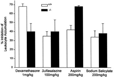
High-dose salicylates (serum levels of 2–5 mM) and glucocorticoids are equally effective antiinflammatory agents not only in acute rheumatism (3) but also in the murine carrageenan air-pouch model (14), and these agents share at least one common therapeutic target. In several in vitro systems, high-dose salicylates and glucocorticoids prevent translocation of NFκB to the nucleus (7–10, 22), thereby impeding release of inflammatory mediators and/or cytokines. To test the hypothesis that the in vivo effects of aspirin and glucocorticoids are also mediated by interference with NFκB-mediated regulation of transcription, we compared their effects in wild-type mice and in animals with targeted disruption of NFκB (p105; B6; 129–Nfkb1fm1Bal; ref. 23). Surprisingly, disruption of NFκB p50 failed to affect the accumulation of leukocytes in air pouches of wild-type mice [(2.6 ± 0.2) × 106 cells per ml vs. (3.1 ± 0.7) × 106 cells per ml; wild type vs. knockout; n = 8; P = not significant]. Nevertheless, aspirin, salicylate, and sulfasalazine retained their antiinflammatory effects in the NFκB p50 knockouts, whereas, as expected, dexamethasone was significantly less antiinflammatory (Fig. 3). Indeed, aspirin inhibited leukocyte accumulation more completely in the NFκB knockout mice than the wild-type mice. This observation supports the venerable notion that there are multiple convergent pathways of inflammation and that elimination of one of these pathways may accentuate the antiinflammatory effects of an agent that blocks another pathway (24). These results provide direct in vivo confirmation that the antiinflammatory actions of glucocorticoids are mediated, at least in part, by modulation of an event involving p105, the precursor of p50 of the NFκB complex. Because the antiinflammatory effects of glucocorticoids, but not salicylates and sulfasalazine, are diminished in the p105 knockouts, it is unlikely that salicylates share with glucocorticoids an antiinflammatory mechanism dependent on IκB-mediated inhibition of nuclear translocation of NFκB.
Figure 3.
Aspirin, sodium salicylate, and sulfasalazine, unlike dexamethasone, are no less antiinflammatory in p105 (NFκB) knockout mice than in wild-type mice. Breeding pairs of NFκB (p105; −/−) knockout mice (B6; 129–Nfkb1fm1Bal) and wild-type controls (+/+; C57BL/6; 129) were obtained from The Jackson Laboratory and bred in the New York University Medical Center animal facility. Air pouches were formed, and the animals were treated with aspirin or sodium salicylate before induction of inflammation and quantitation of leukocytes, as described in Methods and Results. Animals treated with dexamethasone were given a single i.p. dose of dexamethasone (1.5 mg/kg) 1 h before induction of inflammation in the air pouch, and animals treated with sulfasalazine were given sulfasalazine (100 mg/kg) by gastric gavage for 3 days before and on the day of the induction of inflammation. The inhibition of leukocyte accumulation differed significantly between aspirin-treated and dexamethasone-treated knockout and wild-type mice (P < 0.02 for both; Student’s t test). The data are presented as the mean percentages ± SEM of inhibition of leukocyte accumulation in 6–10 mice per group, studied on at least two separate occasions.
Previously, we established that treatment of neutrophils in vitro with pharmacologically relevant concentrations of sodium salicylate diminishes cellular ATP concentrations by 16%, a reduction associated with marked increases in extracellular concentrations of adenosine (13). Adenosine, acting at each of its four known receptors, is a potent endogenous antiinflammatory autacoid both in vitro and in vivo (11). Because we have also found that adenosine, acting at A2 receptors, mediates the antiinflammatory effects of methotrexate and sulfasalazine in vivo (14, 15, 25), we determined concentrations of adenosine in air pouches of control and aspirin-treated animals by HPLC, as we have previously described (14). Adenosine concentrations in exudates from aspirin-treated animals were far greater than those from controls (224 ± 98 nM vs. 13 ± 6 nM, respectively; P < 0.01; n = 6). Elimination of extracellular adenosine from the inflammatory exudate by the addition of ADA to the air pouch completely reversed the antiinflammatory effects of both aspirin and sodium salicylate (Fig. 1 and Table 1), whereas the adenosine A2 receptor antagonist DMPX also reversed the antiinflammatory effect of aspirin (Fig. 1 and Table 1). Reversal of the antiinflammatory effects of both aspirin and sodium salicylate by ADA and DMPX is specific; as we have previously shown, neither of these agents affects the capacity of dexamethasone to diminish inflammation in the air-pouch model (14). Moreover, neither ADA nor DMPX interferes with the capacity of aspirin or sodium salicylate treatment to inhibit prostaglandin accumulation in the inflammatory exudate (Table 1). The dissociation between prostaglandin E2 concentration and inflammation (increased white cell count) indicates that neither aspirin nor sodium salicylate mediates their antiinflammatory effects by inhibiting prostaglandin production and suggests that prostaglandin E2 is not the major mediator of inflammation in this model. Moreover, neither aspirin nor sodium salicylate significantly altered the concentration of another arachidonate-derived inflammatory mediator, leukotriene B4. Finally, removal of adenosine or adenosine receptor blockade failed to lower concentrations of leukotriene B4 in the inflammatory exudates (Table 1). Thus, aspirin and sodium salicylate increased adenosine concentrations in inflammatory exudates, and either enzymatic elimination or pharmacologic antagonism of adenosine at its receptors reversed this antiinflammatory effect of aspirin and sodium salicylate. These results indicate that adenosine, acting at A2 receptors, mediates the effects of aspirin and sodium salicylate on leukocyte accumulation in the murine air-pouch model of inflammation.
DISCUSSION
We report here that aspirin, sodium salicylate, and sulfasalazine share an antiinflammatory mechanism in the murine air-pouch model of inflammation. Our data indicate that adenosine, acting at its receptors on inflammatory cells, mediates the effects of all three of these agents on leukocyte accumulation. Moreover, the inhibition of leukocyte accumulation by salicylates and sulfasalazine, but not dexamethasone, is independent of any effect of these agents on either prostaglandin synthesis or translocation of the p50 component of NFκB to the nucleus.
Although it has long been argued that there is no one model that applies to inflammation in general (24), the readout of inflammation in our experiments was the influx of neutrophils into an air pouch, a more sharply defined measure of inflammation than simple measurements of soft tissue swelling or edema (16). Our data therefore disprove the notion that aspirin-like drugs work in every sort of inflammatory insult by virtue of their direct inhibition of cyclooxygenase(s). The data suggest, indeed, a complementary mechanism of action of clinically relevant (millimolar) doses of aspirin: extracellular release of adenosine at sites of inflammation. Although the studies reported here do not directly establish the mechanism by which aspirin and sodium salicylate promote adenosine release, it has been known for decades that salicylates inhibit oxidative phosphorylation (26–29), thereby diminishing intracellular concentrations of ATP and enhancing the release of adenosine (30). We have previously shown that sodium salicylate diminishes intracellular ATP concentrations and raises extracellular adenosine concentrations (13). Because adenosine has an extremely short half-life in biological fluids (31), its extracellular concentration depends heavily on cellular production, uptake, and use. Impaired oxidative phosphorylation could promote adenosine release by leading to enhanced ATP breakdown and increased release of adenine nucleotides, which are converted extracellularly to adenosine (25), or to diminished uptake and use of adenosine. Thus, it is likely that salicylates promote adenosine release by interfering with oxidative phosphorylation. Whatever the exact mechanism of adenosine release proves to be, the fact that adenosine mediates the antiinflammatory effects in vivo of such diverse agents as aspirin, methotrexate (14), and sulfasalazine (15) suggests another target for drug discovery.
Previously, we established that sulfasalazine promotes adenosine release both in vitro and in vivo and that adenosine, acting at its receptors, mediates the antiinflammatory effects of sulfasalazine in the murine air-pouch model of inflammation (15). There are four known types of adenosine receptors (A1, A2a, A2b, and A3), and our results have indicated that the adenosine A2 receptor, presumably A2a, is the receptor responsible for the antiinflammatory effects of sulfasalazine in this model. Others have suggested that the A3 adenosine receptor mediates much of adenosine’s antiinflammatory effect, because occupancy of adenosine A3 receptors inhibits release of tumor necrosis factor-α, IL-6, and IL-8 (32–37) by stimulated monocyte/macrophages and endothelial cells. The fact that sulfasalazine is as active in the knockout mice as in the wild-type controls provides further evidence against a role for adenosine A3 receptors in the antiinflammatory effects of sulfasalazine in this model; Bowlin and coworkers have reported that occupation of A3 receptors inhibits secretion of tumor necrosis factor-α by inhibiting NFκB translocation to the nucleus (32).
The role of NFκB in the pathogenesis of the inflammatory response has been well documented in experiments performed both in vitro and in vivo (38–40). NFκB binding sites are present in the upstream regulatory sequences of a number of genes encoding such inflammatory proteins as cytokines and adhesion molecules (38–40). Animals rendered deficient for expression of p105, a precursor of one of the subunits of NFκB (p50), manifest a number of immunological defects (23); however, no defects in acute inflammatory responses have been reported to date. We were surprised to observe that acute inflammation in the air-pouch model seemed to be unaffected in the NFκB (p105) knockout mice, although it is possible that p65 homodimers may substitute for p50/p65 heterodimers as transactivators of inflammatory mediators (41, 42). IκB is a member of the heteromeric complex that causes the NFκB complex to remain in the cytosol; phosphorylation and degradation of IκB permits the translocation of the NFκB complex to the nucleus. Aspirin and glucocorticoids are reported to modulate NFκB signaling indirectly via their effects on IκB, a protein that complexes with NFκB heterodimers to inhibit their translocation to the nucleus; aspirin inhibits IκB phosphorylation and catabolism (8, 9), whereas glucocorticoids promote transcription and synthesis of new IκB molecules (43–45) or interact directly with NFκB (46, 47). Sulfasalazine, an antiinflammatory agent chemically related to salicylate, also inhibits NFκB translocation to the nucleus by inhibiting IκB phosphorylation and proteolysis; the antiinflammatory effects of sulfasalazine have been ascribed to diminished NFκB translocation resulting from diminished IκB phosphorylation and degradation (48). The fact that glucocorticoids lose their antiinflammatory effects in the p105 knockouts is consistent with the hypothesis that glucocorticoids prevent NFκB translocation to the nucleus and thereby mediate, in part, their antiinflammatory effects. Neither sulfasalazine nor salicylates lose their antiinflammatory effects in p105 knockouts, an observation that is inconsistent with the hypothesis that sulfasalazine and salicylates share an NFκB-dependent antiinflammatory mechanism with glucocorticoids.
We have found that, in one model of inflammation, aspirin and its metabolite, salicylate, work independently of their inhibition of prostaglandin biosynthesis and their effects on NFκB. Instead, like sulfasalazine, they promote the release of an antiinflammatory purine autacoid, adenosine. It is likely that other properties of these venerable drugs remain to be discovered.
Acknowledgments
We thank Ms. Avani Desai for her outstanding technical assistance. This work was supported by National Institutes of Health Grants AR41911, GM56268, and HL1972, by an Arthritis Challenge Grant from Amersham Pharmacia–Upjohn, by General Clinical Research Center Grant MO1RR00096, and by Kaplan Cancer Center Grant CA160877.
ABBREVIATIONS
- COX-1
cyclooxygenase 1
- COX-2
cyclooxygenase 2
- IκB
inhibitor of NFκB
- ADA
adenosine deaminase
- DMPX
3,7-dimethyl-1-propargylxanthine
References
- 1.Vane J R. Nature (London) 1971;231:232–235. doi: 10.1038/newbio231232a0. [DOI] [PubMed] [Google Scholar]
- 2.Weissmann G. In: Cecil Textbook of Medicine. Wyngaarden J B, Smith L H Jr, Bennett J C, editors. Philadelphia: Saunders; 1992. pp. 114–118. [Google Scholar]
- 3.Abramson S B, Leszczynska-Piziak J, Clancy R M, Philips M R, Weissmann G. Biochem Pharmacol. 1994;47:563–572. doi: 10.1016/0006-2952(94)90189-9. [DOI] [PubMed] [Google Scholar]
- 4.Pillinger M H, Capodici C, Rosenthal P, Kheterpal N, Hanft S, Philips M R, Weissmann G. Proc Natl Acad Sci USA. 1998;95:14540–14545. doi: 10.1073/pnas.95.24.14540. [DOI] [PMC free article] [PubMed] [Google Scholar]
- 5.Yin M J, Yamamoto Y, Gaynor R B. Nature (London) 1998;396:77–80. doi: 10.1038/23948. [DOI] [PubMed] [Google Scholar]
- 6.Grilli M, Pizzi M, Memo M, Spano P. Science. 1996;274:1383–1385. doi: 10.1126/science.274.5291.1383. [DOI] [PubMed] [Google Scholar]
- 7.Kopp E, Ghosh S. Science. 1994;265:956–959. doi: 10.1126/science.8052854. [DOI] [PubMed] [Google Scholar]
- 8.Pierce J W, Read M A, Ding H, Luscinskas F W, Collins T. J Immunol. 1996;156:3961–3969. [PubMed] [Google Scholar]
- 9.Oeth P, Mackman N. Blood. 1995;86:4144–4152. [PubMed] [Google Scholar]
- 10.Claria J, Serhan C N. Proc Natl Acad Sci USA. 1995;92:9475–9479. doi: 10.1073/pnas.92.21.9475. [DOI] [PMC free article] [PubMed] [Google Scholar]
- 11.Schwenger P, Bellosta P, Vietor I, Basilico C, Skolnik E Y, Vilcek J. Proc Natl Acad Sci USA. 1997;94:2869–2873. doi: 10.1073/pnas.94.7.2869. [DOI] [PMC free article] [PubMed] [Google Scholar]
- 12.Cronstein B N. In: Molecular and Cellular Basis of Inflammation. Serhan C N, Ward P A, editors. Totowa, NJ: Humana; 1998. pp. 259–274. [Google Scholar]
- 13.Cronstein B N, Van de Stouwe M, Druska L, Levin R I, Weissmann G. Inflammation. 1994;18:323–335. doi: 10.1007/BF01534273. [DOI] [PubMed] [Google Scholar]
- 14.Cronstein B N, Naime D, Ostad E. J Clin Invest. 1993;92:2675–2682. doi: 10.1172/JCI116884. [DOI] [PMC free article] [PubMed] [Google Scholar]
- 15.Gadangi P, Longaker M, Naime D, Levin R I, Recht P A, Montesinos M C, Buckley M T, Carlin G, Cronstein B N. J Immunol. 1996;156:1937–1941. [PubMed] [Google Scholar]
- 16.Otterness I G, Bliven M L. In: Nonsteroidal Antiinflammatory Drugs. Lombardino J G, editor. New York: Wiley; 1985. pp. 111–252. [Google Scholar]
- 17.Needleman P, Isakson P C. J Rheumatol. 1997;24,Suppl. 49:6–8. [PubMed] [Google Scholar]
- 18.Wallace J L, Bak A, McKnight W, Asfaha S, Sharkey K A, MacNaughton W K. Gastroenterology. 1998;115:101–109. doi: 10.1016/s0016-5085(98)70370-1. [DOI] [PubMed] [Google Scholar]
- 19.Gilroy D W, Tomlinson A, Willoughby D A. Eur J Pharmacol. 1998;355:211–217. doi: 10.1016/s0014-2999(98)00508-1. [DOI] [PubMed] [Google Scholar]
- 20.Dinchuk J E, Car B D, Focht R J, Johnston J J, Jaffee B D, Covington M B, Contel N R, Eng V M, Collins R J, Czerniak P M, et al. Nature (London) 1995;378:406–409. doi: 10.1038/378406a0. [DOI] [PubMed] [Google Scholar]
- 21.Kirtikara K, Morham S G, Raghow R, Laulederkind S J, Kanekura T, Goorha S, Ballou L R. J Exp Med. 1998;187:517–523. doi: 10.1084/jem.187.4.517. [DOI] [PMC free article] [PubMed] [Google Scholar]
- 22.Funder J W. Annu Rev Med. 1997;48:231–240. doi: 10.1146/annurev.med.48.1.231. [DOI] [PubMed] [Google Scholar]
- 23.Sha W C, Liou H C, Tuomanen E I, Baltimore D. Cell. 1995;80:321–330. doi: 10.1016/0092-8674(95)90415-8. [DOI] [PubMed] [Google Scholar]
- 24.Weissmann G. In: Mediators of Inflammation. Weissmann G, editor. New York: Plenum; 1974. pp. 1–7. [Google Scholar]
- 25.Morabito L, Montesinos M C, Schreibman D M, Balter L, Thompson L F, Resta R, Carlin G, Huie M A, Cronstein B N. J Clin Invest. 1998;101:295–300. doi: 10.1172/JCI1554. [DOI] [PMC free article] [PubMed] [Google Scholar]
- 26.Glader B E. N Engl J Med. 1976;294:916–918. doi: 10.1056/NEJM197604222941702. [DOI] [PubMed] [Google Scholar]
- 27.Gutknecht J. J Membr Biol. 1990;115:253–260. doi: 10.1007/BF01868640. [DOI] [PubMed] [Google Scholar]
- 28.Gutknecht J. Mol Cell Biochem. 1992;114:3–8. doi: 10.1007/BF00240290. [DOI] [PubMed] [Google Scholar]
- 29.Miyahara J T, Karler R. Biochem J. 1965;97:194–198. doi: 10.1042/bj0970194. [DOI] [PMC free article] [PubMed] [Google Scholar]
- 30.Newby A C, Holmquist C A, Illingworth J, Pearson J D. Biochem J. 1983;214:317–323. doi: 10.1042/bj2140317. [DOI] [PMC free article] [PubMed] [Google Scholar]
- 31.Moser G H, Schrader J H, Deussen A. Am J Physiol. 1989;256:C799–C806. doi: 10.1152/ajpcell.1989.256.4.C799. [DOI] [PubMed] [Google Scholar]
- 32.Bowlin T L, McWhinney C D, Borcherding D R, Edwards C K, Hoffman P F, Watts L, Wolos J A. Arthritis Rheum. 1995;38:S401. (abstr.). [Google Scholar]
- 33.Hasko G, Szabo C, Nemeth Z H, Kvetan V, Pastores S M, Vizi E S. J Immunol. 1996;157:4634–4640. [PubMed] [Google Scholar]
- 34.McWhinney C D, Dudley M W, Bowlin T L, Peet N R, Schook L, Bradshaw M, De M, Borcherding D R, Edwards C K., III Eur J Pharmacol. 1996;310:209–216. doi: 10.1016/0014-2999(96)00272-5. [DOI] [PubMed] [Google Scholar]
- 35.Meade C J, Mierau J, Leon I, Ensinger H A. J Pharmacol Exp Ther. 1996;279:1148–1156. [PubMed] [Google Scholar]
- 36.Sajjadi F G, Takabayashi K, Foster A C, Domingo R C, Firestein G S. J Immunol. 1996;156:3435–3442. [PubMed] [Google Scholar]
- 37.Bouma M G, van den Wildenberg F A J M, Buurman W A. Am J Physiol. 1996;39:C522–C529. doi: 10.1152/ajpcell.1996.270.2.C522. [DOI] [PubMed] [Google Scholar]
- 38.Baldwin A S., Jr Annu Rev Immunol. 1996;14:649–683. doi: 10.1146/annurev.immunol.14.1.649. [DOI] [PubMed] [Google Scholar]
- 39.Sha W C. J Exp Med. 1998;187:143–146. doi: 10.1084/jem.187.2.143. [DOI] [PMC free article] [PubMed] [Google Scholar]
- 40.Baeuerle P A, Baltimore D. Cell. 1996;87:13–20. doi: 10.1016/s0092-8674(00)81318-5. [DOI] [PubMed] [Google Scholar]
- 41.Shu H B, Agranoff A B, Nabel E G, Leung K, Duckett C S, Neish A S, Collins T, Nabel G J. Mol Cell Biol. 1993;13:6283–6289. doi: 10.1128/mcb.13.10.6283. [DOI] [PMC free article] [PubMed] [Google Scholar]
- 42.Martin T, Cardarelli P M, Parry G C, Felts K A, Cobb R R. Eur J Immunol. 1997;27:1091–1097. doi: 10.1002/eji.1830270508. [DOI] [PubMed] [Google Scholar]
- 43.Scheinman R I, Cogswell P C, Lofquist A K, Baldwin A S., Jr Science. 1995;270:283–286. doi: 10.1126/science.270.5234.283. [DOI] [PubMed] [Google Scholar]
- 44.Auphan N, DiDonato J A, Rosette C, Helmberg A, Karin M. Science. 1995;270:286–290. doi: 10.1126/science.270.5234.286. [DOI] [PubMed] [Google Scholar]
- 45.Shames B D, Meldrum D R, Selzman C H, Pulido E J, Cain B S, Banerjee A, Harken A H, Meng X. Am J Physiol. 1998;275:H1084–H1091. doi: 10.1152/ajpheart.1998.275.3.H1084. [DOI] [PubMed] [Google Scholar]
- 46.McKay L I, Cidlowski J A. Mol Endocrinol. 1998;12:45–56. doi: 10.1210/mend.12.1.0044. [DOI] [PubMed] [Google Scholar]
- 47.Scheinman R I, Gualberto A, Jewell C M, Cidlowski J A, Baldwin A S., Jr Mol Cell Biol. 1995;15:943–953. doi: 10.1128/mcb.15.2.943. [DOI] [PMC free article] [PubMed] [Google Scholar]
- 48.Wahl C, Liptay S, Adler G, Schmid R M. J Clin Invest. 1998;101:1163–1174. doi: 10.1172/JCI992. [DOI] [PMC free article] [PubMed] [Google Scholar]


