Summary
Cavernous haemangioma of the nose is a rare lesion but it has to be added to the differential diagnosis of an intra-nasal bleeding mass. A high index of suspicion, upon computed tomography delineation of the extent of the mass, including the presence of bone remodelling plus histological evaluation can be usefully employed to define an accurate diagnosis. In the present case of an adult female with a huge cavernous haemangioma arising from the mucosa of the left middle nasal meatus, the two most interesting points were the rarity as a site of occurrence of this tumour and the successful extirpation of this lesion with the minimally invasive trans-nasal endoscopic technique. We recommend the minimal invasive trans-nasal endoscopic technique for adequate exposure, sufficient control of bleeding and for complete removal of a nasal haemangioma reaching the nasopharynx and paranasal sinuses.
Keywords: Nose, Benign neoplasms, Cavernous haemangioma, Epistaxis, Surgical treatment
Riassunto
L’emangioma cavernoso del naso è una lesione rara, che necessità però di essere ricordata nella diagnosi differenziale delle neoformazioni intranasali sanguinanti. Un elevato indice di sospetto, la delimitazione alla CT dell’estensione della massa, con evidenza di rimaneggiamento osseo ed infine la valutazione istologica consentono la diagnosi di questa neoplasia. Viene descritto un caso di emangioma cavernoso originante dalla mucosa del meato medio di sinistra in una giovane donna; i punti più interessanti del caso sono la rarità della sede di origine del tumore e i buoni risultati dell’asportazione chirurgica mediante tecnica endoscopica transnasale minimamente invasiva. Noi raccomandiamo questo tipo di tecnica per l’adeguata esposizione, per il sufficiente controllo del sanguinamento e la possibilità di eseguire la completa rimozione dell’emangioma nasale esteso al rinofaringe ed ai seni paranasali.
Introduction
Haemangiomas are benign vascular tumours, which originate in the skin, mucosae and deep structures such as bones, muscles and glands. They are of two major types, capillary and cavernous. When these neoplasms rarely arise in the nasal cavity, they are predominantly capillary and are found attached to the nasal septum. Cavernous haemangiomas, on the other hand, are more likely to be found on the lateral wall of the nasal cavity 1. The case is presented of a haemangioma arising from the mucosa of the middle nasal meatus, in a 48-year-old female, successfully treated by the trans-nasal endoscopic excision technique. To our knowledge, mucous haemangioma of the middle nasal meatus has not previously been reported in the English literature.
Case report
A 48-year-old female was referred to our hospital on account of a 6-month history of recurrent epistaxis and gradually worsening nasal obstruction on the left side.
Anterior rhinoscopy showed a large purplish and easily bleeding mass, coated with white necrotic tissue that totally filled the left nasal fossa. Nasopharynx examination, by flexible endoscopy, through the right nostril revealed the posterior extension of the mass in the nasopharynx. Laboratory examination, complete blood count and routine blood chemistry were within the normal range.
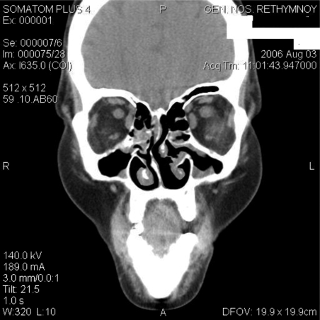
On the contrast-enhanced computed tomography (CT) scan the lesion was consistent with a benign tumour. The nasal mass was well-circumscribed and intensely enhanced with no internal calcification or phleboliths. The nasal septum was not displaced. No erosion or remodelling of the surrounding osseous walls was present. The tumour was located in the nasal cavity with extension to the nasopharynx and into the ethmoid cells on the left side (Fig. 1).
Fig. 1.
Reconstructed coronal CT scan with contrast showing an intensely anhancing mass of left nasal cavity.
The patient was cross-matched for three units of blood and an incisional biopsy was performed which revealed only blood clots and chronic necrotizing inflammation with varying degrees of fibrosis and haemosiderin deposit.
Complete resection of the tumour was achieved by trans-nasal endoscopic surgery and the bleeding was controlled with a coagulation-suction device as well as with a light packing, which was removed 2 days later without recurrence of bleeding. The precise site of origin of this tumour was the mucous membrane of the left middle nasal meatus and was determined only during the procedure.
Histopathological study of the tumour showed large blood-filled spaces lined with flattened endothelium and the tumour was found to be a cavernous haemangioma.
The patient followed a satisfactory post-operative course and was discharged within 3 days of hospitalisation. Physical examination and a CT scan, 40 days later, revealed no residual disease (Fig. 2). There was no recurrence of the lesion at 6 months’ follow-up
Fig. 2.
Coronal CT scan 40 days postoperatively where no macroscopic lesion was shown.
Discussion
Although haemangiomas are common lesions of the head and neck, those of the nasal cavity and paranasal sinuses are rare. Many different classification systems for haemangiomas have appeared in the literature, with histological subtyping being the most widely accepted. Thus, depending on the dominant vessel size at microscopy, haemangiomas are divided into capillary, cavernous and mixed types 1. Cavernous haemangiomas are composed of large endothelium-lined vascular spaces 2. Thrombus within these vascular spaces can occasionally calcify and be identified at CT as phleboliths.
Cavernous haemangiomas with a location at the nose or paranasal sinuses are uncommon. They have been described arising from the inferior turbinate 3 4, vomer 5, Lamina perpendicularis ossi ethmoidalis 6 and sinus maxillaris 7.
Nasal mucosal haemangiomas should be differentiated from haemangiomas that arise from the nasal bones or maxilla, which are primary osseous lesions, the symptoms and surgical approach of which are completely different.
The largest study, by Osborn in 1959, reviewed 51 patients with haemangiomas of the nose seen over an 11-year period. Of these 51 cases, only two were of the cavernous variety 8.
Mean age at presentation of cavernous haemangiomas of the nasal cavity is ~40 years and sex incidence appears equal 9.
This lesion is a unilateral, slowly growing haemorrhagic mass, frequently red or purple, which is sometimes coated with necrotic tissues. Pain is not a characteristic symptom of nasal haemangiomas.
This tumour, when symptomatic, produces recurrent epistaxis or haemoptysis and nasal obstruction 9, 10.
The presence of a bleeding mass in the nasal cavity is consistent with various malignant and benign lesions and the definitive diagnosis is made by histological confirmation of the surgical specimen.
The differential diagnosis of the nasal haemangiomas includes inverted papilloma, olfactory neuroblastoma, lymphoma, haemangiopericytoma, haemangioendothelioma, arteriovenous fistula, lymphangioma, glomangioma, melanoma, adenocarcinoma, squamous cell carcinoma and metastatic malignancies such as renal cell carcinoma.
The pre-operative biopsy of the tumour in order to obtain a histo-pathological differential diagnosis is not an easy task and must be performed with great care to avoid severe bleeding. Imaging investigations should be performed prior to any attempt of biopsy.
CT features of the cavernous haemangioma is a soft tissue density circumscribed mass, enhancing after injection of contrast. Contrast CT scanning usually reveals anatomical location and extension of the tumour. The underlying bone is usually normal but may be remodelled by adjacent long-standing pressure from the expanding mass 10 11. The occurrence of phleboliths is considered to be more typical of cavernous haemangiomas.
MR images confirm the absence of cloted blood, which results in low signal intensity T1-weighted images and very high signal intensity on T2-weighted images. Foci of hypointense signals may represent phleboliths. Characteristically these lesions do not contain large vessels and, therefore, demonstrate none of the signal voids associated with the hypervascularity typical of other vascular malformations 10 11.
If there is any suspicion concerning the nature of these vascular tumours, the risk of bleeding is so high that angiography is always warranted, not only because it can offer the possibility of correct diagnosis but also because, with the aid of trans-arterial embolization, undue haemorrhage during surgical intervention can be avoided 12 13. Exceptionally, in the case of our patient, angiography was not performed because this was not available in our Institution and the patient refused to move to another place on account of social and financial reasons (the patient was a financial immigrant).
Many forms of treatment have been advocated to cure haemangiomas with surgical resection of the tumour, with a cuff of surrounding uninvolved tissue and ligation or cautery to the feeding vessels, being the most successful 9. Other methods of treatment, including cryotherapy, corticosteroid treatment, sclerosing solutions and resection using YAG laser have been used with differing results. An alternative form of management of these tumours, providing there is favourable anatomy, is embolization of the haemangiomas, which is, however, only possible if appropriate angiographic facilities and expertise are available 3.
Choice of the surgical approach depends on the exact location of the tumour. Many surgical approaches have been suggested including the midfacial degloving, lateral rhinotomy, trans-palatal, trans-antral approach and the Le Fort I osteotomy procedure. The trans-nasal endoscopic approach has been proposed as the technique of choice in cases of intra-nasal haemangiomas of the nasal cavity and para-nasal sinuses 9 14.
In our patient, the minimally invasive trans-nasal endoscopic technique has proven to be reliable in terms of direct, fast, adequate exposure and visualization of the lesion, control of bleeding, complete removal of the tumour and avoidance of recurrence in the short term.
References
- 1.Batsakis JG, Rice DH. The pathology of head and neck tumors: vasoformative tumors, part 9A. Head Neck Surg 1981;3:231-9. [DOI] [PubMed] [Google Scholar]
- 2.Enzinger F, Weiss S. Soft tissue tumor. 2nd edition. St Louis: Mosby, 1988. [Google Scholar]
- 3.Webb CG, Porter G, Sissons GRJ. Cavernous hemangioma of the nasal bones: an alternative management option. J Laryngol Otol 2000;114:287-9. [DOI] [PubMed] [Google Scholar]
- 4.Fahmy FF, Back G, Smith CE, Hosni A. Osseous hemangioma of inferior turbinate. J Laryngol Otol 2001;115:417-8. [DOI] [PubMed] [Google Scholar]
- 5.Nakahira M, Kishimoto S, MiuraT, Saito H. Intraosseous hemangioma of the vomer: a case report. Am J Rhinol 1997;11:473-7. [DOI] [PubMed] [Google Scholar]
- 6.Graumüller S, Terpe H, Hingst V, Dommerich S, Pau HW. Intraossäres Hämangiom der Lamina perpendicularis ossis ethmoidalis. HNO 2003;5:142-5. [DOI] [PubMed] [Google Scholar]
- 7.Engels T, Schörner W, Felix R, Witt H, Jahnke V. Kavernöses Hämangiom des Sinus maxillaris. HNO 1990;38:342-4. [PubMed] [Google Scholar]
- 8.Osborn DA. Hemangiomas of the nose. J Laryngol Otol 1959;73:174-9. [DOI] [PubMed] [Google Scholar]
- 9.Iwata N, Hattori K, Tsujimura T. Hemangioma of the nasal cavity: a clinicopathological study. Auris Nasus Larynx 2002;29:335-9. [DOI] [PubMed] [Google Scholar]
- 10.Dillon WP, Som PM, Rosenau W. Hemangioma of the nasal vault: MR and CT features. Radiology 1991;180:761-5. [DOI] [PubMed] [Google Scholar]
- 11.Itoh K, Nishimura K, Togashi K, Fujisawa I, Nakano Y, Itoh H, et al. RM imaging of cavernous hemangioma of face and neck. J Comput Assist Tomogr 1986;10: 831-5. [DOI] [PubMed] [Google Scholar]
- 12.Kim HJ, Kim JH, Kim JH, Hwang EG. Bone erosion caused by sinonasal cavernous hemangioma: CT findings in two patients. AJNR 1995;16:1176-8. [PMC free article] [PubMed] [Google Scholar]
- 13.Hayden RE, Luna M, Goepfert H. Hemangiomas of the sphenoid sinus. Otolaryngol Head Neck Surg 1980;88:136-8. [DOI] [PubMed] [Google Scholar]
- 14.Jungheim M, Chilla R. Der interessante Fall Nr. 64 = The monthly interesting case – case no. 64. cavernous hemangioma. Laryngorhinootologie 2004;83:665-8. [DOI] [PubMed] [Google Scholar]


