Summary
Personal experience in the diagnosis and management of two patients, one adult and one child, with pathologically proven bronchogenic cysts is described. Both patients presented with a solitary neck mass that proved to be bronchogenic cysts on histological examination. Aim of the review is to define the cytology, histopathological and clinical characteristics of bronchogenic cysts and discuss the features that distinguish them from other cervical cysts. Definitive treatment requires surgical excision.
Keywords: Neck, Malformations, Congenital cyst, Bronchogenic cyst
Riassunto
Presentiamo la nostra esperienza riguardante due pazienti, adulto e bambino, affetti da cisti broncogena. Entrambi i pazienti presentavano una massa del collo diagnosticata come cisti broncogena. Lo scopo del presente articolo è quello di definire, mediante la citologia, l’istopatologia e l’imaging, le caratteristiche cliniche della cisti broncogena, discutendo i fattori che la distinguono da altre cisti cervicali. Il trattamento definitivo richiede l’escissione chirurgica.
Introduction
Bronchogenic cysts are rare congenital malformations of ventral foregut development. They are usually located in the mediastinum and intrapulmonary regions, localization in the cervical area is unusual. The majority of cases reported in the literature have been found in the paediatric population, few cases in adults. The purpose of this report is to discuss two different cases never diagnosed before. The second case described refers to an adult with a “high” lateral cervical mass, while the majority of cases, in the literature, concern “low” cervical masses. The second refers to a 13-year-old girl with hyoid mass and active cutaneous fistulae.
Case 1
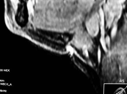
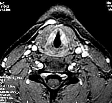
A 13-year-old girl came to our Clinic with a painless recurrent swelling in the midline of the neck associated with previous infection of a fistulous tract for one year. Physical examination revealed a 1x1 cm, soft and mobile cystic mass over the hyoid bone with hyperaemia and fistulous opening. Ultrasonoghaphy (US) showed a cystic mass 8x8 mm consistent with a thyroglossal cyst. Fine needle aspiration biopsy (FNAB) was not performed. Magnetic Resonance Imaging (MRI) of the neck revealed a mass located anteriorly to the hyoid bone with the presence of the fistulous tract (Fig. 1a, b).
Fig. 1a.
(Case 1) GE T1-weighted, FAT SAT, MRI lateral view, showing lesion with fistulous tract.
Fig. 1b.
(Case 1) GE T1-weighted, MRI axial view, showing anteromedial neck lesion with hyperintense signal probably due to presence of proteinaceous material or blood.
Surgical excision of the fistulous tract and associated cyst was performed via an elliptical cervical incision. Both the fistulous tract and cyst were located above the strap muscle and were completely excised in continuity.
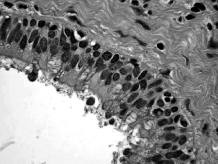
Histopathological examination revealed a bronchogenic cyst (Fig. 2).
Fig. 2.
(Case 1) Histopathology: mucoid cells (H&E, orig. magn. x 400).
The patient has remained without recurrence of the lesion for more than 9 months since surgery.
Case 2
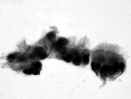
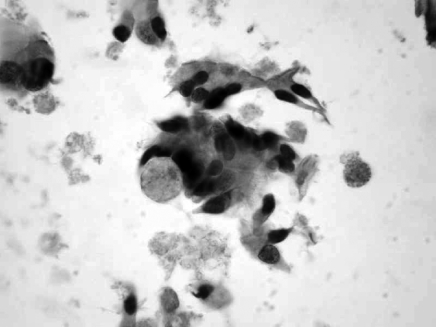
A 39-year-old male patient was referred to our Clinic due to progressive enlargement of a cervical mass. Upon examination, a significant 2x2 cm, soft, smooth, non-pulsatile and mobile cystic mass was detected in the right cervical region. The mass, which had first been observed 5 years earlier, was not associated with dysphagia or previous infection. A FNAB was performed. Cytology showed, in the background, clear serous material and the presence of non-atypical ciliated columnar cells and degenerative fragments of cytoplasm some of which containing pyknotic nuclear debry and tufts of cilia (ciliocytoforia) and mucoid and ciliated cells (Fig. 3a, b).
Fig. 3a.
(Case 2) Cytologic findings: in background of clear serous material, non atypical ciliated columnar cells are present and degenerative fragments of cytoplasm some of which contain pyknotic nuclear debry and tufts of cilia (ciliocytoforia) (H&E, orig. magn. x 400).
Fig. 3b.
(Case 2) Cytologic findings: mucoid and ciliated cells (H&E, orig. magn. x 400).
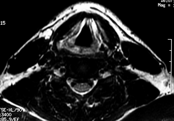
Gadolinium-Contrast Enhanced MRI revealed a 1.3x1.5 cm cystic mass with regular margins located in the right cervical region (not connected to the bronchial tree) inferior to the right submandibular gland, displacing it anteriorly and anterior to sternocleidomastoideus muscle. There were no signs of invasion of the adjacent structures. Radiological features indicated the development of cystic abnormalities (Fig. 4a, b).
Fig. 4a.
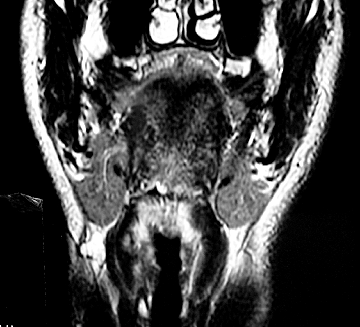
(Case 2) GE T2 weighted, MRI coronal view, shows lesion located in right cervical region, below submandibular region and anterior to sternocleidomastoideus muscle.
Fig. 4b.
(Case 2) GE T2 weighted, MRI axial view, lesion anterior to right sternocleidomastoideus.
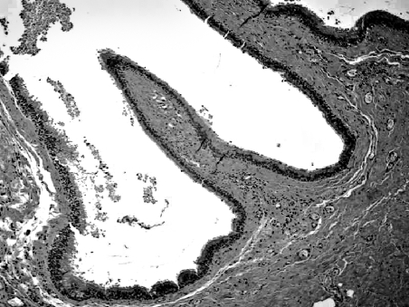
The patient was scheduled for elective surgery through a right lateral cervicotomy. Surgical exploration revealed a tumour located below the right submandibular gland and anterior to the sternocleidomastoideus muscle. The neoformation was excised and sent for histological examination which revealed respiratory epithelium lining the cervical bronchial cyst and mucoid cells (Fig. 5).
Fig. 5.
(Case 2) Histopathology: respiratory epithelium lines cervical bronchial cyst (H&E, orig. magn. x 200).
The patient has been without recurrence of the lesion for more than one year since surgery.
Discussion
The majority of bronchogenic cysts are diagnosed in the paediatric population, the location being either intra- or extra-thoracic 1 2.
Intra-thoracic cysts are usually found in the anterior mediastinum or around the ilum and may remain connected to the tracheobronchial airway by either a fibrous cord or a patent bronchus-like connection 3. In the absence of an attachment, bronchogenic cysts can migrate and occasionally become displaced anteriorly by fusion of the mesenchymal bars forming the sternum. A superficial presternal or suprasternal location is most common, while deep neck or laterally located bronchogenic cysts are comparatively rare. Bronchogenic cysts of the cervical area are usually asymptomatic, but if the cyst is large, symptoms may occur, including dyspnoea, respiratory distress, cough and dysphagia. Very occasionally, secondary infection may occur which results in sinus tract formation and external drainage of purulent material if the cyst is superficial, or with abscess formation if the cyst is deep 4. If a draining sinus is present, in association with the bronchogenic cysts, as in Case 1 of our series, both the sinus tract and the cyst should, ideally, be excised in continuity 5.
In adults, the two most common locations of bronchogenic cyst are the mediastinum and lung parenchyma. Most bronchogenic cysts are unilocular, filled with fluid and are not in communication with the airways. However, when a patent connection exists, symptoms are likely to occur and the risk of developing infectious complications increases 6.
A review of the literature revealed 14 cases of bronchogenic cysts of the neck in adults: the thyroid and paratracheal region were more frequently affected than the supraclavicular and suprasternal notch. In that review, 10 cases of cervical bronchogenic cysts were reported in the midline region, while 4 cases were located in the lateral neck. The male to female ratio was 4:1.
Reports in the English language literature are outlined in Table I.
Table I. Cases of bronchogenic cysts of the neck in adults reported in the literature.
| Reported cases of bronchogenic cysts of neck in adults | ||||
| No. | Authors ref.* | Age (yrs) | Sex | Location |
| 1 | Dubois14(1981) | 24 | M | Supraclavicular region |
| 2 | McManus4 (1984) | 34 | M | Between right carotid sheath and tracheo-oesophageal groove; beginning at cricoid cartilage and extending into mediastinum |
| 3 | Rapado15 (1998) | 54 | M | Supraclavicular triangle extending into superior mediastinum |
| 4 | Barsotti16(1998) | 49 | M | Calcified cystic tumour posterior to the left lobe of thyroid gland |
| 5 | Majlis17 (1999) | 44 | M | Pretracheal anterior cervical region simulating a thyroid lesion |
| 6 | Shimizu18(2000) | 25 | F | Cystic in left lobe through isthmus of thyroid |
| 7 | Tanita13(2002) | 46 | M | Mass on left scapular area |
| 8 | Hadjihannas19 (2003) | 70 | M | Subcutaneous cystic mass on suprasternal notch |
| 9 | Al-Kasspooles3 (2004) | 62 | M | Neck mass communicating with membranous trachea |
| 10 | Newkirk20 (2004) | 20 | F | Cystic mass in right lobe of thyroid gland |
| 11 | Newkirk20 (2004) | 22 | M | Cystic mass extending from inferior pole of right thyroid lobe to superior mediastinum |
| 12 | Sanli21 (2004) | 48 | F | Cyst in right paratracheal region |
| 13 | Ibanez Aguirre9 (2006) | 26 | M | Cervical mass to lower pole of thyroid lobe |
| 14 | Bocciolini22 (2006) | 57 | M | Cyst in right paratracheal region |
First Author
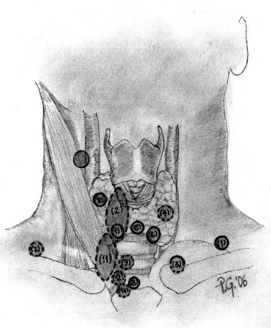
In addition to these cases, our patient is the first to present such a “high” location (Fig. 6). Imaging (MRI and/or CT) and histological findings are essential in establishing the correct pre-operative diagnosis.
Fig. 6.
Distribution of bronchogenic cyst of neck region. Numbers in brackets are related to references reported in Table I.
However, MRI is the study of choice due to its excellent soft-tissue detail without the absolute need of intra-venous contrast administration and in the study of the outline of adjacent vital structures critical in the planning of surgery 7.
In terms of diagnosis, MRI is valuable in demonstrating the size and shape of the cyst and in determining its position in relation to other structures, there are no radiological criteria specific for the bronchogenic cyst offering an indication to diagnosis. Suen et al. found that 5 out of 6 lesions studied by MRI, had a high signal intensity on both T1- and T2-weighted images, believed to be due to the presence of the proteinaceous material or blood. In our cases, both lesions showed high signal intensity on T2-weighted imaging 8. In our cases, MRI revealed a lesion located in the “high” lateral cervical area, below the right submandibular gland and anterior to the sternocleidomastoideus muscle, these radiological features suggested developmental cystic abnormalities.
A definitive diagnosis of bronchogenic cyst requires histopathological confirmation. The lining of bronchogenic cysts is respiratory in origin, ciliated, with a pseudostratified columnar epithelium overlying a fibrous connective tissue wall containing seromucous subcutaneous glands and cartilage plates.
Definitive diagnosis relies on histology of the surgical specimen, with cytology being rarely performed.
The differential diagnosis of cervical bronchogenic cysts includes branchial cleft cysts, thyroglossal cysts, thymic and thyroid cysts, dermoids and lymphangiomas, cystic hygromas, teratomas and cystic neuromas 9.
More common congenital anomalies are known to be initially seen in the upper triangles of the neck: thyroglossal duct cyst and branchial cyst 10.
Clinically, branchial cleft cysts are usually located higher and laterally on the side of the neck, the thyroglossal duct cysts are usually located midline on the anterior aspect of the neck, in close proximity to the hyoid bone.
As far as concerns treatment, surgical excision remains the treatment of choice. Neck exploration and selective dissection via a transcervical approach is ideal for this lesion. Complete excision is imperative to reduce the risk of recurrence.
In the literature, carcinomas arising from bronchogenic cysts have been reported. Tanaka et al. 11, described a case of mucoepidermoid carcinoma in a 59-year-old Japanese female, arising from a bronchogenic cyst of the thymus. Another mucoepidermoid carcinoma of the thyroid, in a 44-year-old female was reported by Mizukami et al. 12. Tanita et al. 13 also reported the case of a 46-year-old Japanese male with a recurrent malignant melanoma that arose from cutaneous bronchogenic cysts in the scapular area.
These carcinomas arising from bronchogenic cysts emphasize the importance of total surgical excision.
Conclusions
The need is stressed for a high index of suspicion in the presence of a lump in the neck on account of the variety of pathological conditions that can occur, which might require different management approaches.
Although ectopic bronchogenic cysts are rare lesions, in the upper lateral neck, it is essential to pay attention also to the upper portion of the neck, since bronchogenic cysts may be found in the same position as more common diseases such as bronchial cyst or thyroglossal duct cyst. In our opinion, the clinical observation of an asymptomatic lateral neck mass, in an adult, should include, in the differential diagnosis, the possibility of a bronchogenic cyst. In fact, the clinical presentation of bronchogenic cysts can be mimicked by branchial cleft cysts or thyroglossal duct cysts.
References
- 1.Halsleton PS. Spencer’s pathology of the lung. 5th ed. New York: McGraw-Hill; 1996. [Google Scholar]
- 2.Pujary K, Pujary P, Shetty R, Hazarika P, Rao L. Congenital cervical bronchogenic cyst. Int J Pediatr Otorhinolaryngol 2001;57:145-8. [DOI] [PubMed] [Google Scholar]
- 3.Al-Kasspooles MF, Alberico RA, Douglas WG, Litwin AM, Wiseman SM, Rigual NR, et al. Bronchogenic cyst presenting as a symptomatic neck mass in an adult: case report and review of the literature. Laryngoscope 2004;114:2214-7. [DOI] [PubMed] [Google Scholar]
- 4.McManus K, Holt R, Aufdemorte TM, Trinkle JK. Bronchogenic cyst presenting as deep neck abscess. Otolaryngol Head Neck Surg 1984;92:109-14. [DOI] [PubMed] [Google Scholar]
- 5.Ustundag E, Iseri M, Keskin G, Yayla B, Muezzinoglu B. Cervical bronchogenic cysts in head and neck region: review of the literature. J Laryngol Otol 2005;119:419-23. [DOI] [PubMed] [Google Scholar]
- 6.Yerman HM, Holinger LD. Bronchogenic cyst with tracheal involvement. Ann Otol Rhinol Laryngol 1990;99:89-93. [DOI] [PubMed] [Google Scholar]
- 7.Mehta RP, Faquin WC, Cunningham MJ. Cervical bronchogenic cysts: a consideration in the differential diagnosis of pediatric cervical masses. Int J Pediatr Otorhinolaryngol 2004;68:563-8. [DOI] [PubMed] [Google Scholar]
- 8.Suen HC, Mathisen DJ, Grillo HC, LeBlanc J, McLoud TC, Moncure AC, et al. Surgical management and radiological characteristic of bronchogenic cysts. Ann Thorac Surg 1993;55:476-81. [DOI] [PubMed] [Google Scholar]
- 9.Ibanez Aguirre J, Marti Cabane J, Bordas Rivas JM, Valenti Ponsa C, Erro Azcarate JM, De Simone P. A lump in the Neck: cervical bronchogenic cyst mimicking a thyroid nodule. Minerva Chir 2006;61:71-2. [PubMed] [Google Scholar]
- 10.Hadi UM, Jammal HN, Hamdan AM, Saad AM, Zaatari GS. Lateral cervical bronchogenic cyst: an unusual cause of a lump in the neck. Head Neck 2001;23:590-3. [DOI] [PubMed] [Google Scholar]
- 11.Tanaka M, Shimokawa R, Matsubara O, Aoki N, Kaymiyama R, Kasuga T, et al. Mucoepidermoid carcinoma of the timic region. Acta Pathol Jpn 1982;32:703-12. [DOI] [PubMed] [Google Scholar]
- 12.Mizukami Y, Matsubara F, Hashimoto T, Haratake J, Terahata S, Noguchi M, et al. Primary mucoepidermoid carcinoma in the thyroid gland. A case report including an ultrastructural and biochemical study. Cancer 1984;52:1741-5. [DOI] [PubMed] [Google Scholar]
- 13.Tanita M, Kikuchi-Numagami K, Ogoshi K, Susuki T, Tabata N, Kudoh K, et al. Malignant melanoma arising from cutaneous bronchogenic cyst of the scapular area. J Am Acad Dermatol 2002;46:19-21. [DOI] [PubMed] [Google Scholar]
- 14.Dubois P, Belanger R, Wellington JL. Bronchogenic cyst presenting as a supraclavicular mass. Can J Surg 1981;24:530-1. [PubMed] [Google Scholar]
- 15.Rapado F, Bennett JDC, Stringfellow JM. Bronchogenic cyst: an unusual cause of lump in the neck. J Laryngol Otol 1998;112:893-4. [DOI] [PubMed] [Google Scholar]
- 16.Barsotti P, Chatzimichalis A, Massard G, Wihlm JM. Cervical bronchogenic cyst mimicking thyroid adenoma. Eur J Cardio-Thoracic Surg 1998;13:612-4. [DOI] [PubMed] [Google Scholar]
- 17.Majlis S, Horvath E, Castro L, Martinez V. Anterior cervical bronchogenic cyst simulating a thyroid lesion. Report of case. Rev Med Chil 1999;127:977-81. [PubMed] [Google Scholar]
- 18.Shimizu J, Kawaura Y, Tatsuzawa Y, Maeda K, Suzuki S. Cervical bronchogenic cyst that presented as a thyroid cyst. Eur J Surg 2000;166:659-61. [DOI] [PubMed] [Google Scholar]
- 19.Hadjihannas E, Ray J, Rhys-Williams S. A cervical bronchogenic cyst in an adult. Eur Arch Otorhinolaryngol 2003;260:216-8. [DOI] [PubMed] [Google Scholar]
- 20.Newkirk KA, Tassler AB, Krowiak EJ, Deeb ZE. Bronchogenic cysts of the neck in adults. Ann Otol Rhinol Laryngol 2004;113:691-5. [DOI] [PubMed] [Google Scholar]
- 21.Sanli A, Onen A, Ceylan E, Yilmaz E, Silistreli E, Acikel U. A case of a bronchogenic cyst in a rare location. Ann Thorac Surg 2004;77:1093-4. [DOI] [PubMed] [Google Scholar]
- 22.Bocciolini C, Dall’Olio D, Cunsolo E, Latini G, Gradoni P, Laudadio P. Cervical bronchogenic cyst: asymptomatic neck mass in an adult male. Acta Otolaryngologica Italica 2006;126:553-6. [DOI] [PubMed] [Google Scholar]


