Abstract
Background
The aim of this article is to make physicians of all specialties aware of the possible variations of clinical course in human cowpox infection. This has been a matter of current interest since the detection of a first cluster of infections among owners of white pet rats in the Krefeld area in the spring of 2008. Two further cases arose in the Krefeld area in November 2008, and there have since been multiple further reports from various regions in Germany and the neighboring countries.
Method
The authors report on the first six documented cases of infection with cowpox virus among young persons owning pet rats, with both typical and atypical clinical courses.
Results
The clinical, molecular biological, and serological findings confirmed cowpox virus infection in all six cases. The DNA sequence of the cowpox virus hemagglutinin gene was identical in all patients. The infections had arisen after direct contact with pet rats.
Conclusions
Molecular genetic analysis of the cases described here suggests that the observed occurrence of cowpox virus infection among human beings and pet rats in multiple geographical areas represents a unitary epidemiological event that has not yet come under control. Further cases can be expected.
Keywords: keeping of animals, zoonosis, skin infection, cowpox, risk of infection
In addition to the variola (smallpox) virus (an orthopoxvirus) the poxvirus family includes many viruses that are potentially pathogenic for humans. These include the molluscipoxvirus (molluscum contagiosum), the parapoxvirus (milker’s node virus, orf virus), and other orthopoxviruses. The latter include the species "cowpox virus" and have been known in Europe since Jenner introduced the smallpox vaccine. For years, however, coxpox virus has not been transmitted by cows but by infected cats (1– 4). In humans, the virus causes mostly self limiting local infection after an incubation period of 8–12 days; however, severe clinical courses have been described, and the death of an immunocompromised patient with atopy has been reported (5).
The reservoir for cowpox virus is symptomatically and asymptomatically infected wild rodents that may infect hunting cats. In addition to cats, cowpox virus related illness has been reported in Germany in several species of zoo animal (6). The literature contains individual case reports of cowpox infections in humans after direct contact with wild rats (Rattus norvegicus) (7, 8).
Since cowpox infections with virus characterization in humans were first scientifically described in the first half of the 20th century (9), less than 200 cases of infection with this causative agent have been registered in Europe. On the basis of relevant gene sequences, all these were individual cases caused by single viral strains.
For the first time, and thus far not described, a cluster of 4 cases of infection with cowpox virus of an identical strain was observed in the Krefeld area in the spring of 2008. In all cases, infection was apparently transmitted by white pet rats (Latin: Rattus norvegicus forma domestica; also known as fancy rat). Two further cases were diagnosed in November 2008. Since December 2008, case reports of this viral strain and transmission mode from various German regions have been on the increase, and individual reports have also been received from neighboring countries.
Case reports and results
Case 1
In March 2008, a 15 year old girl presented to the dermatological department at the HELIOS Klinikum Krefeld, after 5 days of antibiotic treatment prescribed by her primary care physician for several papular lesions with surrounding erythema on her forehead, neck, shoulder, and thumb had not resulted in any improvement. Additionally, the patient complained of general malaise, headache, and fever.
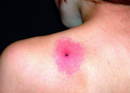
Clinical examination ascertained pronounced cervical lymphadenitis in addition to the lesions, which were starting to ulcerate (figure 1). Laboratory tests were inconspicuous, with the exception of raised C-reactive protein (to 25 mg/L [normal value <3 mg/L]). A wound swab from the forehead lesion grew meticillin sensitive Staphylococcus aureus.
Figure 1.
Figure 1: Case 1, early stage cowpox lesion, onset of ulceration, on patient’s shoulder
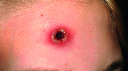
In spite of treatment with ampicillin/sulbactam, the lesions developed into hemorrhagic ulcers with black scabs or crusts and erythematous, edematous margins (figure 2). An additional lesion developed on the girl’s tongue. A biopsy was taken from the shoulder, among others to rule out atypical mycobacteriosis.
Figure 2.
Figure 2: Case 1, fully developed ulcerated cowpox lesion on the forehead, with black scab and inflamed margin (photograph taken about a week after the photograph shown in figure 1)
During a further, detailed medical history, the patient reported having purchased a fancy rat in a pet shop in Duisburg 2 weeks before she had become ill. The rat had developed a respiratory infection and had died within a few days, in spite of antibiotic treatment given by the vet. She reported playing with and petting the rat, but she hadn’t been bitten.
On the basis of this information, a differential diagnosis of infection with orthopoxvirus or parapoxvirus was considered, and a scab specimen and serum were sent for testing to the reference laboratory for poxviruses at the Robert Koch Institute (RKI). Electron microscopic examination and real time polymerase chain reaction (PCR) and sequencing confirmed cowpox virus (10). The serum contained high antibody titers against orthopoxviruses (immunofluorescence test, IgG 1:2560/IgM 1:640).
The lesions gradually healed over several weeks and formed non-inflamed flat scars.
An attempt to exhume the rat for veterinary investigations remained unsuccessful as the rat could not be found.
Case 2
The 16 year old boyfriend of the patient in Case 1 had become ill at the same time, with two lesions on the bridge of his nose and on the middle finger of his left hand. He presented a week after his girlfriend, with ulcerated lesions of up to 1.5 cm diameter with black scabs in the center (as shown in figure 2 [Case 1]) and similar general symptoms. He also had cervical lymphadenopathy. A bacteriological wound swab did not yield any viral strains.
The patient had played with the white rat, together with his girlfriend and did not have any memory of receiving a bite.
At the RKI, cowpox virus was confirmed in the scab specimen, and the serum antibody titers were massively raised (IgG 1:20480; IgM 1:5120).
Case 3
Three weeks after patients 1 and 2, a 32 year old man presented to Krefeld’s dermatology hospital, who had received treatment with cefuroxime from his primary care physician for 5 days for a skin lesion on his left hand. The lesion had developed into an ulcer with a dark scab and inflamed margin (as shown in figure 2 [Case1]) after the patient had had wound surgery to uncover a suspected abscess. His raised temperature of up to 39.3 degrees Celsius had not receded completely.
Meticillin sensitive Staphylococcus aureus and hemolytic group G streptococci were cultured from a wound specimen.
A short while before, the patient had purchased 3 white rats in a pet shop in Mönchengladbach, of which 2 had developed respiratory symptoms and had then died.
Examination of biopsy samples and serum from this patient identified a viral strain that was consistent with the causative agent of the first 2 cases in the DNA sequence of the hemagglutinin gene.
Case 4
At the beginning of May 2008 (2 weeks after patient 3), a 17 year old boy presented to the emergency admissions unit at the HELIOS Klinikum Krefeld with a painful periorbital swelling of his left eye. Owing to a suspected diagnosis of complicated sinusitis he was initially admitted to the ear, nose, and throat department. After he had developed severe conjunctivitis, he was discharged from inpatient treatment with a diagnosis of epidemic keratoconjunctivitis.
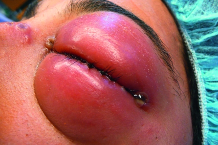
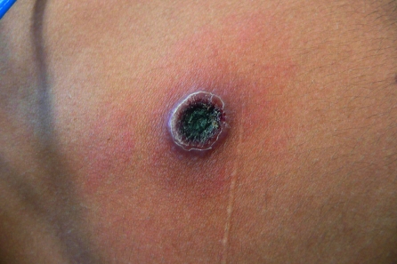
Five days later he presented to the emergency department again, with a massive swelling and erythema of the eyelids of his left eye and small ulcerations in the nasal orbital angle (figure 3). Physical examination found a bland crusty skin lesion, ca. 1 cm in diameter, on the patient’s abdomen (figure 4).
Figure 3.
Figure 3: Case 4, massive swelling of eyelid in cowpox infection, satellite lesion in nasal orbital angle
Figure 4.
Figure 4: Case 4, non-inflamed, scabby, older cowpox lesion on abdominal skin
When asked for animal contact, the patient reported having purchased a white rat in a pet shop in Krefeld several weeks earlier. The rat had developed brown lesions on its paws and had shown reluctance to walk. He had killed the animal, "to spare its pain and shorten its suffering." He had not considered the wound on his abdomen important.
Infection with cowpox virus of the same strain as that observed in the other 3 cases was confirmed by real time PCR/sequencing from eye secretions and scabs from the abdominal lesion and by specific antibodies in the serum (IgG 1:5120, IgM 1:2560).
Since the patient’s left eye seemed at risk, treatment was initiated with cidofovir, an inhibitor of viral DNA polymerase.
The patient’s subsequent treatment is not fully completed; it is likely to include surgical correction of entropium (the inversion or turning in of the border of the eyelids) because of scar formation. After the treatment is concluded, details of the clinical course and the treatment of this particular patient will be reported elsewhere.
Case 5
In early November 2008, the pathology institute at the HELIOS Klinikum Krefeld received an excision biopsy from the cervical soft tissues of an 18 year old woman for assessment because of suspected malignancy. The investigating pathologist remembered histopathological changes—such as blister formation with ballooning degeneration and keratocyte necrosis—which had been noticed in March of that year in specimens taken from patient 1.
Further investigations found a temporal association with 2 white rats purchased shortly beforehand in Wesel, which had become ill and died subsequently. A finding of specific DNA embedded in a paraffin block and further tissue specimens as well as high serum antibody titers again confirmed infection with the known cowpox virus strain.
Because of extensive necrosis formation, the patient needed inpatient treatment with surgical revision.
Four more rats owned by the patient also fell ill and died. All 6 rats tested positive for cowpox virus on serology and molecular biology. The nucleotide sequence of the hemagglutinin gene of the isolated viruses was identical with the human isolates described here.
Case 6
In November 2008, the 23 year old boyfriend of the patient of Case 5 was transferred from a peripheral hospital to the lung center at the HELIOS Klinikum Krefeld with suspected absceding pneumonia. He had had a dry cough for about 10 days and his general condition had deteriorated and he was severely feverish.
Radiologically, bilateral pulmonary round focal infiltrations were found but no sign of abscesses. Bronchoscopy with bronchoalveolar lavage (BAL) found signs of acute bronchitis in the entire bronchial system but no distinct papular or vesicular efflorescences. Microbiological and serological investigations, including blood cultures and diagnostic tests for tuberculosis, remained inconclusive. The patient tested negative for HIV antibodies and was immunocompetent (560 T-lymphocyte helper cells/µL). No signs of a tumor or lymphoma were found.
Treatment with ceftriaxone/clarithromycin was continued at first; later, therapy was swapped to piperacillin/subactam plus doxycycline owing to continued feverish episodes of up to 40 degrees Celsius, especially at night. Subsequently, the fever and the initially notably raised inflammatory parameters in the serum continued to fall.
Because of the known cowpox infection of the patient’s girlfriend, whose rats he had come into contact with, BAL fluid, pharyngeal swab, and EDTA blood were tested. A biopsy specimen taken specifically from a pulmonary focus during a second bronchoscopy was tested for cowpox virus DNA, and the serum was analyzed for specific antibodies.
In the BAL fluid and the biopsy specimen, a small number of copies of cowpox DNA was found, but the pharyngeal swab (to exclude contamination owing to pharyngeal enanthema) and EDTA blood were negative on PCR. Compared with a previous examination the patient had seroconverted (together with his girlfriend, who was already ill at this point in time) (IgG 1:80, IgM 1:20 on 12 November 2008; IgG 1:20480, IgM 1:2560 on 27 November 2008).
Extensive examination of the entire integument by a dermatologist and of the oro-naso-pharyngeal region by a specialist in ear, nose, and throat medicine did not find efflorescences that indicated cowpox.
His radiological findings improved only slowly but gradually, so the patient was treated on an outpatient basis after clinical improvement and 2 and a half weeks’ inpatient treatment. The patient rejected transbronchial biopsy of a remaining radiological focus in early 2009.
Synopsis of the case reports
The clinical findings, together with molecular biological and serological findings, without doubt confirm infection with cowpox virus in the first 5 patients, which was obviously transmitted by white pet rats.
Since the lesions of patients 1 and 2 developed simultaneously and both patients went through the different stages, the authors assume a common source of infection (pet rat) and exclude human to human transmission—something that has not been described in the literature to-date either.
The severe eye infection in patient 4 was most probably caused but contact infection of the chronologically earlier abdominal lesion by the patient himself.
In Case 5, an identical strain of cowpox virus was isolated from the patient and her rats—an unequivocal confirmation of transmission from rat to human.
In all investigated cases, cowpox viruses were isolated that had an identical nucleotide sequence of the complete open reading frame of the hemagglutinine gene (921bp). This only weakly preserved gene is used for typing and molecular-epidemiological differentiation of different isolates of cowpox virus (11, 12).
The immunofluorescence test used by the RKI for serological investigations is genus specific for infections with orthopoxviruses (13). The cross reactive variola viruses, monkeypox viruses, and vaccinia viruses from this genus did not need to be considered for differential diagnosis.
The pulmonary involvement in the 6th patient—which has thus far not been described in the literature—could not be confirmed because of lacking bronchoscopic findings. An etiological contribution of the cowpox virus could not be excluded in this case because:
In spite of comprehensive attempts to find a different cause of the patient’s illness, none was found
The causative strain was confirmed
An epidemiological association existed
Pulmonary involvement has been documented in several animal species, including rats.
Discussion
Illnesses caused by cowpox virus develop in humans typically after an incubation period of 8 to 12 days. They are mostly located on the hands, face, or trunk, with a sequence of papule, vesicle, pustule, hemorrhagic ulceration with dark scabs, and subsequent healing with scar formation, which may take several weeks (figures 1, 2, 4). The pathology is accompanied by regional lymphadenopathy and general symptoms. Bacterial superinfections and involvement of the eyes and oral mucosa can develop; an unusual disease course is not uncommon.
As these cases have shown, the diagnosis is often made with a delay, after several diagnostic and therapeutic detours.
The cases observed in the Krefeld region obviously indicate the start of a current wave of supraregional cases of infection and illness due to cowpox virus in pet rats and their owners. Since the end of 2008 further cases have been reported from the Munich region (14), North Rhine–Westphalia (Aachen, Recklinghausen, and Bielefeld regions), and neighboring countries (France, Netherlands). By the end of February 2009, 21 cases had been confirmed from Germany and at least 6 from neighboring countries. Because of the confirmed genetically identical hemagglutinine gene of the causative strain, events can be assumed to be epidemiologically related.
Because of the usually very close contact of the owners with their pet rats the zoonotic potential has to be categorized as high. The persons who have become ill so far are mainly adolescents and young adults who have not been vaccinated for smallpox since the early 1980s when the vaccination program was halted. This vaccine also confers relative protection against other orthopoxviruses.
In patients with wounds that do not heal easily and are consistent with the described morphology, animal contacts should be assessed and cowpox virus infection should be tested as a differential diagnosis.
The virus can be confirmed by PCR in the acute phase from exudates, contents of pustules, scabs of skin lesions, and other clinical materials.
The treatment of the mostly self limiting illness is symptomatic by covering wound dressings. Surgical interventions may prolong the healing process and should be used sparingly. Antibiotic treatment is required only in patients with a bacterial superinfection. The benefits of antiviral treatment or anti-vaccinial-hyperimmunoglobulin is not well documented in the literature, and these options should be reserved for very severe disease courses. The patients should be alerted to the risk of contact infection, for example, of the eyes. Person to person spread has not been observed thus far.
Because of the current situation described in this article, intense attempts are being made to detect the entry pathway of this infection. Investigations in 3 of the 4 pet shops in 3 neighboring towns that were implicated in the first 4 cases did not confirm with absolute certainty the presumed epidemiological association. However, further investigations from early 2009 have shown that all infected rats that were purchased in different pet shops came from the same wholesaler. This wholesaler receives about 1500 rats per week from a breeder in another European country, who also delivers to other wholesalers.
With regard to the possible trade in asymptomatically infected animals, routine investigation in German wholesalers is not a promising option at this point in time. Currently, further pet shops, breeders, and retailers are being inspected in order to determine the exact origin and further whereabouts of the already sold infected rats and thus avoid further spread of this zoonosis. In the past 4 months, more than 130 000 rats have entered the German market from the breeder implicated in these cases, so that more cases of cowpox have to be expected.
Key messages.
After an initial occurrence in the spring of 2008 there has been an increase in the number of cases of infection with cowpox virus, transmitted by a reservoir of infection that has not been described so far: domestic white pet rats.
The infectious animals were always purchased immediately beforehand and fell ill with respiratory symptoms or lesions to their paws. Most animals were already dead by the time their owner fell ill.
Infection with cowpox virus typically manifests in humans after an incubation period of 8 to 12 days. The resulting lesions are located on the hands, face, or trunk, and develop in a chronological sequence of papule, vesicle, pustule, hemorrhagic ulceration with a dark scab, and subsequent healing over weeks, mostly accompanied by scarring. The pathology is usually accompanied by regional lymphadenopathy and general symptoms.
The diagnosis results from the clinical appearance and the medical history as well as from polymerase chain reaction (PCR) confirmation of virus in exudates, contents of pustules, scabs of skin lesions, and other clinical material.
In spite of intense efforts, the epidemiological events have not been brought under control thus far. Further cases in humans are to be expected.
Acknowledgments
Translated from the original German by Dr Birte Twisselmann.
Footnotes
Conflict of interest statement
The authors declare that no conflict of interest exists according to the guidelines of the International Committee of Medical Journal Editors.
References
- 1.Editorial: What’s new pussycat? Cowpox. Lancet. 1986;328 [PubMed] [Google Scholar]
- 2.Steinborn A, Essbauer S, Marsch W Ch. Kuh-/Katzenpocken-Infektion beim Menschen: Ein potentiell verkanntes Krankheitsbild. Dtsch Med Wochenschr. 2003;128:607–610. doi: 10.1055/s-2003-38051. [DOI] [PubMed] [Google Scholar]
- 3.Pahlitzsch R, Hammarin AL, Widell A. A case of facial cellulitis and necrotizing lymphadenitis due to cowpox infection. Clin Infect Dis. 2006;43:737–742. doi: 10.1086/506937. [DOI] [PubMed] [Google Scholar]
- 4.Bonnekoh B, Falk K, Reckling KF, et al. Cowpox infection transmitted from domestic cat. J Dtsch Dermatol Ges. 2008;6:210–213. doi: 10.1111/j.1610-0387.2007.06546.x. [DOI] [PubMed] [Google Scholar]
- 5.Eis-Hubinger AM, Gerritzen A, Schneweis KE, et al. Fatal cowpox-like virus infection transmitted by cat. Lancet. 1990;336 doi: 10.1016/0140-6736(90)92387-w. [DOI] [PubMed] [Google Scholar]
- 6.Kurth A, Wibbelt G, Gerber H-P, Petschaelis A, Pauli G, Nitsche A. Rat-to-elephant-to-human transmission of cowpox virus. Emerg Infect Dis. 2008;14:670–671. doi: 10.3201/eid1404.070817. [DOI] [PMC free article] [PubMed] [Google Scholar]
- 7.Postma BH, Diepersloot RJ, Niessen GJ, Droog RP. Cowpox-virus-like infection associated with rat bite. Lancet. 1991;337:733–734. doi: 10.1016/0140-6736(91)90317-i. [DOI] [PubMed] [Google Scholar]
- 8.Wolfs TF, Wagenar JA, Niesters HG, Osterhaus AD. Rat-to-human transmission of cowpox infection. Emerg Infect Dis. 2002;8:1495–1496. doi: 10.3201/eid0812.020089. [DOI] [PMC free article] [PubMed] [Google Scholar]
- 9.Baxby D, Bennett M. Cowpox: a re-evaluation of the risks of human cowpox based on new epidemiological information. Arch Virol. 1997;13:1–12. doi: 10.1007/978-3-7091-6534-8_1. [DOI] [PubMed] [Google Scholar]
- 10.Kurth A, Nitsche A. Fast and reliable diagnostic methods for the detection of human poxvirus infections. Future Virology. 2007;2:467–469. [Google Scholar]
- 11.Damaso CR, Esposito JJ, Condit RC, Moussatche N. An emergent poxvirus from humans and cattle in Rio de Janeiro State: Cantagolo virus may derive from Brazilian smallpox vaccine. Virology. 2000;277:439–449. doi: 10.1006/viro.2000.0603. [DOI] [PubMed] [Google Scholar]
- 12.Vorou RM, Papavassiliou VG, Pierroutsakos IN. Cowpox virus infection: an emerging health threat. Curr Opin Infect Dis. 2008;21:153–156. doi: 10.1097/QCO.0b013e3282f44c74. Review. [DOI] [PubMed] [Google Scholar]
- 13.Nakano JH, Esposito JJ. Poxviruses. In: Lennette EH, Schmidt N, editors. Diagnostic procedures for viral, rickettsial and chlamydial infections. Washington DC: American Public Health Association; 1989. pp. 257–308. [Google Scholar]
- 14.Robert Koch-Institut. Kuhpocken: Zu einer Häufung von Infektionen nach Kontakt zu „Schmuseratten“ im Großraum München. Epid Bull. 2009;9:53–56. [Google Scholar]


