Neuroendocrine tumours (NETs) are rare neoplasms originating from the neuroendocrine system. They are classified into 3 different subtypes: well-differentiated NETs (carcinoid), well-differentiated endocrine carcinomas (malignant carcinoid) and poorly differentiated endocrine carcinomas (small-cell carcinomas).1,2 The large-cell neuroendocrine carcinoma has recently been introduced3 and has been described in different locations, including the cervix, larynx, gallbladder, bladder, soft tissues and the ampulla of Vater. We report the case of a patient with an advanced cecal large-cell neuroendocrine carcinoma and present a literature review to focus the attention of surgeons and oncologists on this rare but peculiar type of colon cancer.
Case report
A 51-year-old woman was admitted to hospital for generalized abdominal pain of 12 weeks’ duration, decreased appetite, weight loss and black stools. The abdominal pain, constant and throbbing in nature, radiated to the back and heart. Physical examination showed a diffuse tenderness of the epigastric region and of the right iliac fossa, with guarding throughout the abdomen. Blood tests showed a fall in hemoglobin level (87 g/L) and in mean red blood cell volume (77.3 fL), and raised C-reactive protein (788.6 nmol/L) and alkaline phosphatase (4.9 μkat/L). Abdominal ultrasound showed multiple hyperechoic nodular areas in the liver and a mass in the right ileal fossa, 3 cm in diameter, highly suspicious for a cecal tumour. Chest radiographs were normal.
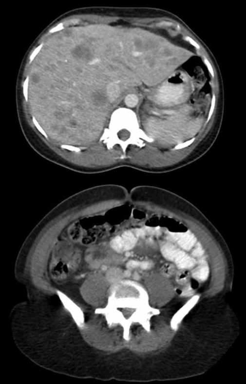
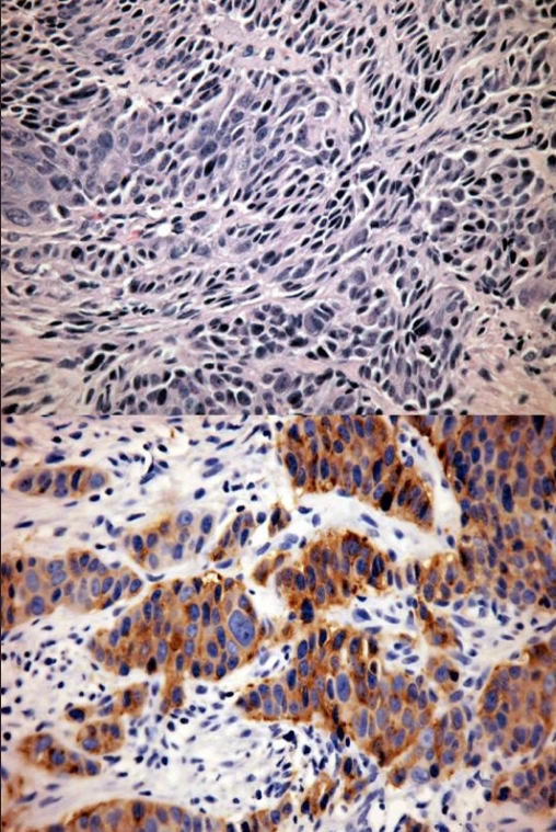
A computed tomography (CT) scan of the abdomen showed a substantially enlarged liver with several metastatic lesions in both lobes and an abnormal thickening of the cecum (Fig. 1). On colonoscopy we found a 3–5 cm polypoid and a friable cecal mass, including the ileocecal valve. Biopsies revealed features consistent with a large-cell neuroendocrine carcinoma (various degrees of dysplasia infiltrated by a neoplasm composed of large cells arranged in solid sheets and with an abundant cytoplasm; mitotic activity 23/10 at high power fields; positivity for CD56 and synaptophisine, but not for chromogranine-A) (Fig. 2). A CT scan of the chest revealed a small right pleural effusion and an atelectasia at the right lung base, without any focal lesion. Bone scan showed no skeletal metastases. We diagnosed advanced, metastatic large-cell neuroendocrine carcinoma.
Fig. 1.
Computed tomography scan with medium contrast showing (top) multiple liver metastases in both lobes of the lungs and (bottom) the cecal mass.
Fig. 2.
Immunohistochemical staining shows (top) fragments of large bowel mucosa with various degrees of epithelial dysplasia infiltrated by malignant large cells. These are arranged in organoid and solid patterns without glandular differentiation and have vesicular nuclei, occasional nucleoli and a prominent cytoplasm. Mitotic activity is 23/10 high-power fields; sporadic necrosis is also present. Staining also shows (bottom) strong and diffuse reactions to synaptophisin. Stains for CD56, bcl2 and p53 were also positive, chromogranin A, CK7(−)/CK20(+) and TTF1 negative; Ki-67 expression was about 80% (not shown). (Original magification for both images ×200).
We started supportive therapy with intravenous fluids and morphine immediately, but during the first days of treatment, the patient deteriorated further owing to persistent nausea, vomiting, diarrhea and per rectum bleeding. Several abdominal radiographs failed to reveal any major sign of obstruction. We conducted a liver ultrasound, after which the patient’s alkaline phosphatase levels rose further (21.8 μkat/L). We found no signs of biliary obstruction. The patient died 3 weeks later while on palliative treatment.
Discussion
Neuroendocrine tumours are a distinct subset of neoplasms that manifest in different organs but share various morphologic, ultrastructural, immunohistochemical and molecular characteristics. The current World Health Organization Classification of tumours2 adopted the histological classification of neuroendocrine cancers of the lung proposed by Travis and colleagues,3 which describes low-grade carcinoid tumours (typical and atypical carcinoids) and high-grade neuroendocrine carcinomas (small- and large-cell neuroendocrine carcinomas). All show various degrees of neuroendocrine morphologic features (organoid nesting, palisading, a trabecular pattern and rosette-like structures) and positive immunochistochemical reactions for neuroendocrine markers.
The cardinal features for a correct differential diagnosis are the mitotic activity and the presence of necrosis. Tumours with carcinoid morphology have up to 2 mitotic figures/2 mm2 and no necrosis; atypical carcinoids have 2–10 mitotic figures/2 mm2 and carcinoid morphology; small-cell carcinomas have more than 11 mitotic figures/2 mm2 and are characterized by relatively small cells with scant cytoplasm. The diagnosis of large-cell neuroendocrine carcinomas is the most troublesome: it requires a mitotic rate higher than 11 mitotic figures/2 mm2, the expression of at least 1 neuroendocrine marker (other than the neuron-specific enolase) and large cell size with abundant cytoplasm.
The first study that introduced the term “large-cell neuroendocrine carcinoma” was by Bernick and colleagues.4 In his series, 0.6% of patients with colorectal cancer were affected by neuroendocrine carcinomas and 0.2% were large-cell neuroendocrine carcinomas. Most tumours were located in the rectum and cecum, presented in an advanced stage and shared a median overall survival of 10.4 (range 0–263.7) months.4 Our patient’s cancer matched the criteria proposed by Travis and colleagues3 and had an aggressive behaviour similar to that described by Bernick and coworkers4 and to large-cell neuroendocrine carcinomas of other organs (e.g., pancreas or gallbladder).2,5
Colonic large-cell neuroendocrine carcinoma is a rare type of cancer that, along with its more common counterparts in other organs, has an aggressive behaviour and a dismal prognosis.
Footnotes
Competing interests: None declared.
References
- 1.Solcia E, Kloppel G, Sobin LH. Histological typing of endocrine tumours. 2nd ed. Heidelberg (Germany): Springer; 2000. [Google Scholar]
- 2.Hamilton ST, Aaltonen LA, editors. WHO Classification of tumours, Vol 2 Pathology and genetics of tumours of the digestive system. Lyon (France): IARC Press; 2000. [Google Scholar]
- 3.Travis WD, Linnoila RI, Tsokos MG, et al. Neuroendocrine tumors of the lung with proposed criteria for large-cell neuroendocrine carcinoma. An ultrastructural, immunohistochemical, and flow cytometric study of 35 cases. Am J Surg Pathol. 1991;15:529–53. doi: 10.1097/00000478-199106000-00003. [DOI] [PubMed] [Google Scholar]
- 4.Bernick PE, Klimstra DS, Shia J, et al. Neuroendocrine carcinomas of the colon and rectum. Dis Colon Rectum. 2004;47:163–9. doi: 10.1007/s10350-003-0038-1. [DOI] [PubMed] [Google Scholar]
- 5.Papotti M, Cassoni P, Sapino A, et al. Large cell neuroendocrine carcinoma of the gallbladder: report of two cases. Am J Surg Pathol. 2000;24:1424–8. doi: 10.1097/00000478-200010000-00014. [DOI] [PubMed] [Google Scholar]


