Abstract
Genome integrity in eukaryotes depends on licensing mechanisms that prevent loading of the minichromosome maintenance complex (MCM2-7) onto replicated DNA during S phase. Although the principle of licensing appears to be conserved across all eukaryotes, the mechanisms that control it vary, and it is not clear how licensing is regulated in plants. In this work, we demonstrate that subunits of the MCM2-7 complex are coordinately expressed during Arabidopsis (Arabidopsis thaliana) development and are abundant in proliferating and endocycling tissues, indicative of a role in DNA replication. We show that endogenous MCM5 and MCM7 proteins are localized in the nucleus during G1, S, and G2 phases of the cell cycle and are released into the cytoplasmic compartment during mitosis. We also show that MCM5 and MCM7 are topologically constrained on DNA and that the MCM complex is stable under high-salt conditions. Our results are consistent with a conserved replicative helicase function for the MCM complex in plants but not with the idea that plants resemble budding yeast by actively exporting the MCM complex from the nucleus to prevent unauthorized origin licensing and rereplication during S phase. Instead, our data show that, like other higher eukaryotes, the MCM complex in plants remains in the nucleus throughout most of the cell cycle and is only dispersed in mitotic cells.
Eukaryotic genomes can be very large (up to 1011 nucleotides; Gregory et al., 2007) and are typically organized into multiple linear chromosomes. To replicate an entire genome, DNA replication forks initiate from hundreds to thousands of sites known as origins of replication. This parallel processing strategy enables efficient replication but also demands that strict regulatory mechanisms are in place to ensure that each piece of the genome is replicated precisely once per cell division cycle. DNA replication origins are restricted to a single firing event per cell division cycle by a “licensing” mechanism that separates initiation into two discrete steps: origin selection and origin activation. While the principle of licensing is conserved across eukaryotes, the key regulatory proteins and mechanisms that control it vary. It is not clear how this process is regulated in plants.
A generalized eukaryotic licensing model based on yeast and animal systems has been proposed (Dutta and Bell, 1997; Waga and Stillman, 1998; Bell and Dutta, 2002). In this model, origin selection occurs in late M and early G1 phases of the cell cycle when the six-subunit origin recognition complex (ORC1-6) binds to origin DNA. DNA-bound ORC recruits the CDC6 (for cell division cycle 6) and CDT1 (for cdc10-dependent transcript 1) proteins in early G1, and together, these proteins facilitate the loading of multiple copies of the putative replicative helicase complex (minichromosome maintenance complex [MCM2-7]) onto the origin (Randell et al., 2006; Ranjan and Gossen, 2006; Bochman and Schwacha, 2008).
The MCM complex consists of six different subunits (MCM2–MCM7) that interact to form a ring-shaped heterohexamer with a central channel large enough to encircle DNA (Adachi et al., 1997; Kearsey and Labib, 1998; Pape et al., 2003; Liu et al., 2008). After loading onto DNA, the MCM complex interacts with CDC45 and GINS to unwind DNA at the replication fork (Gambus et al., 2006; Moyer et al., 2006; Pacek et al., 2006; Chang et al., 2007). Each of the MCM2 to MCM7 proteins contains several highly conserved sequence features, including an ATP hydrolysis domain, an Arg finger motif, and a zinc-binding domain (Maiorano et al., 2006). Based on these features, putative homologs of the MCM2 to MCM7 proteins have been identified in diverse organisms including fungi, animals, archaea, and plants (Forsburg, 2004; Shultz et al., 2007).
Once the MCM complex has been loaded, origins are licensed to replicate and any site containing the MCM complex has the potential to form an active DNA replication fork (Bell and Dutta, 2002). As the replication fork progresses, the MCM complex is released into the nucleoplasm and must be prevented from reloading onto nascent DNA, as unauthorized loading can result in rereplication (Fujita et al., 1996; Tsuruga et al., 1997; Kearsey and Labib, 1998; Namdar and Kearsey, 2006). Distinct mechanisms have evolved to prevent MCM reloading during S phase in budding yeast and animals.
In budding yeast, multiple mechanisms regulate licensing. Two ORC subunits, ORC2 and ORC6, are inactivated by cyclin-dependent kinase-mediated phosphorylation in S phase (Nguyen et al., 2001; Vas et al., 2001). ORC6 is subject to additional regulation by direct interaction with the S phase cyclin CLB5 (Wilmes et al., 2004). CDC6 levels are reduced by transcriptional repression and ubiquitin-targeted destruction, and any remaining CDC6 is phosphorylated and inactivated by interaction with the mitotic cyclin CLB2 (Moll et al., 1991; Drury et al., 1997, 2000; Mimura et al., 2004). In addition, CDT1 and the MCM complex are actively exported from the nucleus during S phase, which prevents their association with replicated DNA (Tanaka and Diffley, 2002; Liku et al., 2005). After mitosis is complete, MCM is imported gradually back into the nucleus and reloaded onto the DNA in preparation for the next S phase. Simultaneous deregulation of each of these mechanisms is required for detectable rereplication (Nguyen et al., 2001; Vas et al., 2001).
In animal systems, the MCM complex remains in the nucleus during S phase but its loading is prevented by inactivation of the MCM loading factor, CDT1 (Diffley, 2004; Blow and Dutta, 2005; Kerns et al., 2007). As animal cells enter mitosis, the MCM complex is briefly dispersed into the cytoplasm followed by reassociation with chromatin in late anaphase (Todorov et al., 1994; Coue et al., 1996; Schulte et al., 1996; Su and O'Farrell, 1997). CDT1 is the primary target for licensing control in animals, and deregulation of CDT1 can induce rereplication (Melixetian et al., 2004; Nishitani et al., 2004; Thomer et al., 2004; Teer and Dutta, 2008). CDT1 activity is regulated by direct interaction with the Geminin protein and by ubiquitin-targeted proteolysis (Wohlschlegel et al., 2000; Fujita, 2006; Kim and Kipreos, 2007). Geminin levels fluctuate through the cell cycle, reaching a maximum in S, G2, and M phases when licensing is prohibited (Wohlschlegel et al., 2000).
MCM subunits have been identified in diverse plant genomes, including Arabidopsis (Arabidopsis thaliana; Springer et al., 1995; Stevens et al., 2002; Masuda et al., 2004; Dresselhaus et al., 2006; Shultz et al., 2007), rice (Oryza sativa; Shultz et al., 2007), maize (Zea mays; Sabelli et al., 1996, 1999; Bastida and Puigdomenech, 2002; Dresselhaus et al., 2006), and tobacco (Nicotiana tabacum; Dambrauskas et al., 2003). Consistent with a role in DNA replication, MCM genes from Arabidopsis (Springer et al., 1995, 2000; Holding and Springer, 2002; Stevens et al., 2002) and maize (Sabelli et al., 1996; Bastida and Puigdomenech, 2002; Dresselhaus et al., 2006) are preferentially expressed in young tissues that contain a high number of replicating cells. Homozygous mutants of the Arabidopsis MCM7 homolog, prolifera (PRL), are embryonic lethal (Springer et al., 2000), and heterozygous mutants display improper cytokinesis that is likely related to defects in S phase progression (Holding and Springer, 2002). Several reports have suggested that MCM dynamics in plants resemble those in budding yeast (Springer et al., 2000; Dresselhaus et al., 2006; Takahashi et al., 2008). In this report, we analyzed the expression, subcellular location, and chromatin-binding properties of MCM subunits to determine whether active MCM export during S phase plays a role in regulating licensing in plants.
RESULTS
Arabidopsis MCM Complex Subunits Are Developmentally Regulated
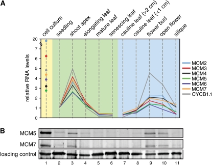
Because the MCM2-7 complex is predicted to function as a heterohexamer at the replication fork, we examined the expression profile for each of the six subunits across various stages of Arabidopsis vegetative and floral development. Relative transcript levels were determined by real-time quantitative reverse transcription (RT)-PCR and normalized using the seedling values (Fig. 1A). Seedling values were chosen for normalization because expression levels were in the middle of the detected range. The pattern of MCM gene expression generally followed the pattern of Arabidopsis Cyclin B 1;1 (CYCB1;1; Fig. 1A), which encodes a B-type cyclin that is a marker for cell proliferation (Ferreira et al., 1994). MCM and CYCB1;1 mRNAs were most abundant in cultured cells, shoot apices, and flower buds, which contain mitotic and endocycling cells (Galbraith et al., 1991; Barow, 2006). The cell culture was sampled during the exponential stage of growth and, based on CYCB1;1 expression, contained a comparable fraction of replicating cells as the microdissected shoot apical region. The lowest signals were detected in mature and senescing leaves, consistent with the absence of DNA replication in these tissues.
Figure 1.
Expression of the Arabidopsis MCM2-7 complex is coregulated during development. A, Quantitative RT-PCR analysis of MCM2-7 mRNA abundance in Arabidopsis vegetative (shaded green) and floral (shaded blue) tissues and in suspension cell culture (shaded yellow). The tissue types listed at the top are described in “Materials and Methods.” Each reaction was performed in triplicate, and se calculations are reported in Supplemental Table S3. All values were normalized to Ubiquitin-Conjugating Enzyme (At5g25760), and relative levels are scaled to expression in seedlings. CYCB1;1 (At4g37490) was used as a marker for cell proliferation. The abundance of MCM2 transcripts in cultured cells was verified in multiple experiments. B, Immunoblot analysis of MCM5 and MCM7 protein abundance in equivalent tissues to those used in A. Total protein extract (50 μg) from cultured cells (lane 1), seedling (2 weeks; lane 2), shoot apex (lane 3), elongating leaf (<1 cm; lane 4), mature leaf (>2 cm; lane 5), senescing leaf (lane 6), cauline leaf (>2 cm; lane 7), cauline leaf (<1 cm; lane 8), flower bud (lane 9), open flower (lane 10), and silique (lane 11) was resolved by SDS-PAGE, and the blots were probed with polyclonal antibodies specific for Arabidopsis MCM5 (top) and MCM7 (middle) proteins. A nonspecific band that reacted with the secondary antibody was used as a loading control (bottom).
Comparison of the relative transcript levels across the various plant tissues revealed correlations ranging from 0.99 (MCM3 versus MCM5) to 0.91 (MCM4 versus MCM6; Supplemental Table S4), indicating that expression of the MCM2 to MCM7 genes is tightly coordinated. The one exception was MCM2 expression in cultured cells, which was 20-fold higher than in seedlings, while MCM3 to MCM7 expression was only 3- to 6-fold higher (Fig. 1A; Supplemental Table S3). This difference was not observed in shoot apices, where expression of the MCM genes, including MCM2, were 3- to 5-fold higher than in seedlings. The high levels of MCM2 mRNA in cultured cells were reproducibly detected in separate experiments, suggesting that MCM2 expression is deregulated in the cultured Arabidopsis cells. We do not know whether deregulation of MCM2 transcription has functional implications, but there are numerous reports of MCM overexpression in cancer cells (Lei, 2005; Honeycutt et al., 2006; Mukherjee et al., 2007; Winnepenninckx and Van den Oord, 2007; Scarpini et al., 2008).
We asked if MCM mRNA and protein levels change in parallel during Arabidopsis development. For these experiments, we generated polyclonal antibodies against recombinant Arabidopsis MCM5 and MCM7 proteins. The antibodies specifically recognized recombinant MCM5 and MCM7 on immunoblots despite significant sequence homology in their AAA+ domains (Supplemental Fig. S1A). The antibodies also detected single bands of the expected sizes on immunoblots of total protein extracts from Arabidopsis (Supplemental Fig. S1B, lanes 2 and 5) and tobacco (Supplemental Fig. S1B, lanes 3 and 6) cultured cells. The antibodies were used to examine endogenous MCM5 and MCM7 levels in total protein extracts from Arabidopsis tissue samples harvested at developmental stages equivalent to those used for the mRNA studies. Consistent with the mRNA profiles, MCM5 and MCM7 proteins were most abundant in cultured cells (Fig. 1B, lane 1), the shoot apical region (Fig. 1B, lane 3), and flower buds (Fig. 1B, lanes 9 and 10). They were not detected in mature tissues (Fig. 1B, lanes 5–7).
Together, the RNA and protein data demonstrated that components of the MCM complex are developmentally regulated in Arabidopsis. The MCM genes are expressed and their proteins are detected primarily in proliferating tissues, consistent with a role in DNA replication. The similarity between protein and mRNA abundance in various tissues suggested that transcriptional regulation is an important determinant of MCM protein abundance at the tissue level.
MCM5 and MCM7 Display Similar Localization Patterns
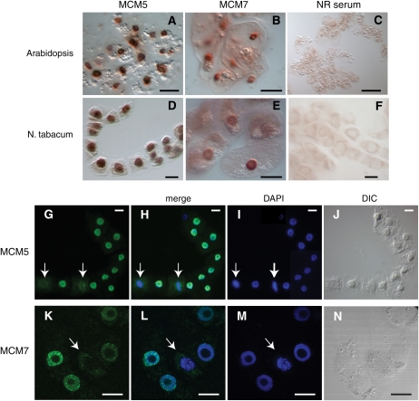
To better understand the functional organization of the MCM complex in plants, we investigated the subcellular localization of endogenous MCM5 and MCM7 proteins. Immunoperoxidase staining was used to visualize MCM5 and MCM7 proteins in cultured cells derived from Arabidopsis (Fig. 2, A–C) and tobacco (Fig. 2, D–F). The localization patterns of MCM5 and MCM7 were consistent with nuclear compartmentalization in the majority of cells from both species, and the pattern was clearly different from the diffuse background staining obtained using normal rabbit control serum (Fig. 2, C and F). In a small fraction of cells, a clear nuclear signal could not be discerned. Because MCM dynamics are related to cell cycle stage in yeast and animal systems, we used immunofluorescence microscopy to visualize DNA in conjunction with the MCMs in tobacco cells (Fig. 2, G–N). Consistent with the immunoperoxidase results, both MCM5 (green in Fig. 2, G and H) and MCM7 (green in Fig. 2, K and L) colocalized with 4′,6-diamidino-phenylindole (DAPI)-stained DNA (blue) in the majority of cells. In a small fraction of the cells, the MCM5 and MCM7 signals were diffuse throughout the cell and did not colocalize with DNA (marked by arrows). In these cells, condensed chromosomes characteristic of mitotic cells were visible. The correlation between the presence of condensed chromosomes and the diffuse MCM5 and MCM7 signals was observed in many cells through multiple experiments, suggesting that MCM is dispersed during mitosis.
Figure 2.
Localization of endogenous MCM5 and MCM7 proteins in cultured plant cells. A to F, Immunoperoxidase staining demonstrated that MCM5 (A and D) and MCM7 (B and E) displayed localization patterns consistent with nuclear compartmentalization in both Arabidopsis and tobacco cultured cells. The discrete staining patterns for MCM5 and MCM7 were distinct from the diffuse background staining obtained using normal rabbit (NR) control serum (C and F). G to N, Immunofluorescence microscopy revealed that MCM5 (green in G and H) and MCM7 (green in K and L) colocalized with DAPI-stained DNA (blue) in most tobacco cells but not when condensed chromosomes were visible (arrows). Differential interference contrast (DIC) images are shown in J and N. The images are typical, and these patterns were observed in many cells over multiple experiments. Bars = 10 μm (A and B), 100 μm (C), and 15 μm (D–N).
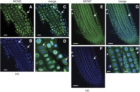
We then examined the subcellular localization patterns of MCM5 and MCM7 in Nicotiana benthamiana root tips, which contain proliferating cell populations (Fig. 3). N. benthamiana was chosen because the root tips are large enough to generate 30-μm longitudinal vibratome sections. For these experiments, histone H1 (blue fluorescence) was used as a marker for chromatin rather than DAPI staining the DNA. Because histone H1 is detected using an antibody as opposed to a small dye molecule, this strategy allowed us to conclude that an absence of MCM signal on condensed chromosomes was not the result of inaccessibility to the antibody complex. As in cultured cells, both MCM5 (Fig. 3, A–D) and MCM7 (3, E–H) colocalized with chromatin in most root tip cells, but not when mitotic figures were visible (mitotic figures are marked with arrows and asterisks). The dim H1 signal in the central core of the root tip in Figure 3B is characteristic of poor antibody penetration, most likely due to incomplete vibratome sectioning. At high magnification (Fig. 3, D and H), the two patterns of MCM5 and MCM7 localization were easily distinguishable and exclusion of MCM from condensed chromatin was apparent. These results again suggested that MCM5 and MCM7 are in the nucleus throughout most of the cell cycle and are displaced for only a brief period during mitosis. In addition, the strong similarity between our results for root tips and suspension cells shows that the results in Figure 2 do not arise from a cell culture artifact.
Figure 3.
Localization of endogenous MCM5 and MCM7 proteins in N. benthamiana root tips. Confocal microscopy was used to visualize immunofluorescence-labeled MCM5 (green in A, C, and D), MCM7 (green in E, G, and H), and histone H1 (blue) in longitudinal sections of root tips (approximately 30 μm thick). MCM5 (C) and MCM7 (G) colocalized with DNA in most cells. When condensed chromosomes were visible, MCM5 (arrows in A–D) and MCM7 (arrows and asterisks in E–H) did not colocalize with DNA. At high magnification (D and H), nuclear and nonnuclear localization patterns are clearly distinct. Bars = 20 μm (A–C), 10 μm (D), 50 μm (E–G), and 10 μm (H).
MCM5 and MCM7 Are in G1, S, and G2/M Nuclei
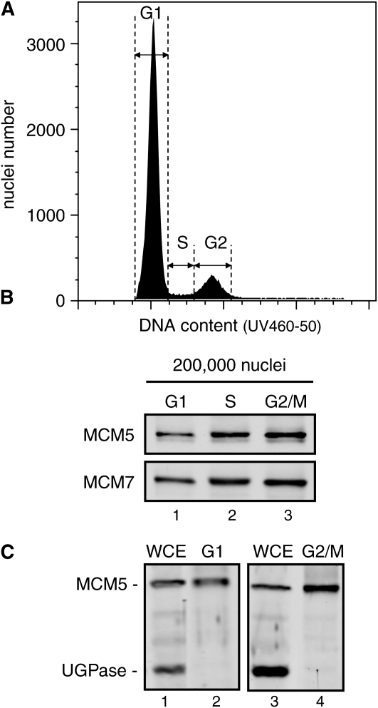
To further investigate the point in the cell cycle at which MCM5 and MCM7 shift from the nuclear to the nonnuclear compartments, we used fluorescence-activated cell sorting (FACS) to isolate Arabidopsis nuclei with G1, S, or G2/M DNA content (Fig. 4A). The FACS technology does not distinguish between cells in G2 and early mitosis, both of which have 4C DNA content. Nuclei were isolated from formaldehyde-cross-linked, 4-d-old Arabidopsis suspension cells, stained with DAPI, and sorted using a UV laser (355 nm). After reversal of the cross-linking, the protein equivalent of 200,000 nuclei was resolved by SDS-PAGE for each sample and MCM levels were monitored by immunoblot analysis. MCM5 and MCM7 were detected in the G1 (Fig. 4B, lane 1), S (Fig. 4B, lane 2), and G2/M (Fig. 4B, lane 3) nuclei. We used a commercial antibody against the cytoplasmic marker, UDP-Glc pyrophosphorylase (UGPase), to assess the purity of the nuclear extracts. The UGPase antibody detected a band of the expected size in whole cell extracts (Fig. 4C, lanes 1 and 3) but not in G1 (Fig. 4C, lane 2) or G2/M (Fig. 4C, lane 4) nuclear extracts, while the MCM5 antibody cross-reacted with a band in both extracts. These results established that MCM5 and MCM7 proteins are in nuclei with 4C as well as 2C DNA content. They are also consistent with our immunohistochemical experiments showing that the MCM proteins are in the nuclei of most cells and that their release is associated with chromosomal condensation characteristic of mitosis. Interestingly, the MCM5 and MCM7 signals increased from G1 through S, and the G2 signals were nearly double those in G1 nuclei (Fig. 4B, compare lanes 1 and 3). This observation suggested a direct relationship between MCM abundance and DNA content, which also doubles from G1 to G2.
Figure 4.
Arabidopsis MCM5 and MCM7 proteins are nuclear in G1, S, and G2/M cells. A, Nuclei were isolated from formaldehyde-cross-linked Arabidopsis suspension cells and sorted into G1, S, and G2/M populations based on DNA content by FACS. B, After cross-link reversal, proteins from 200,000 nuclei in G1 (lane 1), S (lane 2), and G2/M (lane 3) phases of the cell cycle were resolved by SDS-PAGE, and the blots were probed with anti-MCM5 (top) and anti-MCM7 (bottom) antibodies. C, Cytoplasmic contamination did not account for the presence of MCM5 and MCM7 in G2/M phase nuclei. Proteins from FACS-sorted G1 (lane 2) and G2/M (lane 4) nuclei and from whole cell extract (WCE; lanes 1 and 3) were resolved by SDS-PAGE. Blots were probed with anti-MCM5 (top) and anti-UGPase (bottom) antibodies.
Association of MCM and ORC Proteins with Chromatin
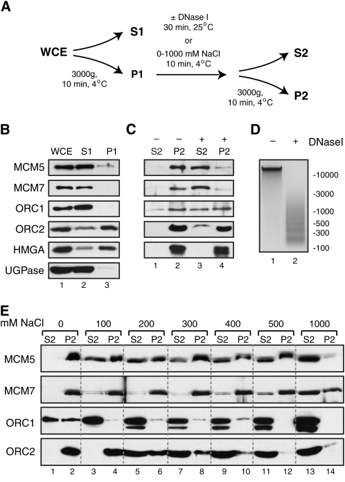
The predominantly nuclear localization pattern of MCM5 and MCM7 prompted us to examine MCM-chromatin interactions. Whole cell extracts were prepared from asynchronous Arabidopsis cultured cells and separated into soluble (S1) and pellet (P1) fractions by low-speed centrifugation (Fig. 5A). The S1 fraction includes Triton-soluble proteins from the nucleoplasm and the cytoplasmic compartment, while the P1 fraction contains proteins bound to chromatin and the nuclear matrix. Immunoblot analysis (Fig. 5B) indicated that the cytoplasmic marker protein UGPase partitioned to the S1 sample, while the chromatin marker protein HMGA (for high mobility group A) was most abundant in the P1 sample, thereby validating the quality of the fractionation. ORC2 displayed a similar distribution between the S1 and P1 fractions as HMGA, while MCM5, MCM7, and ORC1 were found primarily in the S1 fraction (Fig. 5B, lane 2). The whole cell extract and S1 signals were of similar intensities for MCM5, MCM7, and ORC1 (Fig. 5B, compare lanes 1 and 2), indicating that most of these proteins are not bound to chromatin in asynchronous cells.
Figure 5.
Chromatin binding of DNA replication initiation proteins in asynchronous Arabidopsis cultured cells. A, Scheme of the biochemical fractionation methods. B, An asynchronous culture of Arabidopsis cells was subjected to the fractionation described in A. Proteins (50 μg) from the whole cell extract (WCE) and volume equivalents from the non-chromatin-associated (S1) and chromatin-bound (P1) fractions were resolved by SDS-PAGE, and the blots were probed with the indicated antibodies. C, The purified chromatin fraction (P1) was incubated with or without 1 unit of DNase I for 30 min at 25°C and fractionated as in A. Volume equivalents from the soluble (S2) and pellet (P2) fractions were resolved by SDS-PAGE, and blots were probed with the indicated antibodies. D, DNA was extracted from samples processed as in C, resolved on a 1% agarose gel, and visualized with ethidium bromide staining. E, The purified chromatin fraction (P1) was treated with the indicated concentrations of NaCl for 10 min at 4°C and fractionated as in A. Volume equivalents from the soluble (S2) and pellet (P2) fractions were resolved by SDS-PAGE, and blots were probed with the indicated antibodies.
In other eukaryotes, the MCM2-7 complex forms a heterohexameric ring that is topologically constrained on the DNA (Maiorano et al., 2006). To determine whether Arabidopsis MCM proteins are similarly constrained, we treated the P1 fraction with DNase I (Fig. 5, C and D). Soluble (S2) and nuclease-resistant (P2) proteins were then separated by centrifugation and visualized on immunoblots. DNase I treatment resulted in limited DNA digestion (Fig. 5D, lane 2) and did not release the chromatin protein HMGA (Fig. 5C). The majority of ORC2 and approximately 50% of ORC1 also remained in the nuclease-resistant P2 fraction (Fig. 5C, lane 4). In contrast, MCM5 and MCM7 were nearly completely released upon DNase I treatment (Fig. 5C). This result indicated that MCM is retained in the P1 fraction by a topological interaction with DNA rather than by binding to insoluble chromatin or nuclear matrix proteins.
Next, we investigated the stability of the DNA replication initiation complex by treating the P1 fraction with increasing concentrations of NaCl (Fig. 5A). Again, soluble (S2) and salt-resistant (P2) proteins were then separated by centrifugation and visualized on immunoblots (Fig. 5E). Significant fractions of both MCM5 and MCM7 were highly salt resistant, remaining in the P2 fraction at 500 mm NaCl (Fig. 5E, lane 12). Approximately 50% of MCM5 that copurified with the P1 fraction was released at 100 mm NaCl (Fig. 5E, compare lanes 3 and 4). Given that no additional MCM5 was released in the 200 to 500 mm treatments (Fig. 5E, lanes 5–12), this labile fraction likely represents nonspecific interactions that persist during the very low-salt conditions of the initial extraction. At high salt concentrations, MCM5 is slightly more labile than MCM7. MCM5 was fully extracted at 1 m NaCl (Fig. 5E, lanes 13 and 14), while a significant fraction of MCM7 remained stably associated with the pellet in this treatment. In contrast, nearly all of the ORC1 protein was extracted by the 100 mm NaCl treatment (Fig. 5E, lanes 3 and 4) and ORC2 was removed at 200 to 300 mm NaCl (Fig. 5E, lanes 5–8). Together, these results demonstrated that the stabilities of the Arabidopsis MCM and ORC chromatin complexes differ significantly. The very tight association of MCM5 and MCM7 proteins is indicative of the formation of a topologically constrained MCM/DNA complex in Arabidopsis, as has been observed in other eukaryotes.
DISCUSSION
In previous work, we demonstrated that the core eukaryotic DNA replication machinery is conserved in plants (Shultz et al., 2007). In this study, we characterized the expression profile, localization pattern, and chromatin-binding properties of the putative replicative helicase complex, MCM2-7. Our results show that the Arabidopsis MCM2-7 complex is preferentially expressed in proliferating tissues and that two subunits, MCM5 and MCM7, are topologically loaded onto DNA. This behavior supports a conserved role for the MCM complex in licensing and DNA replication in plants. Our experiments also demonstrate that the MCM5 and MCM7 proteins are located in the nucleus throughout the plant cell division cycle except during chromosome condensation in mitosis, when they are dispersed throughout the cell. These results strongly suggest that, unlike budding yeast, plants do not regulate origin licensing by actively exporting the MCM complex from the nucleus during S phase. Instead, the plant MCM localization patterns resemble those in animals, which rely on other mechanisms to control licensing.
In yeast and animals, the MCM2 to MCM7 proteins function as a heterohexameric complex at the DNA replication fork (Maiorano et al., 2006; Bochman and Schwacha, 2008; Kanter et al., 2008), and each subunit is present in approximately stoichiometric quantities (Crevel et al., 2001; Forsburg, 2004; Namdar and Kearsey, 2006). Previous studies in Arabidopsis and maize showed that MCM3 (Sabelli et al., 1996; Stevens et al., 2002), MCM6 (Dresselhaus et al., 2006), and MCM7 (Springer et al., 1995, 2000; Bastida and Puigdomenech, 2002; Holding and Springer, 2002) are expressed in young tissues that contain actively replicating cells, but it has not been clear whether the entire complex is coordinately expressed. We showed that the relative mRNA abundance for each of the six MCM subunits was highly correlated with replication potential and that the entire complex was coordinately expressed across 11 tissue types representing various stages of Arabidopsis vegetative and floral development (Fig. 1; Supplemental Table S4). Furthermore, we have demonstrated that MCM5 and MCM7 protein accumulation patterns mirror their steady-state mRNA levels. Together, these results suggest that MCM2-7 expression is coordinated during plant development, possibly at the level of transcription.
The E2F/DP transcription factor family regulates many genes involved in DNA replication and cell cycle control (De Veylder et al., 2002; Ramirez-Parra et al., 2003). Arabidopsis MCM2 to MCM5 and MCM7 genes contain E2F consensus sites in their promoters, and their transcripts are elevated in plants ectopically expressing E2FA/DPA (Stevens et al., 2002; Vandepoele et al., 2005). We found an E2F consensus motif in the Arabidopsis MCM6 promoter (R.W. Shultz, unpublished data), suggesting that it is also regulated by the E2F/DP pathway. Interestingly, our data indicated that MCM2, but not the other MCM genes, is overexpressed in cultured cells. We validated this result with multiple experiments and by mining an independently conducted microarray data set that analyzed transcript levels in the same Arabidopsis cell line (Tanurdzic et al., 2008). MCM2 overexpression suggested that other mechanisms may also affect the regulation of MCM transcript abundance and underscored that caution must be used when cultured plant cells are employed as models for studying plant DNA replication.
In budding yeast, MCM activity is modulated in part by active export from the nucleus during S phase (Sherman and Forsburg, 1998; Forsburg, 2004), while in animals it is dispersed only upon nuclear envelope breakdown in mitosis (Forsburg, 2004). We generated several lines of evidence indicating that MCM dynamics in plants are more similar to those in animals. First, immunofluorescence microscopy of endogenous MCM5 and MCM7 proteins in both Arabidopsis and Nicotiana tissues revealed that the vast majority of cells display an exclusively nuclear localization pattern (Figs. 2 and 3). The amount of time spent in each phase of the cell cycle is remarkably constant across different tissues and plant species, with G2 and M constituting approximately one-third of the entire cycle (Kidd et al., 1987; Baskin, 2000; Menges et al., 2005). If the MCM complex were exported during S phase and excluded from the nucleus in G2 and M, at least one-third of the cell population should show dispersed localization, which is inconsistent with our results. Second, we observed a high correlation between mitotic figures and nonnuclear localization of MCM, as would be predicted for passive release during mitosis. Finally, immunoblot analysis of Arabidopsis nuclei sorted into G1, S, and G2/M populations demonstrated that MCM5 and MCM7 are present in G2/M nuclei at levels comparable to those in G1 nuclei (Fig. 4). It is difficult to reconcile these findings with a model where MCM subunits are exported during S phase.
Several reports have suggested that MCM dynamics in plants resemble those in budding yeast. However, most of these studies are based on ectopic expression of MCM proteins using strong promoters (Dresselhaus et al., 2006; Takahashi et al., 2008). Nuclear import of the MCM2-7 complex in other eukaryotes is mediated by a bipartite nuclear localization signal that is split between the MCM2 and MCM3 subunits (Kimura et al., 1996; Liku et al., 2005), so overexpression of a single subunit without a nuclear localization signal may result in its artifactual accumulation in the cytoplasm. Hence, it is essential to study MCM dynamics without perturbing the stoichiometry of the complex. Indeed, when the endogenous MCM7 promoter was used to drive the expression of a MCM7-GFP fusion in Arabidopsis, the majority of cells showed nucleus-localized GFP (Springer et al., 2000). When these cells were followed with time-lapse photography, the MCM7-GFP signal dispersed from the nucleus within a single 4-min interval (Springer et al., 2000). This rapid transition from the nucleus to the cytoplasm is consistent with passive release rather than active export. Patterns of endogenous MCM3 localization in maize root tips are also consistent with the patterns we observed for MCM5 and MCM7 in dicots, suggesting that MCM dynamics are conserved across higher plants (Sabelli et al., 1999). However, an analysis of endogenous MCM6 localization in maize Black Mexican Sweet (BMS) cells revealed that maize MCM6 was present in the nucleus during G1 and early S and absent in G2, similar to the pattern of MCM localization in budding yeast (Dresselhaus et al., 2006). This raises the intriguing possibility that export of the MCM6 subunit during S phase may serve a regulatory role in preventing licensing during S and G2. It is also possible that MCM regulation has been altered in the cultured BMS cells. Additional experiments will be required to distinguish between these possibilities and determine MCM6 dynamics in intact plants.
Current models based on data from yeast and animal systems propose that the MCM complex is loaded onto origin DNA prior to the onset of DNA replication and is progressively released as DNA replication forks proceed during S phase. Thus, in an asynchronous population of cells, there are two MCM populations: one that is topologically constrained on DNA and one that is not. Our analysis of the chromatin-binding properties of MCM5 and MCM7 in asynchronous Arabidopsis cells also showed two distinct MCM populations. The majority of total cellular MCM5 and MCM7 are not bound to chromatin (Fig. 5B). However, a significant fraction of MCM5 and MCM7 copurify with chromatin and are released by limited DNase I digestion, indicative of a complex that is topologically bound to DNA (Fig. 5C). Both MCM5 and MCM7 remained stably associated with chromatin at 500 mm NaCl, but MCM7 was more resistant to high-salt extraction than MCM5 (Fig. 5E). Similar results have been reported in yeast, mouse, and frog, where MCM4, MCM6, and MCM7 form a “core” complex that is more tightly associated with chromatin than the remaining subunits (Ishimi et al., 1996; Coue et al., 1998; Holthoff et al., 1998; Sherman and Forsburg, 1998; Sherman et al., 1998; Lee and Hurwitz, 2000; Prokhorova and Blow, 2000; Davey et al., 2003).
It is intriguing that we detected nearly all of the total cellular MCM5 and MCM7 proteins in the non-chromatin-bound fraction (Fig. 5B). In parallel assays, the majority of the chromatin marker HMGA remained associated with the pellet fraction, establishing that our extraction protocol does not result in widespread dissociation of chromatin and cannot account for the bulk of MCM5 and MCM7 in the soluble fraction. In asynchronous, immortalized human cells, the non-chromatin-bound fraction of MCM ranges from slightly more than half (Holthoff et al., 1998; Mendez and Stillman, 2000) to approximately 90% of the total cellular protein (Burkhart et al., 1995). A significant fraction of the non-chromatin-bound MCM in the Arabidopsis culture may have originated from cells that have exited the cell cycle. In human and mouse cell lines, MCM subunits remain abundant but are displaced from chromatin in early quiescent cells (Madine et al., 2000; Kingsbury et al., 2005). In spite of the parallels with animal systems, it is possible that the very large fraction of non-chromatin-bound MCM has biological relevance in plants, which needs to be addressed in future experiments.
We have clearly demonstrated that plant MCM5 and MCM7 subunits remain in the nucleus during S and G2 phases of the cell cycle, which raises the question of how MCM activity is regulated to prevent rereplication. In animals, a CDT1-Geminin switch regulates this process. Computational analyses of plant genomes have so far failed to identify a candidate Geminin homolog. A yeast two-hybrid screen for Arabidopsis CDT1-interacting proteins identified Glabra2-Expression Modulator as a candidate plant Geminin homolog (Caro et al., 2007). The potential role of Glabra2-Expression Modulator in cell proliferation is interesting, but it remains to be determined whether this protein has a direct role in licensing. The yeast-like dynamics of MCM6 reported in maize BMS cells (Dresselhaus et al., 2006) raises the possibility that one or more MCM subunits may contribute to licensing regulation in plants. In addition, our result showing that, unlike ORC2, Arabidopsis ORC1 is weakly associated with chromatin is reminiscent of mammalian ORC1, which serves a regulatory function and is not part of the core origin recognition complex (Li and DePamphilis, 2002; Ohta et al., 2003). Recent evidence suggested that plant ORC1 proteins can activate transcription by triggering localized histone acetylation (Sanchez and Gutierrez, 2009), and it is possible that ORC1 also triggers chromatin alterations that regulate DNA replication initiation. Future studies are needed to delineate the precise timing of MCM release during the cell cycle and elucidate the mechanisms that prevent MCM reloading onto replicated DNA in S and G2 phases in plants.
MATERIALS AND METHODS
Plant Materials and Growth Conditions
An Arabidopsis (Arabidopsis thaliana ecotype Columbia-0) suspension cell culture was maintained by subculturing 5 mL of 7-d-old cells into 50 mL of fresh medium weekly. Cells were grown in 250-mL baffled flasks at 160 rpm under constant fluorescent light at 21°C. Each liter of medium contained 3.2 g of Gamborg's B5 basal medium with minimal organics, 0.5 g of MES, 1.1 mg of 2,4-dichlorophenoxyacetic acid, and 30 g of Suc and was adjusted to pH 5.7 using 1 m KOH before autoclaving. The tobacco (Nicotiana tabacum) NT-1 cell line was maintained as described previously (Abranches et al., 2005).
For vegetative tissue samples, Arabidopsis plants were grown in soil for 5 weeks at 20°C with an 8-h/16-h light/dark cycle under a light intensity of 15,000 lux. For floral tissue samples, plants grown as above were shifted to a 16-h/8-h light/dark cycle to induce flowering. Seedlings were grown on petri dishes containing 1× Murashige and Skoog salts, 1× Gamborg's B5 vitamins, 1% (w/v) Suc, and 0.7% (w/v) agar for 2 weeks. All tissue samples were frozen in liquid nitrogen prior to RNA and protein extraction. Nicotiana benthamiana plants used for immunolocalization were grown in magenta boxes containing 1× Murashige and Skoog salts, 1× Gamborg's B5 vitamins, 3% (w/v) Suc, and 0.7% (w/v) agar for 6 weeks under constant light at 25°C.
The tissues analyzed in Figure 1 are as follows: cell culture, 4-d-old suspension cells; seedling, 2-week-old whole seedlings; shoot apex, the shoot apex was extracted from all visible leaves and leaf primordia using a dissecting microscope; elongating leaf, leaves between 0.5 and 1 cm in length; mature leaf, fully expanded with no signs of senescence; senescing leaf, mostly green with some yellowing; cauline leaf (>2 cm), large cauline leaves from the base of the inflorescence; cauline leaf (<1 cm), small cauline leaves from the top of the inflorescence; flower bud, unopened; open flower, fully opened; silique, fully formed but still green.
Real-Time RT-PCR
Total RNA was extracted from 100 mg of frozen tissue powder using the NucleoSpin RNA Plant kit (Clontech). cDNA was produced from total RNA (3 μg) using PowerScript reverse transcriptase (Clontech) and oligo(dT)15 primer followed by RNase A (Qiagen) treatment. For quantitative RT-PCR, the cDNA was diluted 100-fold with water and 2.5 mL was used in a 25-mL reaction containing 400 nm each primer and 1× SYBR Green PCR master mix (Applied Biosystems). Samples were analyzed in triplicate in a MX3000P qPCR thermocycler (Stratagene). Relative expression levels were calculated according to the 2−ΔΔCT method (Livak and Schmittgen, 2001) using Ubiquitin-conjugating enzyme (At5g25760) as the internal control. Primers used for these analyses are listed in Supplemental Table S2.
Cloning of Arabidopsis ORC and MCM Subunits
Genes were cloned using the TOPO cloning system (Invitrogen), verified by sequencing, and transferred to expression vectors by Gateway recombination (Invitrogen). Manufacturer protocols were followed unless otherwise noted. Clones and primers are described in Supplemental Table S1.
Recombinant Protein Purification and Antibody Production
His6-tagged AtORC1, AtORC2, AtMCM5, and AtMCM7 polypeptides were expressed in Escherichia coli BL21 (DE3) cells from the pDEST17- and pET-DEST42-based vectors (Supplemental Table S1), purified by nickel-nitrilotriacetic acid agarose chromatography (Qiagen), and used as antigens for antibody production in rabbits (Cocalico Biologicals) The resulting polyclonal sera were used directly for immunoblot analysis or affinity purified for immunolocalization studies. For affinity purification, recombinant proteins were coupled to HiTrap NHS-activated HP columns (GE Healthcare). Polyclonal sera (1 mL) were passed over the column, and antibodies were eluted with low-pH buffer (100 mm Gly, pH 2.5). The specificity of the affinity-purified sera was validated by immunoblot analysis (Supplemental Fig. S1). The HMGA antiserum has been described previously (Spiker and Everett, 1987). UGPase antibodies were from Agrisera, and the histone H1 antiserum was from Upstate.
Protein Extraction and Immunoblotting
Plant material was ground under liquid nitrogen, and proteins were extracted by a trichloroacetic acid-acetone (TCA-A) method (Shultz et al., 2005). Proteins (50 μg) were resolved on 10% (w/v) gels by SDS-PAGE and transferred to nitrocellulose membranes. Membranes were blocked with 5% (w/v) nonfat milk in Tris-buffered saline (TBS; 25 mm Tris-HCl, pH 7.4, 140 mm NaCl, and 3 mm KCl) and probed with primary antibody (1:5,000 dilution) in blocking solution with 0.1% Tween (TBST) for 1 h at room temperature. After washing in TBST, membranes were probed with secondary antibodies diluted 1:10,000 in TBST blocking solution, washed in TBST, and detected using the Li-Cor Odyssey infrared imaging system. Secondary antibodies were Rockland IRDye 800-conjugated goat anti-rabbit antibody and Molecular Probes Alexa 680-conjugated goat anti-rabbit antibody.
Immunohistochemical Staining and Immunofluorescence Microscopy
Cells from 4-d-old Arabidopsis or NT-1 suspension cultures were fixed with 1% (w/v) formaldehyde in phosphate-buffered saline (PBS; 137 mm NaCl, 2.7 mm KCl, 10 mm Na2HPO4, and 2 mm KH2PO4, pH 7.4) for 15 min at room temperature and dried onto poly-Lys-coated slides. N. benthamiana roots were fixed in 4% (w/v) formaldehyde for 1 h and embedded in 5% (w/v) agarose. Longitudinal root tip sections (approximately 30 μm) were cut with a vibratome and dried onto poly-Lys-coated slides. Cells or sections were permeabilized with 0.5% (w/v) cellulase for 10 min and blocked with 5% (w/v) bovine serum albumin in PBS for 1 to 4 h. Tissue was probed with affinity-purified anti-AtMCM5 or anti-AtMCM7 antibody at 1:250 dilution in blocking solution overnight at 4°C. Anti-histone H1 antibody (Upstate) at 1:250 dilution was included in the root section incubations. Slides were washed with PBS plus 0.1% (v/v) Tween 20 and probed with secondary antibodies. The Vectastain elite ABC kit (Vector Laboratories) was used for immunohistochemical detection. Alexa Fluor 488 goat anti-rabbit IgG (Invitrogen) and Alexa Fluor 350 goat anti-mouse IgG (Invitrogen) were used for immunofluorescence detection. Tissue was visualized with a Nikon Eclipse E800 microscope and a confocal microscope system (Leica SP; Leica).
FACS
Four-day-old asynchronous Arabidopsis suspension cells were fixed in 1% paraformaledyde for 15 min. After washing with 1× PBS buffer three times, the cell pellet was frozen in liquid nitrogen and chopped in lysis buffer (15 mm Tris-HCl, pH 7.5, 2 mm EDTA, 80 mm KCl, 20 mm NaCl, 15 mm β-mercaptoethanol, 2 μg mL−1 DAPI, and 0.1% Triton X-100) using a single-edge blade for 5 min and incubated at 4°C for 5 min. The chopped cell suspension was filtered through a four-tiered nylon mesh (100, 50, 30, and 20 μm), stained with DAPI (1 μg mL−1), and sorted by an Influx cell sorter (Cytopeia) equipped with a 355-nm UV laser. 1× PBS buffer (pH 7.5) was used as a sheath fluid. FACS analysis was conducted using FlowJo (version 8.7.1).
Chromatin Purification
Arabidopsis 4-d-old cultured cells (8 g wet weight) were ground under liquid nitrogen, added to 40 mL of buffer A (ice cold; 10 mm PIPES, pH 6.8, 10 mm KCl, 1.5 mm MgCl2, 340 mm Suc, 10% [v/v] glycerol, 1 mm dithiothreitol, 1 mm ATP, 0.5% [v/v] Triton X-100, and 1× Plant Protease Inhibitor [Sigma]), and incubated on ice for 10 min. After removal of debris by filtration through Miracloth, proteins from 1 mL of this whole cell extract were extracted by TCA-A. A second aliquot of whole cell extract (1 mL) was centrifuged at 3,000g for 10 min at 4°C to separate non-chromatin-bound proteins (S1) from chromatin-bound proteins (P1) and extracted by TCA-A. The remaining whole cell extract (38 mL) was centrifuged as above, and the chromatin pellet was resuspended in 5 mL of buffer A, divided into aliquots in new tubes, and centrifuged again. The purified chromatin pellets were resuspended in 1 mL of buffer A supplemented with the indicated concentrations of NaCl, incubated on ice for 10 min or resuspended in 100 μL of buffer A supplemented with 1 unit of DNase I (New England Biolabs), and incubated at 25°C for 30 min. Soluble (S2) and insoluble (P2) proteins were resolved by SDS-PAGE and visualized by immunoblot. For DNA extraction, 20 mm EDTA, 0.5% (w/v) SDS, and 10 μg of RNase A (Fermentas) were added and samples were incubated at 37°C for 30 min. Then, 20 μg of Proteinase K (New England Biolabs) was added, samples were incubated at 37°C for 2 h, and DNA was purified with a PCR cleanup column (Qiagen).
Supplemental Data
The following materials are available in the online version of this article.
Supplemental Figure S1. Validation of the MCM5, MCM7, ORC1, and ORC2 antibodies.
Supplemental Table S1. Description of clones generated for this study.
Supplemental Table S2. List of primers used for real-time quantitative RT-PCR.
Supplemental Table S3. Relative expression values with error calculations for Figure 1A.
Supplemental Table S4. Correlation coefficients for MCM gene expression values.
Supplementary Material
Acknowledgments
The Arabidopsis (Columbia-0) suspension cell culture was kindly provided by Dr. Iris Meier (Ohio State University). We also thank Patricia Springer (University of California, Riverside) for a plasmid containing the AtMCM7 coding sequence, Steven Spiker (North Carolina State University) for the HMGA antisera, and Pete Pascuzzi (North Carolina State University) for helpful discussions.
This work was supported by the National Science Foundation Plant Genome Research Program (grant no. 0421651).
The author responsible for distribution of materials integral to the findings presented in this article in accordance with the policy described in the Instructions for Authors (www.plantphysiol.org) is: Randall W. Shultz (rwshultz@ncsu.edu).
The online version of this article contains Web-only data.
Open Access articles can be viewed online without a subscription.
References
- Abranches R, Shultz RW, Thompson WF, Allen GC (2005) Matrix attachment regions and regulated transcription increase and stabilize transgene expression. Plant Biotechnol J 3 535–543 [DOI] [PubMed] [Google Scholar]
- Adachi Y, Usukura J, Yanagida M (1997) A globular complex formation by Nda1 and the other five members of the MCM protein family in fission yeast. Genes Cells 2 467–479 [DOI] [PubMed] [Google Scholar]
- Barow M (2006) Endopolyploidy in seed plants. Bioessays 28 271–281 [DOI] [PubMed] [Google Scholar]
- Baskin TI (2000) On the constancy of cell division rate in the root meristem. Plant Mol Biol 43 545–554 [DOI] [PubMed] [Google Scholar]
- Bastida M, Puigdomenech P (2002) Specific expression of ZmPRL, the maize homolog of MCM7, during early embryogenesis. Plant Sci 162 97–106 [Google Scholar]
- Bell SP, Dutta A (2002) DNA replication in eukaryotic cells. Annu Rev Biochem 71 333–374 [DOI] [PubMed] [Google Scholar]
- Blow JJ, Dutta A (2005) Preventing re-replication of chromosomal DNA. Nat Rev Mol Cell Biol 6 476–486 [DOI] [PMC free article] [PubMed] [Google Scholar]
- Bochman ML, Schwacha A (2008) The Mcm2-7 complex has in vitro helicase activity. Mol Cell 31 287–293 [DOI] [PubMed] [Google Scholar]
- Burkhart R, Schulte D, Hu B, Musahl C, Gohring F, Knippers R (1995) Interactions of human nuclear proteins P1mcm3 and P1cdc46. Eur J Biochem 228 431–438 [PubMed] [Google Scholar]
- Caro E, Castellano MM, Gutierrez C (2007) A chromatin link that couples cell division to root epidermis patterning in Arabidopsis. Nature 447 213–217 [DOI] [PubMed] [Google Scholar]
- Chang YP, Wang G, Bermudez V, Hurwitz J, Chen XS (2007) Crystal structure of the GINS complex and functional insights into its role in DNA replication. Proc Natl Acad Sci USA 104 12685–12690 [DOI] [PMC free article] [PubMed] [Google Scholar]
- Coue M, Amariglio F, Maiorano D, Bocquet S, Mechali M (1998) Evidence for different MCM subcomplexes with differential binding to chromatin in Xenopus. Exp Cell Res 245 282–289 [DOI] [PubMed] [Google Scholar]
- Coue M, Kearsey SE, Mechali M (1996) Chromatin binding, nuclear localization and phosphorylation of Xenopus cdc21 are cell-cycle dependent and associated with the control of initiation of DNA replication. EMBO J 15 1085–1097 [PMC free article] [PubMed] [Google Scholar]
- Crevel G, Ivetic A, Ohno K, Yamaguchi M, Cotterill S (2001) Nearest neighbour analysis of MCM protein complexes in Drosophila melanogaster. Nucleic Acids Res 29 4834–4842 [DOI] [PMC free article] [PubMed] [Google Scholar]
- Dambrauskas G, Aves SJ, Bryant JA, Francis D, Rogers HJ (2003) Genes encoding two essential DNA replication activation proteins, Cdc6 and Mcm3, exhibit very different patterns of expression in the tobacco BY-2 cell cycle. J Exp Bot 54 699–706 [DOI] [PubMed] [Google Scholar]
- Davey MJ, Indiani C, O'Donnell M (2003) Reconstitution of the Mcm2-7p heterohexamer, subunit arrangement, and ATP site architecture. J Biol Chem 278 4491–4499 [DOI] [PubMed] [Google Scholar]
- de la Paz Sanchez M, Gutierrez C (2009) Arabidopsis ORC1 is a PHD-containing H3K4me3 effector that regulates transcription. Proc Natl Acad Sci USA 106 2065–2070 [DOI] [PMC free article] [PubMed] [Google Scholar]
- De Veylder L, Beeckman T, Beemster GT, de Almeida Engler J, Ormenese S, Maes S, Naudts M, Van Der Schueren E, Jacqmard A, Engler G, et al (2002) Control of proliferation, endoreduplication and differentiation by the Arabidopsis E2Fa-DPa transcription factor. EMBO J 21 1360–1368 [DOI] [PMC free article] [PubMed] [Google Scholar]
- Diffley JF (2004) Regulation of early events in chromosome replication. Curr Biol 14 R778–R786 [DOI] [PubMed] [Google Scholar]
- Dresselhaus T, Srilunchang KO, Leljak-Levanic D, Schreiber DN, Garg P (2006) The fertilization induced DNA replication factor MCM6 of maize shuttles between cytoplasm and nucleus, and is essential for plant growth and development. Plant Physiol 140 512–527 [DOI] [PMC free article] [PubMed] [Google Scholar]
- Drury LS, Perkins G, Diffley JF (1997) The Cdc4/34/53 pathway targets Cdc6p for proteolysis in budding yeast. EMBO J 16 5966–5976 [DOI] [PMC free article] [PubMed] [Google Scholar]
- Drury LS, Perkins G, Diffley JF (2000) The cyclin-dependent kinase Cdc28p regulates distinct modes of Cdc6p proteolysis during the budding yeast cell cycle. Curr Biol 10 231–240 [DOI] [PubMed] [Google Scholar]
- Dutta A, Bell SP (1997) Initiation of DNA replication in eukaryotic cells. Annu Rev Cell Dev Biol 13 293–332 [DOI] [PubMed] [Google Scholar]
- Ferreira PC, Hemerly AS, Engler JD, van Montagu M, Engler G, Inze D (1994) Developmental expression of the Arabidopsis cyclin gene cyc1At. Plant Cell 6 1763–1774 [DOI] [PMC free article] [PubMed] [Google Scholar]
- Forsburg SL (2004) Eukaryotic MCM proteins: beyond replication initiation. Microbiol Mol Biol Rev 68 109–131 [DOI] [PMC free article] [PubMed] [Google Scholar]
- Fujita M (2006) Cdt1 revisited: complex and tight regulation during the cell cycle and consequences of deregulation in mammalian cells. Cell Div 1 22. [DOI] [PMC free article] [PubMed] [Google Scholar]
- Fujita M, Kiyono T, Hayashi Y, Ishibashi M (1996) hCDC47, a human member of the MCM family: dissociation of the nucleus-bound form during S phase. J Biol Chem 271 4349–4354 [DOI] [PubMed] [Google Scholar]
- Galbraith DW, Harkins KR, Knapp S (1991) Systemic endopolyploidy in Arabidopsis thaliana. Plant Physiol 96 985–989 [DOI] [PMC free article] [PubMed] [Google Scholar]
- Gambus A, Jones RC, Sanchez-Diaz A, Kanemaki M, van Deursen F, Edmondson RD, Labib K (2006) GINS maintains association of Cdc45 with MCM in replisome progression complexes at eukaryotic DNA replication forks. Nat Cell Biol 8 358–366 [DOI] [PubMed] [Google Scholar]
- Gregory TR, Nicol JA, Tamm H, Kullman B, Kullman K, Leitch IJ, Murray BG, Kapraun DF, Greilhuber J, Bennett MD (2007) Eukaryotic genome size databases. Nucleic Acids Res 35 D332–D338 [DOI] [PMC free article] [PubMed] [Google Scholar]
- Holding DR, Springer PS (2002) The Arabidopsis gene PROLIFERA is required for proper cytokinesis during seed development. Planta 214 373–382 [DOI] [PubMed] [Google Scholar]
- Holthoff HP, Baack M, Richter A, Ritzi M, Knippers R (1998) Human protein MCM6 on HeLa cell chromatin. J Biol Chem 273 7320–7325 [DOI] [PubMed] [Google Scholar]
- Honeycutt KA, Chen Z, Koster MI, Miers M, Nuchtern J, Hicks J, Roop DR, Shohet JM (2006) Deregulated minichromosomal maintenance protein MCM7 contributes to oncogene driven tumorigenesis. Oncogene 25 4027–4032 [DOI] [PubMed] [Google Scholar]
- Ishimi Y, Ichinose S, Omori A, Sato K, Kimura H (1996) Binding of human minichromosome maintenance proteins with histone H3. J Biol Chem 271 24115–24122 [DOI] [PubMed] [Google Scholar]
- Kanter DM, Bruck I, Kaplan DL (2008) Mcm subunits can assemble into two different active unwinding complexes. J Biol Chem 283 31172–31182 [DOI] [PMC free article] [PubMed] [Google Scholar]
- Kearsey SE, Labib K (1998) MCM proteins: evolution, properties, and role in DNA replication. Biochim Biophys Acta 1398 113–136 [DOI] [PubMed] [Google Scholar]
- Kerns SL, Torke SJ, Benjamin JM, McGarry TJ (2007) Geminin prevents rereplication during Xenopus development. J Biol Chem 282 5514–5521 [DOI] [PubMed] [Google Scholar]
- Kidd AD, Francis D, Bennett MD (1987) Replicon size, mean rate of DNA-replication and the duration of the cell-cycle and its component phases in 8 monocotyledonous species of contrasting DNA C values. Ann Bot (Lond) 59 603–609 [Google Scholar]
- Kim Y, Kipreos ET (2007) Cdt1 degradation to prevent DNA re-replication: conserved and non-conserved pathways. Cell Div 2 18. [DOI] [PMC free article] [PubMed] [Google Scholar]
- Kimura H, Ohtomo T, Yamaguchi M, Ishii A, Sugimoto K (1996) Mouse MCM proteins: complex formation and transportation to the nucleus. Genes Cells 1 977–993 [DOI] [PubMed] [Google Scholar]
- Kingsbury SR, Loddo M, Fanshawe T, Obermann EC, Prevost AT, Stoeber K, Williams GH (2005) Repression of DNA replication licensing in quiescence is independent of geminin and may define the cell cycle state of progenitor cells. Exp Cell Res 309 56–67 [DOI] [PubMed] [Google Scholar]
- Lee JK, Hurwitz J (2000) Isolation and characterization of various complexes of the minichromosome maintenance proteins of Schizosaccharomyces pombe. J Biol Chem 275 18871–18878 [DOI] [PubMed] [Google Scholar]
- Lei M (2005) The MCM complex: its role in DNA replication and implications for cancer therapy. Curr Cancer Drug Targets 5 365–380 [DOI] [PubMed] [Google Scholar]
- Li CJ, DePamphilis ML (2002) Mammalian Orc1 protein is selectively released from chromatin and ubiquitinated during the S-to-M transition in the cell division cycle. Mol Cell Biol 22 105–116 [DOI] [PMC free article] [PubMed] [Google Scholar]
- Liku ME, Nguyen VQ, Rosales AW, Irie K, Li JJ (2005) CDK phosphorylation of a novel NLS-NES module distributed between two subunits of the Mcm2-7 complex prevents chromosomal rereplication. Mol Biol Cell 16 5026–5039 [DOI] [PMC free article] [PubMed] [Google Scholar]
- Liu W, Pucci B, Rossi M, Pisani FM, Ladenstein R (2008) Structural analysis of the Sulfolobus solfataricus MCM protein N-terminal domain. Nucleic Acids Res 36 3235–3243 [DOI] [PMC free article] [PubMed] [Google Scholar]
- Livak KJ, Schmittgen TD (2001) Analysis of relative gene expression data using real-time quantitative PCR and the 2(-Delta Delta C(T)) method. Methods 25 402–408 [DOI] [PubMed] [Google Scholar]
- Madine MA, Swietlik M, Pelizon C, Romanowski P, Mills AD, Laskey RA (2000) The roles of the MCM, ORC, and Cdc6 proteins in determining the replication competence of chromatin in quiescent cells. J Struct Biol 129 198–210 [DOI] [PubMed] [Google Scholar]
- Maiorano D, Lutzmann M, Mechali M (2006) MCM proteins and DNA replication. Curr Opin Cell Biol 18 130–136 [DOI] [PubMed] [Google Scholar]
- Masuda HP, Ramos GB, de Almeida-Engler J, Cabral LM, Coqueiro VM, Macrini CM, Ferreira PC, Hemerly AS (2004) Genome based identification and analysis of the pre-replicative complex of Arabidopsis thaliana. FEBS Lett 574 192–202 [DOI] [PubMed] [Google Scholar]
- Melixetian M, Ballabeni A, Masiero L, Gasparini P, Zamponi R, Bartek J, Lukas J, Helin K (2004) Loss of Geminin induces rereplication in the presence of functional p53. J Cell Biol 165 473–482 [DOI] [PMC free article] [PubMed] [Google Scholar]
- Mendez J, Stillman B (2000) Chromatin association of human origin recognition complex, Cdc6, and minichromosome maintenance proteins during the cell cycle: assembly of prereplication complexes in late mitosis. Mol Cell Biol 20 8602–8612 [DOI] [PMC free article] [PubMed] [Google Scholar]
- Menges M, de Jager SM, Gruissem W, Murray JAH (2005) Global analysis of the core cell cycle regulators of Arabidopsis identifies novel genes, reveals multiple and highly specific profiles of expression and provides a coherent model for plant cell cycle control. Plant J 41 546–566 [DOI] [PubMed] [Google Scholar]
- Mimura S, Seki T, Tanaka S, Diffley JF (2004) Phosphorylation-dependent binding of mitotic cyclins to Cdc6 contributes to DNA replication control. Nature 431 1118–1123 [DOI] [PubMed] [Google Scholar]
- Moll T, Tebb G, Surana U, Robitsch H, Nasmyth K (1991) The role of phosphorylation and the CDC28 protein kinase in cell cycle-regulated nuclear import of the S. cerevisiae transcription factor SWI5. Cell 66 743–758 [DOI] [PubMed] [Google Scholar]
- Moyer SE, Lewis PW, Botchan MR (2006) Isolation of the Cdc45/Mcm2-7/GINS (CMG) complex, a candidate for the eukaryotic DNA replication fork helicase. Proc Natl Acad Sci USA 103 10236–10241 [DOI] [PMC free article] [PubMed] [Google Scholar]
- Mukherjee G, Muralidhar B, Bafna UD, Laskey RA, Coleman N (2007) MCM immunocytochemistry as a first line cervical screening test in developing countries: a prospective cohort study in a regional cancer centre in India. Br J Cancer 96 1107–1111 [DOI] [PMC free article] [PubMed] [Google Scholar]
- Namdar M, Kearsey SE (2006) Analysis of Mcm2-7 chromatin binding during anaphase and in the transition to quiescence in fission yeast. Exp Cell Res 312 3360–3369 [DOI] [PubMed] [Google Scholar]
- Nguyen VQ, Co C, Li JJ (2001) Cyclin-dependent kinases prevent DNA re-replication through multiple mechanisms. Nature 411 1068–1073 [DOI] [PubMed] [Google Scholar]
- Nishitani H, Lygerou Z, Nishimoto T (2004) Proteolysis of DNA replication licensing factor Cdt1 in S-phase is performed independently of geminin through its N-terminal region. J Biol Chem 279 30807–30816 [DOI] [PubMed] [Google Scholar]
- Ohta S, Tatsumi Y, Fujita M, Tsurimoto T, Obuse C (2003) The ORC1 cycle in human cells. II. Dynamic changes in the human ORC complex during the cell cycle. J Biol Chem 278 41535–41540 [DOI] [PubMed] [Google Scholar]
- Pacek M, Tutter AV, Kubota Y, Takisawa H, Walter JC (2006) Localization of MCM2-7, Cdc45, and GINS to the site of DNA unwinding during eukaryotic DNA replication. Mol Cell 21 581–587 [DOI] [PubMed] [Google Scholar]
- Pape T, Meka H, Chen S, Vicentini G, van Heel M, Onesti S (2003) Hexameric ring structure of the full-length archaeal MCM protein complex. EMBO Rep 4 1079–1083 [DOI] [PMC free article] [PubMed] [Google Scholar]
- Prokhorova TA, Blow JJ (2000) Sequential MCM/P1 subcomplex assembly is required to form a heterohexamer with replication licensing activity. J Biol Chem 275 2491–2498 [DOI] [PMC free article] [PubMed] [Google Scholar]
- Ramirez-Parra E, Frundt C, Gutierrez C (2003) A genome-wide identification of E2F-regulated genes in Arabidopsis. Plant J 33 801–811 [DOI] [PubMed] [Google Scholar]
- Randell JC, Bowers JL, Rodriguez HK, Bell SP (2006) Sequential ATP hydrolysis by Cdc6 and ORC directs loading of the Mcm2-7 helicase. Mol Cell 21 29–39 [DOI] [PubMed] [Google Scholar]
- Ranjan A, Gossen M (2006) A structural role for ATP in the formation and stability of the human origin recognition complex. Proc Natl Acad Sci USA 103 4864–4869 [DOI] [PMC free article] [PubMed] [Google Scholar]
- Sabelli PA, Burgess SR, Kush AK, Young MR, Shewry PR (1996) cDNA cloning and characterisation of a maize homologue of the MCM proteins required for the initiation of DNA replication. Mol Gen Genet 252 125–136 [DOI] [PubMed] [Google Scholar]
- Sabelli PA, Parker JS, Barlow PW (1999) cDNA and promoter sequences for MCM3 homologues from maize, and protein localization in cycling cells. J Exp Bot 50 1315–1322 [Google Scholar]
- Scarpini C, White V, Muralidhar B, Patterson A, Hickey N, Singh N, Mullerat J, Winslet M, Davies RJ, Phillips ML, et al (2008) Improved screening for anal neoplasia by immunocytochemical detection of minichromosome maintenance proteins. Cancer Epidemiol Biomarkers Prev 17 2855–2864 [DOI] [PubMed] [Google Scholar]
- Schulte D, Richter A, Burkhart R, Musahl C, Knippers R (1996) Properties of the human nuclear protein p85Mcm: expression, nuclear localization and interaction with other Mcm proteins. Eur J Biochem 235 144–151 [DOI] [PubMed] [Google Scholar]
- Sherman DA, Forsburg SL (1998) Schizosaccharomyces pombe Mcm3p, an essential nuclear protein, associates tightly with Nda4p (Mcm5p). Nucleic Acids Res 26 3955–3960 [DOI] [PMC free article] [PubMed] [Google Scholar]
- Sherman DA, Pasion SG, Forsburg SL (1998) Multiple domains of fission yeast Cdc19p (MCM2) are required for its association with the core MCM complex. Mol Biol Cell 9 1833–1845 [DOI] [PMC free article] [PubMed] [Google Scholar]
- Shultz RW, Settlage SB, Hanley-Bowdoin L, Thompson WF (2005) A trichloroacetic acid-acetone method greatly reduces infrared autofluorescence of protein extracts from plant tissue. Plant Mol Biol Rep 23 405–409 [Google Scholar]
- Shultz RW, Tatineni VM, Hanley-Bowdoin L, Thompson WF (2007) Genome-wide analysis of the core DNA replication machinery in the higher plants Arabidopsis and rice. Plant Physiol 144 1697–1714 [DOI] [PMC free article] [PubMed] [Google Scholar]
- Spiker S, Everett KM (1987) Blotting index of dissimilarity: use to study immunological relatedness of plant and animal high mobility group (HMG) chromosomal proteins. Plant Mol Biol 9 431–442 [DOI] [PubMed] [Google Scholar]
- Springer PS, Holding DR, Groover A, Yordan C, Martienssen RA (2000) The essential Mcm7 protein PROLIFERA is localized to the nucleus of dividing cells during the G(1) phase and is required maternally for early Arabidopsis development. Development 127 1815–1822 [DOI] [PubMed] [Google Scholar]
- Springer PS, McCombie WR, Sundaresan V, Martienssen RA (1995) Gene trap tagging of PROLIFERA, an essential MCM2-3-5-like gene in Arabidopsis. Science 268 877–880 [DOI] [PubMed] [Google Scholar]
- Stevens R, Mariconti L, Rossignol P, Perennes C, Cella R, Bergounioux C (2002) Two E2F sites in the Arabidopsis MCM3 promoter have different roles in cell cycle activation and meristematic expression. J Biol Chem 277 32978–32984 [DOI] [PubMed] [Google Scholar]
- Su TT, O'Farrell PH (1997) Chromosome association of minichromosome maintenance proteins in Drosophila mitotic cycles. J Cell Biol 139 13–21 [DOI] [PMC free article] [PubMed] [Google Scholar]
- Takahashi N, Lammens T, Boudolf V, Maes S, Yoshizumi T, De Jaeger G, Witters E, Inze D, De Veylder L (2008) The DNA replication checkpoint aids survival of plants deficient in the novel replisome factor ETG1. EMBO J 27 1840–1851 [DOI] [PMC free article] [PubMed] [Google Scholar]
- Tanaka S, Diffley JF (2002) Interdependent nuclear accumulation of budding yeast Cdt1 and Mcm2-7 during G1 phase. Nat Cell Biol 4 198–207 [DOI] [PubMed] [Google Scholar]
- Tanurdzic M, Vaughn MW, Jiang H, Lee TJ, Slotkin RK, Sosinski B, Thompson WF, Doerge RW, Martienssen RA (2008) Epigenomic consequences of immortalized plant cell suspension culture. PLoS Biol 6 2880–2895 [DOI] [PMC free article] [PubMed] [Google Scholar]
- Teer JK, Dutta A (2008) Human Cdt1 lacking the evolutionarily conserved region that interacts with MCM2-7 is capable of inducing re-replication. J Biol Chem 283 6817–6825 [DOI] [PubMed] [Google Scholar]
- Thomer M, May NR, Aggarwal BD, Kwok G, Calvi BR (2004) Drosophila double-parked is sufficient to induce re-replication during development and is regulated by cyclin E/CDK2. Development 131 4807–4818 [DOI] [PubMed] [Google Scholar]
- Todorov IT, Pepperkok R, Philipova RN, Kearsey SE, Ansorge W, Werner D (1994) A human nuclear protein with sequence homology to a family of early S phase proteins is required for entry into S phase and for cell division. J Cell Sci 107 253–265 [DOI] [PubMed] [Google Scholar]
- Tsuruga H, Yabuta N, Hosoya S, Tamura K, Endo Y, Nojima H (1997) HsMCM6: a new member of the human MCM/P1 family encodes a protein homologous to fission yeast Mis5. Genes Cells 2 381–399 [DOI] [PubMed] [Google Scholar]
- Vandepoele K, Vlieghe K, Florquin K, Hennig L, Beemster GTS, Gruissem W, Van De Peer Y, Inze D, De Veylder L (2005) Genome-wide identification of potential plant E2F target genes. Plant Physiol 139 316–328 [DOI] [PMC free article] [PubMed] [Google Scholar]
- Vas A, Mok W, Leatherwood J (2001) Control of DNA rereplication via Cdc2 phosphorylation sites in the origin recognition complex. Mol Cell Biol 21 5767–5777 [DOI] [PMC free article] [PubMed] [Google Scholar]
- Waga S, Stillman B (1998) The DNA replication fork in eukaryotic cells. Annu Rev Biochem 67 721–751 [DOI] [PubMed] [Google Scholar]
- Wilmes GM, Archambault V, Austin RJ, Jacobson MD, Bell SP, Cross FR (2004) Interaction of the S-phase cyclin Clb5 with an “RXL” docking sequence in the initiator protein Orc6 provides an origin-localized replication control switch. Genes Dev 18 981–991 [DOI] [PMC free article] [PubMed] [Google Scholar]
- Winnepenninckx V, Van den Oord JJ (2007) Gene expression profiling of primary cutaneous melanoma. Verh K Acad Geneeskd Belg 69 23–45 [PubMed] [Google Scholar]
- Wohlschlegel JA, Dwyer BT, Dhar SK, Cvetic C, Walter JC, Dutta A (2000) Inhibition of eukaryotic DNA replication by geminin binding to Cdt1. Science 290 2309–2312 [DOI] [PubMed] [Google Scholar]
Associated Data
This section collects any data citations, data availability statements, or supplementary materials included in this article.


