Abstract
We sought to identify proteins in the Bacillus anthracis spore, conserved in other strains of the closely related Bacillus cereus group, that elicit an immune response in mammals. Two high throughput approaches were used. First, an in silico screening identified 200 conserved putative B. anthracis spore components. A total of 192 of those candidate genes were expressed and purified in vitro, 75 of which reacted with the rabbit immune sera generated against B. anthracis spores. The second approach was to screen for cross-reacting antigens in the spore proteome of 10 diverse B. cereus group strains. Two-dimensional electrophoresis resolved more than 200 protein spots in each spore preparation. About 72% of the protein spots were found in all the strains. 18 of these conserved proteins reacted against anti-B. anthracis spore rabbit immune sera, two of which (alanine racemase, Dal-1 and the methionine transporter, MetN) overlapped the set of proteins identified using the in silico screen. A conserved repeat domain protein (Crd) was the most immunoreactive protein found broadly across B. cereus sensu lato strains. We have established an approach for finding conserved targets across a species using population genomics and proteomics. The results of these screens suggest the possibility of a multiepitope antigen for broad host range diagnostics or therapeutics against Bacillus spore infection.
The anthrax causing bacterium Bacillus anthracis is a member of the Bacillus cereus sensu lato (s.l.)1 group, a term given to the polyphyletic species consisting of Bacillus thuringiensis, Bacillus cereus, Bacillus mycoides, Bacillus weihenstephanensis, and Bacillus pseudomycoides (1). Genomics studies of B. cereus s.l. strains have shown a similar chromosomal gene composition within this group (2–7). Many phenotypes that distinguish B. cereus s.l. members, such as crystalline toxin production (8), emesis in humans (9), and anthrax virulence (10), are encoded by genes on large plasmids. Experimental conjugative transfer of plasmids between B. cereus s.l. strains has been demonstrated in vitro, in complex media, and in vector species (11–13). Therefore there is a concern about transfer of virulence genes between genetic backgrounds creating new pathogen lineages. In this regard, there is an emerging evidence of natural dissemination of the pXO1 and pXO2 plasmids that encode the anthrax lethal toxin and capsule, respectively. For example, B. cereus G9241 carries a pXO1 plasmid and lethal toxin genes almost identical to those in B. anthracis (6), and a B. cereus strain, which causes anthrax-like illness in African great apes, apparently contains both pXO1 and pXO2 plasmids (14).
The infectious agent of most if not all human B. cereus s.l. diseases is the spore. The spore is a dormant, environmentally resistant structure that persists in nutrient- or water-limiting conditions. Anthrax infection occurs after introduction of the B. anthracis spore into a skin abrasion or via inhalation or ingestion (10). The spore germinates inside host cells, and the resulting vegetative bacteria express toxins and capsules that elicit an immune response (10, 15, 16). Formation of the B. cereus spore involves asymmetric cell division during which a copy of the genome is partitioned into each of the sister cells. The smaller cell (prespore) develops into mature endospore, and the larger cell (mother cell) contributes to the differentiation process but undergoes autolysis following its completion to release the endospore into the surrounding medium. Synthesis of cortex, coat, and exosporium are a function mainly of the mother cell. The cortex and coat layers are in close proximity to one another, whereas the exosporium tends to appear as an irregularly shaped, loosely attached, balloon-like layer (17–20). The coat and the exosporium contribute to the remarkable resistance of spores to extreme physical and chemical stresses including the exposure to extraterrestrial conditions (21, 22). Recent work on the structure, composition, assembly, and function of the spore coat and exosporium of pathogenic organisms like B. anthracis and B. cereus have highlighted the crucial link that exists between the origin of these layers (19, 23). There are differences in the appearance and thickness of the coat layers among the spores of various strains and species. In some B. thuringiensis strains, the inner coat is laminated but consists of a patchwork of striated packets, appearing either stacked or comblike, and the outer coat is granular (24), whereas in B. anthracis and other B. cereus s.l. isolates the coat appears compact (25–27). The coat layers comprise about 30% of the total proteins present in the spore (19, 28). Intraspecies variation in the structure and composition of the spore surface layers may reflect the environmental conditions under which these spores are formed (29–31).
Because the spore is crucial to infection and persistence of B. anthracis and its close relatives, we undertook an investigation of its protein profile variability across the B. cereus s.l. group. Our goal in this study was to identify conserved antigenic spore proteins that may be transitioned in the future as candidates for immunodiagnostics, therapeutics, or vaccines. We used two high throughput approaches: genome-based bioinformatics analysis and comparative proteomics analysis of spores of B. cereus s.l. to select conserved targets. Our analysis revealed a list of conserved spore proteins within B. cereus but relatively few cross-reacting antigens. Two of these spore conserved antigens (Crd and MetN) have not been described previously for B. anthracis.
EXPERIMENTAL PROCEDURES
Bacterial Cultures—
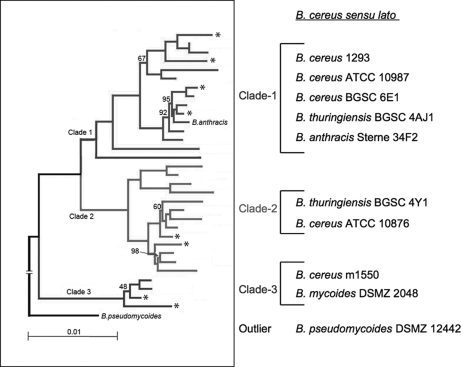
For comparative proteomics analysis, 10 strains from the B. cereus group were selected: B. anthracis Sterne 34F2 (a pXO2− laboratory strain); B. thuringiensis BGSC 4AJ1 and BGSC 4Y1; five strains of B. cereus, ATCC 10987, ATCC 10876, BGSC 6E1, m1293 (Oslo, Norway), and m1550; B. mycoides DSMZ 2048; and B. pseudomycoides DSMZ 12442 (sources of the strains are shown in Supplement 1). Strains for analysis were selected on the basis that they represented distinct branches on a phylogeny of the B. cereus group based on a whole genome sequencing study (Fig. 1) (67). Whole genome shotgun sequences of these strains (except B. anthracis Sterne 34F2 (32) and B. cereus ATCC 10987 (4)) were obtained using 454 pyrosequencing technology to an average coverage of ∼25 reads per base (33) as part of a study on genome variation in this group (67).
Fig. 1.
Phylogeny of B. cereus s.l. genomes. A neighbor-joining tree was created from a concatenation of 373 conserved, single copy proteins from 33 B. cereus group genomes (67). Each branch has 100% recovery in 100 bootstrap replicates unless indicated on the tree. The branches of the strains selected for this study are marked with asterisks and correspond to the strains listed from top to bottom.
Sequence Accession Numbers—
Submission of the whole genome shotgun data to GenBank™ is currently in process. The NCBI Genome Project IDs are given in parentheses: B. thuringiensis, BGSC 4AJ1 (29709) and BGSC 4Y1 (29713); four strains of B. cereus, ATCC 10876 (29671), BGSC 6E1 (29651), m1293 (29657), and m1550 (29649); B. mycoides DSMZ 2048 (29701); and B. pseudomycoides DSMZ 12442 (29707).
Bacterial Spore Preparation—
The growth curves of each of the Bacillus strains in brain-heart infusion broth were determined to estimate the time of late logarithmic phase for each of the strains (Supplement 2). One colony from a blood agar plate was used to inoculate 5 ml of brain-heart infusion broth. At late logarithmic phase (∼6 h of shaking at 250 rpm at 37 °C) 1.0 ml was transferred into 50 ml of modified G medium (34) and incubated at 37 °C for about 72 h at 250 rpm to induce sporulation. The end point of sporulation was determined when the proportion of spores observed under a phase-contrast microscope was more than 20 times that of the vegetative cells. The spore suspensions were heated at 65 °C for 30 min to kill the remaining vegetative cells. The heat-treated spore suspensions were centrifuged at 6000 rpm (4,350 × g) at 25 °C for 30 min and washed extensively with cold (4 °C) Milli-Q water. Spore pellets were purified through a 50% renograffin layer (Bracco Diagnostics) (35) prepared in Milli-Q water and centrifuged at 4,350 × g for 30 min at 25 °C to remove cell debris. The spore pellets were further washed three times with cold Milli-Q water to remove traces of renograffin. The purified spore suspensions were stored in Milli-Q water at 4 °C in 500-μl aliquots. Spores were quantified microscopically with a hemocytometer.
Transmission Electron Microscopy (TEM) Analysis—
Bacillus spore samples were prepared for transmission electron microscopy examination as described previously (36) with slight modification. In brief, spores were fixed in 1% osmium tetroxide and 2% potassium permanganate in 0.1 m phosphate buffer for 2 h at room temperature. Following fixation, spores were thoroughly rinsed, enrobed in agar, and trimmed into 1 mm3. The enrobed material was stained in the block with uranyl acetate, dehydrated by passage through an ethanol series, and embedded in LR White. Sections were cut on a Reichert Jung ultramicrotome Ultracut E, stained with lead citrate, and examined in an FEI Tecnai T12 transmission electron microscope at an accelerating voltage of 80 kV (36). TEM images of the spore samples were examined to verify that (i) whole spore preparations contained both endospores with intact exosporium and (ii) exosporium preparations were not contaminated with endospores (and vice versa).
Extraction of Spore Proteins—
The protein extraction method was adapted from several reported protocols (29, 35, 37). Spore suspensions were centrifuged at 13,200 rpm (16,100 × g) at 25 °C for 10 min, and the pellets were resuspended in 500 μl of lysis buffer containing ZOOM 2D protein solubilizing buffer-1 (Invitrogen) and protease inhibitor mixture (Roche Applied Science). The samples were pulse-sonicated for 15 s for four times at level 4 with a Model 100 cell dismembrator (Fisher Scientific) followed by addition of 4.0 μl of sample buffer (50 mm MgCl2, 1 unit of DNase I, 3 units of RNase A, and 0.5 m Tris-HCl) and incubation in an ice bath for 10 min to eliminate DNA and RNA contamination. The samples were stored at −80 °C in 10-μl aliquots.
Two-dimensional Gel Electrophoresis Separation of Spore Proteins—
The spore protein extracts were mixed with IPG rehydration buffer (8 m urea, 4% (w/v) CHAPS, 0.04 m Tris base, 0.065 m dithiothreitol, and 0.01% (w/v) bromphenol blue) in a final volume of 260 μl. To generate an electrophoretic map of each of the spore preparations 50 μg of protein extract was mixed with IPG rehydration buffer, loaded onto Immobiline™ DryStrip (ReadyStrip™ IPG strips; pH 4–7, 6–11, or 3–10; strip length, 11 cm; Bio-Rad), allowed to rehydrate overnight, and isoelectrofocused to 20,000 V-h in a PROTEAN® IEF cell (Bio-Rad). The following focusing parameters were applied. A rapid advance voltage ramping method was used in which 250 V for 15 min was used for ramping in the first step followed by 8,000 V for 2.5 h in the second step, and finally 20,000 V for 6 h was used for the final focusing step. A hold step was programmed to maintain the voltage at 500 V until the run was stopped. The maximum current limit per gel was set at 50 μA. After focusing was completed, IPG strips were equilibrated with 2% (w/v) dithiothreitol in equilibration base buffer containing 6 m urea, 2% SDS, 20% (v/v) glycerol, 0.375 m Tris-HCl, and 0.001% bromphenol blue for 15 min followed by another equilibration step with 2.5% (w/v) iodoacetamide in equilibration base buffer for 15 min. The second dimension was carried out with Criterion XT precast 4–12% bis-Tris, 11.0-cm slab gels in a Criterion Dodeca multicell system (Bio-Rad) at 4 °C for 1 h at 200 volts in 1× NuPAGE MOPS-SDS buffer (Invitrogen) with 2.5% NuPAGE antioxidant (Invitrogen) in the upper electrode buffer. Gels were fixed in 40% ethanol and 10% acetic acid for 30 min followed by staining with a SilverQuest silver staining kit (Invitrogen). Images were scanned in the ChemiDoc XRS system (Bio-Rad). This imaging equipment is fully integrated to the ProteomeWorks™ system (Bio-Rad) with a high resolution image acquisition interface with the PDQuest software.
Image Analysis—
Protein spots were analyzed for differential expression patterns using PDQuest software version 7.2 (Bio-Rad). For quantification, a match set of all the gels was generated, and the spots were normalized to the total quantity of the valid spots in each of the gel images in the match set. After normalization, each spot in the master gel was matched with the same spot in each of the gels as mentioned previously (38, 39). For statistical analysis, a matching summary of all the spots in all the gels was generated.
Protein Spot Identification and Data Analysis—
Protein spots of interest were excised from silver-stained gels. In-gel digestion was performed using a modified method of Havlis et al. (40). Extracts from the digest were separated from the matrix using C18 ZipTips. Eluent from the ZipTips was lyophilized to dryness and suspended in 10 μl of 2% acetonitrile and 0.1% trifluoroacetic acid. Electrospray ionization mass spectrometry was carried out using a ThermoFinnigan LTQ (linear trap quadrupole) mass spectrometer equipped with a surveyor liquid chromatography pump. The samples were injected using a Finnigan microautosampler. About 6.4 μl was injected onto a 75-μm-inner diameter nanospray column. The column had a 15-μm opening and was packed with 10 cm of BioBasic C18. A 120-min gradient was used with a two-component solvent system with 0.1% formic acid as solvent A and 0.1% formic acid in acetonitrile as solvent B. The gradient was as follows: 0–40 min, 2% solvent B; 40–65 min, 60% solvent A and 40% solvent B; 65–70 min, 10% solvent A and 90% solvent B; 70–75 min, 98% solvent A and 2% solvent B; and 82–110 min, 98% solvent A and 2% solvent B. The flow rate was 250 μl/min presplit. The flow rate was 800 nl/min at the tip.
The mass spectrometer was operated in double play mode with one full-scan mass spectrum followed by nine data-dependent MS/MS scans. The minimum signal required for a data-dependent scan was 100. Dynamic exclusion was enabled with a repeat count of 3 and a repeat duration of 30 min. Automatic gain control target values were 3000 for full-scan MS and 1000 for MS/MS. Normalized collision energy for collision-induced dissociation was 35. Capillary voltage was 49 V, and spray voltage was 2.0 kV.
Extract_msn in Bioworks 4.0 software (version 3.3) was used to generate the peak list. The SEQUEST algorithm was used to interpret MS/MS data (41). The report format of the SEQUEST database (a part of Bioworks version 3.3) search lists the proteins in order of probability. The protein probability score is based on the probability that a peptide is a random match to the spectral data. The best peptide probability value was used as the probability value for protein identification. If a protein was identified by four or more unique peptides with SEQUEST scores that met or exceeded the acceptance criteria, the protein was considered to be present in the sample analyzed. The search parameters included up to two missed trypsin cleavages with methionine oxidation as variable modification and no fixed modifications. The mass tolerance for precursor ions and fragment ions were considered as 2 and 1 AMU, respectively. Patterns of measured masses were also matched against theoretical masses of proteins found in the NCBI, Swiss-Prot, and TrEMBL databases accessible in the ExPASy Molecular Biology server. We also searched a subset database created from the NCBI non-redundant database using a search term of “Bacillus” proteins and an in-house Biological Defense Research Directorate genome database of predicted proteins from genomes that were sequenced and run through our in-house annotation pipeline. This pipeline consists of a collection of common bioinformatics applications including Glimmer3 (42) for ab initio gene prediction and BLAST-based homology search against a reference genome for gene function assignment, in this case B. anthracis Ames ancestor strain (NCBI RefSeq accession numbers NC_007530, NC_007322, and NC_007323). Sequence coverage >30% was considered as a positive match. Distinct protein spots in the Western blots that could not be identified by LC-MS (resulting from an insufficient amount of protein in the spot, and/or a clearly resolved spot was not obtained in the silver-stained electrophoretic map) were identified by a bioinformatics approach using the TagIdent tool. The molecular weight and pI of such spots were estimated from the analytical gels and applied to TagIdent with very stringent parameters and specified error margins (pI range = 0.25 and molecular weight range = 10%) to match the proteins from the database (Swiss-Prot/TrEMBL/NCBI) near to the defined range of molecular weight and pI with a p value of <0.05.
In Vitro Transcription and Translation—
A total of 200 target ORFs were amplified and expressed using the high throughput cell-free Transcriptionally Active PCR (TAP) fragment system developed by Gene Therapy Systems Inc., San Diego, CA (43). Gene-specific primers for 200 genes were generated using the B. anthracis Sterne 34F2 genome sequence. Primers complementary to the 5′- and 3′-ends of the gene of interest were synthesized. The 5′ and 3′ primers were mixed and added to plates containing Taq polymerase (Clontech-BD Biosciences) and B. anthracis Sterne 34F2 genomic DNA. The PCR was carried out in a thermocycler PCR machine (MWG Biotech, AG) for 30 cycles. Upon completion, PCRs were transferred to a Millipore Montage 96-well cleanup plate and filter-purified using a vacuum manifold. About 10–20 ng of DNA from each PCR from the previous step was used as a template in the second step PCR that also included the DNA fragments containing T7 promoter, His6 sequences, and T7 terminator sequences. This step generated a DNA fragment that contained the gene of interest with the added 5′- and 3′-TAP universal end sequences. The PCR, subsequent purification, and quantification of the PCR product was carried out as detailed above. Promoter and terminator sequences were added onto the TAP primary fragment to generate His6-tagged proteins. The synthesized His6-tagged proteins from the previous step were purified by nickel-nitrilotriacetic acid Superflow 96 kit (Qiagen) according to the manufacturer's instructions. TAP fragments were subsequently filter-purified to remove free nucleotides and other impurities.
The resulting transcriptionally active products were used as a template for cell-free in vitro translation reactions using the Rapid Translational System (RTS) ProteoMaster by Roche Applied Science, and high levels of protein were recovered and then purified. The level of protein obtained was typically 75–100 mg/50-ml reaction. To verify the identity and purity of each of the synthesized proteins, the proteins were resolved by one-dimensional SDS-PAGE and stained with Coomassie Blue dye. The identities of the proteins were confirmed with a QSTAR Hybrid LC/MS/MS system.
ELISA—
The basic sandwich ELISA technique was conducted with the proteins generated by in vitro TAP and RTS approaches. Each of the proteins (100 μl containing 1 μg/ml diluted protein) were coated onto the 96-well plate and incubated overnight at 4 °C. The plates were washed with 2× PBS with Tween 20, and the unbound antigens (proteins) were discarded by flicking the contents of the plate. Each of the wells was blocked for 90 min at 37 °C by adding 200 μl of 3% skim milk prepared in phosphate-buffered saline with 0.05% Tween 20 (PBST). The blocking buffer was discarded, and the plates were tapped dry on a paper towel. This was followed by adding 100 μl of rabbit serum sample diluted in blocking buffer (for generation of rabbit immune sera, see below) and incubating for 1 h at 37 °C. Then the washing steps were repeated followed by addition of an affinity-purified antibody, peroxidase-labeled goat anti-rabbit IgG heavy + light, and incubating for 1 h at 37 °C. The plates were finally washed with PBST and stained with 3,3′,5,5′-tetramethylbenzidine membrane peroxidase substrate until the chemiluminescent immunoreactive spots appeared in the wells. The 96-well plates were read on an ELISA plate reader at a wavelength of 405 nm.
Generation of Rabbit Immune Sera—
Rabbit immune sera were generated against B. anthracis 34F2 Sterne full germination-deficient (ΔgerH) (44) whole spores and purified exosporium. The spores of ΔgerH strain were prepared as mentioned above under “Bacterial Spore Preparation” except that the spores used to immunize the rabbits were not purified by renograffin. Exosporium fragments were recovered by pulse-sonicating B. anthracis 34F2 Sterne spores (preparation mentioned above) for 15 s for four times at level 4 with a Model 100 cell dismembrator (Fisher Scientific). The sample was subjected to low speed centrifugation at 25 °C for 5 min. The supernatant containing the exosporium was filtered (0.45 μm) to remove all the remaining spore materials and stored at −20 °C for further use. We confirmed by TEM examination that this procedure removed the majority of the exosporium fragments without detectable damage to the remaining spore (data not shown). Three outbred rabbits were immunized per antigen by intradermal injection. To generate immune sera each rabbit received either 100 μl of 1 × 10⁷ of ΔgerH spores or 100 μg of filtered exosporium. Nine boosts per antigen were given to each of the rabbits, i.e. a biweekly boost schedule for the first 6 months followed by a monthly boost for the next 3 months. The immune sera obtained from three rabbits in both the groups, against B. anthracis 34F2 exospore and ΔgerH whole spore, were pooled, respectively, for the Western blot analysis. For controls, corresponding antisera were collected from unimmunized rabbits that were injected with phosphate-buffered saline.
To compare the cross-reactivity of spore proteins of the 10 Bacillus strains between immune sera generated against B. anthracis 34F2 Sterne ΔgerH whole spores, exosporium, and killed ungerminated B. anthracis spores, we used rabbit immune sera generated against irradiated (killed) ungerminated B. anthracis Ames spores, a generous gift from Dr. Arthur Freidlander (United States Army Medical Research Institute for Infectious Diseases) (45). For the purpose of this study the four sera have been abbreviated as follows: rabbit anti-gerH whole spore sera (RAGSS), rabbit anti-exosporium sera (RAESS), rabbit anti-whole spore (ungerminated) sera (RAWSS), and rabbit anti-naïve sera (RANS).
Western Blot Analysis of 2D Separated Proteins—
Proteins on 2D gels of all 10 Bacillus strains were transferred to PVDF membranes using Towbin buffer (25 mm Trizma (Tris base), 190 mm glycine, and 20% methanol) at 100 V for 30 min. After the transfer, the PVDF membrane was blocked with 3% skim milk prepared in PBST for 1 h followed by three 5-min washes with PBST. The blots were then probed for 1 h with 1:50,000 dilutions of RAGSS, RAESS, RAWSS, or RANS prepared in 3% skim milk. After probing with the primary antibody, the blots were washed three times with PBST and probed with a 1:25,000 dilution of an affinity-purified antibody, peroxidase-labeled goat anti-rabbit IgG heavy + light, for 1 h. The blots were finally washed with PBST and stained with 3,3′,5,5′-tetramethylbenzidine membrane peroxidase substrate until the chemiluminescent immunoreactive spots appeared on the blot. The blots were dried, and the images were captured using a ChemiDoc XRS system (Bio-Rad). Three replicate blots for each of the antigens for all 10 strains were generated.
RESULTS
Bioinformatics-based Screening of Conserved B. anthracis Spore Proteins—
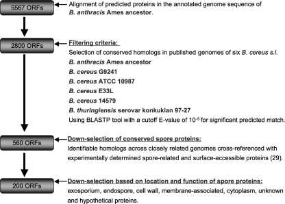
To identify conserved proteins we aligned the 5,567 predicted proteins in the annotated genome sequence of B. anthracis Ames ancestor (RefSeq accession numbers NC_007530, NC_007323, and NC_007322) against a database of the predicted proteomes of the published B. cereus s.l. genomes available at the start of the study in 2004 (B. cereus G9241 (6), B. cereus ATCC 10987 (4), B. cereus E33L (5), B. cereus 14579 (3), and B. thuringiensis serovar konkukian 97-27 (3)) using the BLASTP tool (46, 47). Using a cutoff E-value of 10−5 for the significance of the predicted match, 2,800 proteins were found to be conserved in all six B. cereus group genomes (Fig. 2). We then cross-referenced this set against more than 750 endospore and exosporium constituent proteins of B. anthracis identified by multidimensional chromatography and tandem mass spectroscopy (29) to obtain a subset of 560 proteins. The proteins in this list were then ranked manually as likely antigenic candidates based on (i) annotation and literature references that indicated known spore structural components or late stage sporulation proteins, (ii) bioinformatics-based prediction of subcellular localization using PSORTb (48) that indicated a membrane-associated or exported protein, and (iii) a peptide length greater than 100 amino acids. The ORFs encoding the top 200 candidate proteins were selected for in vitro gene-specific expression (Fig. 2).
Fig. 2.
Filtering strategy for down-selection of spore-associated conserved ORFs from B. cereus s.l.
The 200 target ORFs were then amplified and expressed using the high throughput cell-free TAP fragment system. The resulting transcriptionally active products were used as a template for cell-free in vitro translation reactions using the RTS ProteoMaster by Roche Applied Science, and high levels of protein were recovered and then purified. Of the 200 ORFs, 192 proteins were synthesized successfully, and the identities were verified by mass spectroscopy.
These 192 synthesized proteins were subjected to ELISA against RAGSS, RAESS, and RANS. 117 of the synthesized proteins did not react with any one of the immune sera (Table I). Of the 75 proteins that did react, 23 proteins reacted with both RAGSS and RAESS. None of these 23 proteins had been previously identified as B. anthracis spore surface proteins but were instead mainly ABC transporters, proteins of unknown function (annotated as “hypothetical,” e.g. GBAA2334, GBAA1237, and GBAA1334), sporulation regulators (SpoIIIAG and SpoVAD), and housekeeping proteins. 37 proteins reacted with sera generated from RAGSS, suggesting that these proteins are present either in the spore coat, cortex, and/or core (Table I). Only 15 proteins reacted with RAESS. RAESS-reactive proteins included small acid-soluble spore protein B (SspB), coat protein JB (CotJB), and alanine racemases (Dal-1, also known as Alr), which have previously been identified as spore constituents (Table I) (2, 14, 23, 32). There were three proteins of undetermined function (GBAA5641, GBAA2292, and GBAA1172) that reacted only with RAESS (Table I).
Table I.
Conserved spore proteins of B. anthracis and closely related strains selected by genome-based in silico analysis showing reaction with rabbit immune sera
Results were derived from subtraction of control serum (unimmunized, 0.005 at A405) from anti-GerH whole spores and anti-exosporium sera. −, <0.1; +, 0.1–0.2; ++, 0.2–0.3; +++, >0.3. PTS, phosphotransferase system.
| Genes selected | ORFs | Molecular mass | ELISA with rabbit immune sera |
|
|---|---|---|---|---|
| Anti-spore | Anti-exosporium | |||
| bp | kDa | |||
| Sporulation | ||||
| gi47505340; GBAAT34016.1; small, acid-soluble spore protein B | 198 | 7.3 | − | ++ |
| gi47504727; GBAAT33403.1; stage V sporulation protein AF | 1,473 | 54.0 | + | − |
| gi47504853; GBAAT33529.1; stage III sporulation protein AG | 663 | 24.3 | ++ | + |
| gi47505104; GBAAT33780.1; forespore-specific protein, putative | 627 | 23.0 | + | − |
| gi47504730; GBAAT33406.1; stage V sporulation protein AD | 1,017 | 37.3 | + | + |
| gi47505076; GBAAT33752.1; prespore-specific transcriptional regulator rsfA, putative | 642 | 23.5 | + | − |
| gi47505119; GBAAT33795.1; spo0B-associated GTP-binding protein | 1,287 | 47.2 | + | − |
| gi47503187; GBAAT31863.1; spore cortex lytic enzyme prepeptide | 762 | 27.9 | + | − |
| gi47501241; GBAAT29917.1; cotJB protein | 276 | 10.1 | − | ++ |
| Cell division | ||||
| gi47500466; GBAAT29142.1; cell division protein FtsH | 1,902 | 69.7 | +++ | + |
| gi47504423; GBAAT33099.1; signal recognition particle-docking protein FtsY | 990 | 36.3 | + | − |
| gi47504476; GBAAT33152.1; cell division initiation protein DivIVA | 507 | 18.6 | + | − |
| gi47504487; GBAAT33163.1; cell division protein FtsA | 1,302 | 47.7 | ++ | − |
| gi47504408; GBAAT33084.1; gid protein | 1,305 | 47.9 | − | ++ |
| ABC transporter | ||||
| gi47500766; GBAAT29442.1; iron compound-binding protein ABC transporter | 918 | 33.7 | +++ | + |
| gi47505671; GBAAT34347.1; ABC transporter, ATP-binding protein | 786 | 28.8 | − | + |
| gi47505676; GBAAT34352.1; ABC transporter, ATP-binding protein | 1,026 | 37.6 | + | − |
| gi47500801; GBAAT29477.1; ABC transporter, substrate-binding protein, putative | 999 | 36.6 | ++ | − |
| gi47551555; GBAAT29896.2; Na/Pi-cotransporter family protein | 1,656 | 60.7 | − | + |
| gi47501472; GBAAT30148.1; ABC transporter, ATP-binding protein EcsA | 744 | 27.3 | + | + |
| gi47501606; GBAAT30282.1; oligopeptide ABC transporter, ATP-binding protein | 1,044 | 38.3 | − | ++++ |
| gi47501607; GBAAT30283.1; oligopeptide ABC transporter, ATP-binding protein | 936 | 34.3 | + | ++ |
| gi47501710; GBAAT30386.1; spermidine/putrescine ABC transporter, ATP-binding protein | 996 | 36.5 | + | − |
| gi47501714; GBAAT30390.1; spermidine/putrescine-binding ABC transporter protein | 1,038 | 38.1 | + | + |
| gi47502979; GBAAT31655.1; ABC transporter, ATP-binding protein | 669 | 24.5 | + | − |
| gi47503226; GBAAT31902.1; glycine betaine/l-proline ABC transporter, permease protein | 837 | 30.7 | ++ | + |
| Hypothetical | ||||
| gi47502779; GBAAT31455.1; hypothetical protein | 537 | 19.7 | ++++ | ++++ |
| gi47505416; GBAAT34092.1; hypothetical protein | 420 | 15.4 | + | − |
| gi47501650; GBAAT30326.1; hypothetical protein | 504 | 18.5 | ++ | + |
| gi47501749; GBAAT30425.1; hypothetical protein | 342 | 12.5 | + | + |
| gi47504707; GBAAT33383.1; hypothetical protein | 363 | 13.3 | ++ | + |
| Conserved hypothetical | ||||
| gi47506115; GBAAT34791.1; conserved hypothetical protein | 438 | 16.1 | − | + |
| gi47501825; GBAAT30501.1; conserved hypothetical protein | 555 | 20.4 | + | + |
| gi47502061; GBAAT30737.1; conserved hypothetical protein | 249 | 9.1 | + | + |
| gi47551733; GBAAT31416.2; conserved hypothetical protein | 756 | 27.7 | − | +++ |
| gi47551862; GBAAT32537.2; conserved hypothetical protein | 1,062 | 38.9 | ++++ | +++ |
| gi47504112; GBAAT32788.1; conserved domain protein | 264 | 9.7 | + | +++ |
| gi47504632; GBAAT33308.1; conserved hypothetical protein | 213 | 7.8 | ++ | − |
| gi47503999; GBAAT32675.1; conserved hypothetical protein | 1,467 | 53.8 | + | − |
| gi47504358; GBAAT33034.1; conserved hypothetical protein | 912 | 33.4 | + | − |
| gi47504360; GBAAT33036.1; conserved hypothetical protein | 249 | 9.1 | ++ | − |
| gi47504363; GBAAT33039.1; conserved hypothetical protein | 1,275 | 46.8 | + | − |
| gi47504372; GBAAT33048.1; conserved hypothetical protein | 750 | 27.5 | + | − |
| gi47504390; GBAAT33066.1; conserved hypothetical protein | 273 | 10.0 | + | − |
| gi47501585; GBAAT30261.1; conserved hypothetical protein | 198 | 7.3 | − | ++ |
| Lipoprotein | ||||
| gi47505654; GBAAT34330.1; lipoprotein, putative | 576 | 21.1 | +++ | + |
| gi47500656; GBAAT29332.1; lipoprotein, putative | 1,014 | 37.2 | ++ | − |
| gi47503185; GBAAT31861.1; lipoprotein, putative | 597 | 21.9 | + | − |
| gi47502474; GBAAT31150.1; adhesion lipoprotein | 951 | 34.9 | ++ | − |
| Spore germination | ||||
| gi47500552; GBAAT29228.1; spore germination protein GerD | 618 | 22.7 | ++ | − |
| gi47502385; GBAAT31061.1; bacterial luciferase family protein | 1,056 | 38.7 | + | − |
| DNA binding | ||||
| gi47504294; GBAAT32970.1; DNA-binding protein HU | 273 | 10.0 | + | ++ |
| gi47500486; GBAAT29162.1; DNA-binding protein, putative | 1,074 | 39.4 | + | − |
| Amino acid binding | ||||
| gi47552115; GBAAT34692.2; ATP synthase F1, γ subunit | 861 | 31.6 | − | + |
| gi47504102; GBAAT32778.1; thioredoxin-like protein | 198 | 7.3 | + | +++ |
| gi47504630; GBAAT33306.1; polypeptide deformylase | 555 | 20.4 | + | +++ |
| Energy metabolism | ||||
| gi47552124; GBAAT35466.1; 4-oxalocrotonate tautomerase | 186 | 6.8 | − | + |
| gi47504281; GBAAT32957.1; PTS system, fructose-specific IIABC component | 1,881 | 69.0 | + | − |
| gi47504349; GBAAT33025.1; pyruvate ferredoxin oxidoreductase, α subunit, putative | 1,758 | 64.5 | + | − |
| Cell wall | ||||
| gi47551930; GBAAT33049.2; metallo-β-lactamase family protein | 1,671 | 61.3 | + | − |
| gi47503556; GBAAT32232.1; metallo-β-lactamase family protein | 846 | 31.0 | +++ | − |
| gi47500657; GBAAT29333.1; alanine racemase | 1,170 | 42.9 | − | ++ |
| gi47502504; GBAAT31180.1; chaperone CsaA | 330 | 12.1 | − | + |
| Protein degradation | ||||
| gi47504362; GBAAT33038.1; zinc protease, insulinase family | 1,287 | 47.2 | + | − |
| gi47504380; GBAAT33056.1; zinc protease, insulinase family | 1,242 | 45.5 | + | − |
| gi47504406; GBAAT33082.1; ATP-dependent protease hsIV | 543 | 19.9 | + | − |
| gi47504350; GBAAT33026.1; renal dipeptidase family protein | 924 | 33.9 | + | − |
| Cellular process | ||||
| gi47504391; GBAAT33067.1; N-utilization substance protein A | 1,107 | 40.6 | + | − |
| gi47504101; GBAAT32777.1; glycosyl hydrolase, family 18 | 1,293 | 47.4 | + | + |
| gi47504599; GBAAT33275.1; cytochrome c oxidase, subunit I | 1,836 | 67.3 | + | − |
| Translation | ||||
| gi47500532; GBAAT29208.1; ribosomal protein L30 | 183 | 6.7 | − | + |
| gi47500548; GBAAT29224.1; ribosomal protein S9 | 393 | 14.4 | ++ | ++ |
| gi47504277; GBAAT32953.1; host factor-I protein | 225 | 8.3 | + | ++ |
| gi47504383; GBAAT33059.1; ribosomal protein S15 | 270 | 9.9 | − | + |
| gi47502550; GBAAT31226.1; ATP-dependent RNA helicase, DEAD/DEAH box family | 1,170 | 42.9 | + | − |
| Control | ||||
| Exosporium preparation | ++++ | ++++ | ||
Microvariation in B. cereus s.l. Spore Morphology—
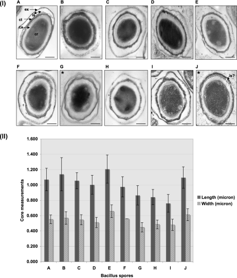
In a parallel screening effort, spores from 10 B. cereus group strains were produced by growth under similar conditions (Supplement 2) for comparative proteomics analysis. The spore preparations were examined by TEM as a quality control step (see “Experimental Procedures”; Fig. 3, panel I) presenting an opportunity for comparative analysis of spore ultrastructure. On average, 127 spores per Bacillus strain were examined. Only longitudinally sectioned spores were measured to obtain the core and interspace dimensions. A representative figure of the calculations is shown in Supplement 3. All Bacillus spores examined display an elongated olive shape with an extension at each pole. Most of the spores in clade 1 were structurally similar with their core measurements ranging from 1.003 to 1.207 μm in length and from 0.511 to 0.658 μm in width (Fig. 3, panel II). The cores of the Bacillus spores of clades 2 and 3 were on average smaller than spores in clade 1, although the variance of the measurements was too high to draw statistically significant conclusions. We observed other structural variations. In spores of B. thuringiensis BGSC 4Y1 (Fig. 3, panel I, G) the exosporium and the coat have a peculiar ribbon-like structure, which was not observed in any of the other strains. Furthermore on higher magnification, as expected, B. thuringiensis BGSC 4Y1 spores showed the presence of crystalline structures in the interspace region. However, we did not see crystals in the spores of B. thuringiensis BGSC 4AJ1. In B. pseudomycoides DSMZ 12442 (Fig. 3, panel I, J) the interspace (IS) region is almost absent as the exosporium wraps tightly around the spore coat even at the pole region. The average IS length in B. pseudomycoides DSMZ 12442 is 0.045 μm with a maximum of 0.192 μm at the elongated portion of the spores. More images of spores of B. thuringiensis BGSC 4Y1 and B. pseudomycoides DSMZ 12442 are shown in Supplement 4. The average IS length in other Bacillus spores ranges from 0.095 to 0.166 μm at the width with the maximum IS ranging from 0.325 to 0.649 μm at the elongated portion of the spores. B. cereus m1293, B. cereus ATCC 10987, and B. mycoides DSMZ 2048 showed larger IS regions compared with other Bacillus spores.
Fig. 3.
Panel I, TEM showing ultrastructural analysis of B. anthracis and closely related strains. A, B. anthracis Sterne 34F2; B, B. thuringiensis BGSC 4AJ1; C, B. cereus m1293; D, B. cereus BGSC 6E1; E, B. cereus ATCC 10987; F, B. cereus ATCC 10876; G, B. thuringiensis BGSC 4Y1; H, B. cereus m1550; I, B. mycoides DSMZ 2048; J, B. pseudomycoides DSMZ 12442. ex, exosporium; is, interspace; ct, coat; cx, cortex; cr, core. The bar is 100 nm at magnification ×60,000. All the spores were prepared using the modified G medium at 37 °C up to 72 h at 250 rpm and purified using a 50% renograffin gradient. Panel II, core dimensions of the spores of closely related Bacillus strains. A–J show the same strains listed for panel I. Error bars represent standard deviation in microns.
Comparative Proteomics Analysis of B. cereus s.l. Spores—
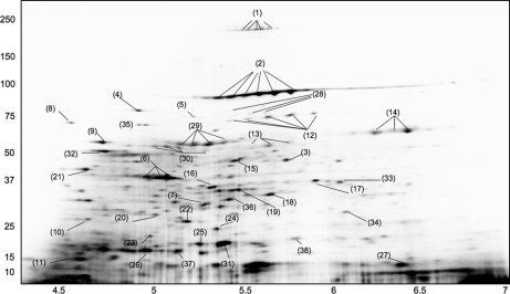
We determined the spore proteome of all 10 Bacillus strains by generating two-dimensional electrophoresis maps of the proteins extracted from renograffin-purified whole spores grown in modified G medium at 37 °C for about 72 h under shaker condition (Fig. 4). At least four spore preparations of each strain followed by 2D electrophoresis were conducted to confirm the results. A total of 38 proteins from 10 clusters of orthologous groups (cell wall/surface, translation, protein folding and assembly, energy metabolism, amino acid biosynthesis, sporulation, nucleotide metabolism, cell signaling, cellular process, and hypothetical proteins) were identified by mass spectroscopy from spots excised from the B. anthracis 34F2 Sterne 2D gel (Table II and Fig. 4).
Fig. 4.
Spore proteome of B. anthracis 34F2 Sterne. Protein identifications associated with the spot numbers (1–38) are shown in Table II. Representative gels from four experiments are shown.
Table II.
Proteins identified from B. anthracis 34F2 Sterne spore
The spot number indicates the corresponding protein spot in the 2D electrophoretic map shown in Fig. 4.
| Spot no. | Locus tag (GBAA) | Accession no. (gi) | Protein | No. of peptides matched | Sequence coverage | Theoretical molecular mass (kDa)/pI |
|---|---|---|---|---|---|---|
| % | ||||||
| Cell wall/surface | ||||||
| 1 | 1094 | 47526368 | Wall-associated protein, putative (RhsA) | 16 | 32 | 249.01/5.97 |
| 2 | 0887 | 47526173 | S-layer protein EA1 precursor (Eag) | 21 | 31 | 91.36/5.70 |
| 3 | 0252 | 47525509 | Alanine racemase (Dal-1/Alr) | 12 | 36 | 43.66/5.51 |
| Translation | ||||||
| 4 | 0107 | 47525363 | Translation elongation factor G (FusA) | 25 | 36 | 76.33/4.91 |
| 5 | 3950 | 47529240 | Translational initiation factor (InfB) | 23 | 33 | 75.75/5.03 |
| 6 | 0108 | 47525364 | Translation elongation factor Tu (Tuf) | 28 | 53 | 42.93/4.93 |
| 7 | 3964 | 47529254 | Translation elongation factor Ts (Tsf) | 22 | 45 | 32.30/5.25 |
| Protein folding assembly | ||||||
| 8 | 4539 | 47529836 | Molecular chaperone DnaK | 16 | 40 | 65.76/4.65 |
| 9 | 0267 | 47525527 | Chaperonin GroEL | 23 | 31 | 57.43/4.79 |
| 10 | 4540 | 47529837 | Hsp70 cofactor, GrpE protein | 11 | 61 | 21.52/4.56 |
| 11 | 0266 | 47525525 | Co-chaperonin GroES | 7 | 37 | 10.06/4.69 |
| Energy metabolism | ||||||
| 12 | 4843 | 47530138 | Pyruvate kinase (PykA-2) | 14 | 55 | 62.10/5.20 |
| 13 | 0309 | 47500720 | Δ1-Pyrroline-5-carboxylate dehydrogenase | 22 | 31 | 56.22/5.43 |
| 14 | 0008 | 47525264 | Inosine-5-monophosphate dehydrogenase (GuaB) | 17 | 47 | 52.37/6.30 |
| 15 | 4385 | 47529680 | Dihydrolipoamide dehydrogenase (BfmbC) | 33 | 51 | 50.77/5.53 |
| 16 | 5367 | 47530678 | Phosphoglycerate kinase (Pgk) | 21 | 61 | 42.29/5.14 |
| 17 | 5716 | 47531053 | Adenylosuccinate synthetase (PurA) | 18 | 60 | 47.42/5.57 |
| 18 | 4184 | 47529480 | Pyruvate dehydrogenase, E1α (PdhA) | 14 | 63 | 41.44/5.52 |
| 19 | 5369 | 47530679 | Glyceraldehyde-3-PO4 dehydrogenase (Gap-2) | 10 | 35 | 35.82/5.37 |
| 20 | 4183 | 47529479 | Pyruvate dehydrogenase, E1β (PdhB) | 12 | 32 | 35.22/4.75 |
| 21 | 5364 | 47530674 | Enolase (Eno) | 13 | 43 | 46.41/4.66 |
| Amino acid biosynthesis | ||||||
| 22 | 5387 | 47778397 | Thioredoxin reductase B (TrxB) | 9 | 47 | 34.66/5.16 |
| 23 | 0131 | 47525387 | Adenylate kinase (Adk) | 6 | 35 | 23.74/4.91 |
| Sporulation | ||||||
| 24 | 0803 | 47526092 | Coat protein JC (CotJC) | 8 | 51 | 21.65/5.15 |
| 25 | 0053 | 47525307 | Stage V sporulation protein T (SpoVT) | 21 | 38 | 19.69/5.09 |
| 26 | 1238 | 47526503 | Spore coat protein Z (CotZ-2) | 13 | 31 | 16.84/4.71 |
| 27 | 4898 | 47530192 | Small acid-soluble spore protein B (SspB) | 6 | 53 | 6.80/6.13 |
| Nucleotide metabolism | ||||||
| 28 | 3944 | 47529234 | Polyribonucleotide nucleotidyltransferase (Pnp) | 11 | 36 | 78.20/5.16 |
| 29 | 5583 | 47530904 | CTP synthase (CtrA) | 25 | 34 | 59.75/5.28 |
| 30 | 5549 | 47530869 | ATP synthase, F1 (AtpA) | 25 | 39 | 54.64/5.30 |
| Cell signaling | ||||||
| 31 | 4499 | 47529794 | Mn-superoxide dismutase (SodA-1) | 7 | 58 | 22.66/5.31 |
| Cellular process | ||||||
| 32 | 0344 | 47525611 | Alkyl hydroperoxide reductase, F (AhpF) | 24 | 35 | 54.81/4.93 |
| 33 | 2541 | 47502979 | ABC transporter, ATP-binding protein | 8 | 40 | 25.00/5.93 |
| 34 | 5222 | 47505676 | ABC transporter, ATP-binding protein | 15 | 42 | 37.26/6.18 |
| Hypothetical protein | ||||||
| 35 | 1267 | 47526534 | Hypothetical protein | 23 | 31 | 73.25/5.29 |
| 36 | 0029 | 47525285 | Hypothetical protein | 11 | 39 | 31.76/5.38 |
| 37 | 0020 | 47525275 | Hypothetical protein | 9 | 59 | 11.86/5.17 |
| 38 | 0145 | 47525401 | Hypothetical protein | 10 | 45 | 17.45/5.82 |
Comparative protein composition analysis of the spores of all 10 Bacillus strains was performed using PDQuest software by generating a match set of all the gels. After normalizing the spots in the gels, each spot in the master gel (B. anthracis Sterne 34F2) was matched with the same spot in each of the gels generating a summary of all the spots in all the gels (Table III). More than 300 spots were resolved in B. anthracis Sterne 34F2, B. thuringiensis BGSC 4AJ1, B. cereus m1293, and B. mycoides DSMZ 2048, and more than 200 spots were resolved in the 2D gels of the six remaining Bacillus strains (Table III). Protein spots of strains in clade 1 had a match rate of ≥80% with the protein spots of B. anthracis Sterne 34F2 (reference strain). Protein spots of strains in clades 2 and 3 matched ≥78%, and the outlier B. pseudomycoides DSMZ 12442 matched up to 75% with the reference strain. About 72% of B. anthracis Sterne 34F2 protein spots matched with every strain.
Table III.
Matching summary of protein spots expressed in two-dimensional electrophoretic maps of spores of B. anthracis and closely related strains
Spots matched to every gel, 72%.
| Strains | Clades | Spot count | Spots matched | Match rate |
|---|---|---|---|---|
| % | ||||
| B. anthracis Sterne 34F2 | 1 | 370 | 370 | 100 |
| B. thuringiensis BGSC 4AJ1 | 1 | 390 | 322 | 83 |
| B. cereus 1293 | 1 | 337 | 269 | 80 |
| B. cereus BGSC 6E1 | 1 | 255 | 212 | 83 |
| B. cereus ATCC 10987 | 1 | 231 | 190 | 82 |
| B. cereus ATCC 10876 | 2 | 236 | 184 | 78 |
| B. thuringiensis BGSC 4Y1 | 2 | 294 | 229 | 78 |
| B. cereus m1550 | 3 | 228 | 185 | 81 |
| B. mycoides DSMZ 2048 | 3 | 378 | 298 | 79 |
| B. pseudomycoides DSMZ 12442 | Outlier | 246 | 184 | 75 |
Western Blot Analysis against Rabbit Immune Sera—
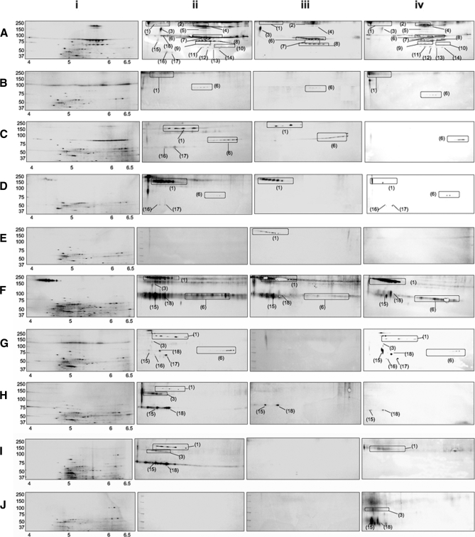
Western blots derived from 10 different spore preparations were probed with RAGSS, RAESS, RAWSS, and RANS (see “Experimental Procedures”). Comparison of the silver-stained 2D electrophoretic maps of each of the 10 Bacillus strains was referenced with corresponding Western immunoblots, allowing us to identify sera-reactive spore proteins (Fig. 5). Only the portions of the 2D gels that showed the appearance of sera-reactive protein spots are shown in Fig. 5. Although the limited dynamic range of the proteomics approach imposes severe restrictions on the repertoire of proteins detected, 18 sera-reactive proteins in total were detected from the Bacillus strains (Table IV). The spot number indicates the corresponding protein spot in the 2D electrophoretic Western blots shown in Fig. 5.
Fig. 5.
Western blot analysis of spore proteins of B. anthracis and closely related strains against rabbit immune sera generated against B. anthracis. Only portions of the gels that showed positive reaction to immune sera are shown. Panel i, silver-stained gel; panel ii, Western blot of spore proteins against rabbit sera to B. anthracis ΔgerH whole spore (RAGSS); panel iii, Western blot of spore proteins against rabbit sera to B. anthracis Sterne 34F2 exosporium (RAESS); panel iv, Western blot of spore proteins against rabbit sera to B. anthracis Ames irradiated ungerminated whole spore (RAWSS). A, B. anthracis Sterne 34F2; B, B. thuringiensis BGSC 4AJ1; C, B. cereus 1293; D, B. cereus BGSC 6E1; E, B. cereus ATCC 10987; F, B. cereus ATCC 10876; G, B. thuringiensis BGSC 4Y1; H, B. cereus m1550; I, B. mycoides DSMZ 2048; J, B. pseudomycoides DSMZ 12442. Protein identifications associated with the spot numbers (1–18) are shown in Table IV.
Table IV.
Spore proteins identified in B. anthracis and closely related strains from Western blot analysis against rabbit immune sera
The spot number indicates the corresponding protein spot in the 2D electrophoretic Western blots shown in Fig. 5.
| Spot no. | Locus tag | Accession no. | Protein | Theoretical molecular mass (kDa)/pI |
|---|---|---|---|---|
| 1 | GBAA3601 | gi47528887 | Conserved repeat domain protein (Crd) | 245.09/4.17 |
| 2 | GBAA1094 | gi47526368 | Wall-associated protein, putative (RhsA) | 249.01/5.97 |
| 3 | GBAA3677 | gi47528961 | Aconitate hydratase 1 (AcnA) | 99.01/4.8 |
| 4 | GBAA0052 | gi47525306 | Transcription-repair coupling factor (Mfd) | 134.15/5.36 |
| 5 | GBAA4157 | gi47529455 | Pyruvate decarboxylase (Pyc) | 128.57/6.0 |
| 6 | GBAA0887 | gi47526173 | S-layer protein EA1 precursor (Eag) | 91.36/5.70 |
| 7 | GBAA3944 | gi47529234 | Polyribonucleotide nucleotidyltransferase (Pnp) | 78.20/5.16 |
| 8 | GBAA4843 | gi47530138 | Pyruvate kinase (PykA-2) | 62.10/5.20 |
| 9 | GBAA5583 | gi47530904 | CTP synthase (CtrA) | 59.75/5.28 |
| 10 | GBAA0309 | gi47500720 | Δ1-Pyrroline-5-carboxylate dehydrogenase | 56.22/5.43 |
| 11 | GBAA3609 | gi47528895 | Aldehyde dehydrogenase (DhaS) | 53.74/5.4 |
| 12 | GBAA4184 | gi47529480 | Pyruvate dehydrogenase, E1α (PdhA) | 41.44/5.52 |
| 13 | GBAA0252 | gi47525509 | Alanine racemase (Dal-1/Alr) | 43.66/5.51 |
| 14 | GBAA5222 | gi47505676 | ABC transporter, ATP-binding protein (MetN) | 37.26/6.18 |
| 15 | GBAA4539 | gi47529836 | Molecular chaperone DnaK | 65.76/4.65 |
| 16 | GBAA0267 | gi47525527 | Chaperonin GroEL | 57.43/4.79 |
| 17 | GBAA0108 | gi47525364 | Translation elongation factor Tu (Tuf) | 42.93/4.93 |
| 18 | GBAA0107 | gi47525363 | Translation elongation factor G (FusA) | 76.33/4.91 |
All 18 protein spots were detected in Western immunoblot of RAGSS (Fig. 5A, panel ii), and 14 of the spots were detected against RAWSS (Fig. 5A, panel iv). The only difference between the immunoblots against RAGSS and RAWSS was that the four housekeeping proteins, molecular chaperone (DnaK), chaperone GroEL, translation elongation factor Tu (Tuf), and translation elongation factor G (FusA), were not detected in the immunoblot against RAWSS (Fig. 5A, panels ii and iv). Western blots from six of the 10 different spore preparations, i.e. B. anthracis Sterne 34F2, B. thuringiensis strains BGSC 4AJ1 and BGSC 4Y1, and B. cereus strains BGSC 6E1, ATCC 10987, and ATCC 10876, showed very similar results when probed with RAGSS and RAWSS (Fig. 5, A, B, and D–G, panels ii and iv), although the clade 1 strain B. cereus ATCC 10987 did not show any reaction against either sera (Fig. 5E, panels ii and iv).
For the remaining strains, we obtained somewhat inconsistent results probing against the two anti-spore sera. In the Western blots of B. cereus ATCC 10876, aconitate hydratase 1 (AcnA) was not detected against RAWSS but was detected against RAGSS (Fig. 5F, panels ii and iv). B. pseudomycoides DSMZ 12442 spore proteins did not cross-react with RAGSS, but three proteins, AcnA, DnaK, and FusA, were detected in the immunoblot against RAWSS (Fig. 5J, panels ii and iv). In the remaining three spore preparations (B. cereus m1293 and m1550 and B. mycoides DSMZ 2048) the proteins detected in the immunoblots against RAGSS and RAWSS showed different patterns (Fig. 5, C, H, and I, panels ii and iv). Neither of the three strains, B. thuringiensis BGSC 4Y1, B. mycoides DSMZ 2048, and B. pseudomycoides DSMZ 12442, reacted with the sera generated against the exosporium fraction of B. anthracis Sterne 34F2 (Fig. 5, G, I, and J, panel iii). In the other seven strains, one to seven proteins reacted against RAESS (Fig. 5, A–F and H, panel iii).
Comparison of all the proteins in the Western immunoblots obtained from all 10 Bacillus strains revealed six proteins that were sera-reactive in most of the Bacillus strains: a conserved repeat domain protein family, given the name in this work as “Crd,” S-layer protein EA1 precursor (Eag), DnaK, GroEL, Tuf, and FusA (Table V). Two of the proteins identified by the proteomics screen, Dal-1 and an ABC transporter, ATP-binding protein (MetN), overlapped the list of potential targets from the bioinformatics-based screening (Tables I and V).
Table V.
Sera-reactive conserved Bacillus spore proteins
+, spots present in Western immunoblot against anti-B. anthracis 34F2 ΔgerH spore; •, spots present in Western immunoblot against anti-B. anthracis 34F2 exospore; ▴, spots present in Western immunoblot against anti-whole spore (irradiated ungerminated B. anthracis Ames). Ba, B. anthracis; Bt, B. thuringiensis; Bc, B. cereus; Bm, B. mycoides; Bp, B. pseudomycoides.
| Spot no.a | Locus (GBAA) | Protein | Theoretical molecular mass (kDa)/pI | Ba 34F2 | Bt 4AJ1 | Bc 1293 | Bc 6E1 | Bc 10987 | Bc 10876 | Bt 4Y1 | Bc 1550 | Bm 2048 | Bp 12442 |
|---|---|---|---|---|---|---|---|---|---|---|---|---|---|
| 1 | 3601 | Conserved repeat domain protein (Crd) | 245.09/4.17 | + • ▴ | + ▴ | + • | + • ▴ | • | + • ▴ | + ▴ | + | + ▴ | |
| 2 | 1094 | Wall-associated protein, putative (RhsA) | 249.01/5.97 | + • ▴ | |||||||||
| 3 | 3677 | Aconitate hydratase 1 (AcnA) | 99.01/4.8 | + • ▴ | + | + ▴ | + | + | ▴ | ||||
| 4 | 0052 | Transcription-repair coupling factor (Mfd) | 134.15/5.36 | + • ▴ | |||||||||
| 5 | 4157 | Pyruvate decarboxylase (Pyc) | 128.57/6.0 | + • ▴ | |||||||||
| 6 | 0887 | S-layer protein EA1 precursor (Eag) | 91.36/5.70 | + • ▴ | + • ▴ | + • ▴ | + ▴ | + • ▴ | + ▴ | ||||
| 7 | 3944 | Polyribonucleotide nucleotidyltransferase (Pnp) | 78.20/5.16 | + • ▴ | |||||||||
| 8 | 4843 | Pyruvate kinase (PykA-2) | 62.10/5.20 | + • ▴ | |||||||||
| 9 | 5583 | CTP synthase (CtrA) | 59.75/5.28 | + ▴ | |||||||||
| 10 | 0309 | Δ1-Pyrroline-5-carboxylate dehydrogenase | 56.22/5.43 | + ▴ | |||||||||
| 11 | 3609 | Aldehyde dehydrogenase (DhaS) | 53.74/5.4 | + ▴ | |||||||||
| 12 | 4184 | Pyruvate dehydrogenase, E1α (PdhA) | 41.44/5.52 | + ▴ | |||||||||
| 13 | 0252 | Alanine racemase (Dal-1/Alr) | 43.66/5.51 | + ▴ | |||||||||
| 14 | 5222 | ABC transporter, ATP-binding protein (MetN) | 37.26/6.18 | + ▴ | |||||||||
| 15 | 4539 | Molecular chaperone DnaK | 65.76/4.65 | + | + | + ▴ | + • ▴ | + ▴ | + • ▴ | + | ▴ | ||
| 16 | 0267 | Chaperonin GroEL | 57.43/4.79 | + | + ▴ | ||||||||
| 17 | 0108 | Translation elongation factor Tu (Tuf) | 42.93/4.93 | + | |||||||||
| 18 | 0107 | Translation elongation factor G (FusA) | 76.33/4.91 | + | + | + ▴ | + • ▴ | + • ▴ | + | ▴ |
The spot number indicates the corresponding protein spot in the 2D electrophoretic map in Fig. 5.
Amino Acid Sequence Conservation of Cross-reacting Spore Antigens—
We performed a translated BLAST search against the genome sequences of the B. cereus strains to determine whether failure to cross-react to the antisera derived from B. anthracis spore challenge in other B. cereus group strains was caused by gene loss or sequence divergence (Table VI and Supplement 5). In some cases strains lack an ortholog of the B. anthracis protein. For instance, cell wall-associated protein (RhsA) only has an ortholog with sequence similarity greater than 75% in B. cereus ATCC 10987, BGSC 6E1, and m1550 although RhsA from either of these three strains showed no cross-reaction with any of the sera (Fig. 5, A, D, E, and H). The diverged B. pseudomycoides DSMZ 12442 genome lacks apparent orthologs to a number of B. anthracis spore proteins (Table VI). On the other hand, some proteins that were 100% identical in amino acid sequence to B. anthracis proteins did not exhibit detectable cross-reaction, for example B. thuringiensis BGSC 4AJ1 AcnA, PykA-2, Δ1-pyrroline-5-carboxylate dehydrogenase, DhaS, PdhA, MetN, DnaK, Tuf, and FusA (Table VI and Supplement 5).
Table VI.
Amino acid sequence similarity of orthologs in B. cereus s.l.
Cutoff, at least 50% protein identity over 100 amino acids. Protein identity is represented in percent. The length of the amino acid sequence hit is presented in parentheses. The bold portions represent the correlation of sera-reactive conserved Bacillus spore proteins in Table V. aa, amino acids; Ba, B. anthracis; Bt, B. thuringiensis; Bc, B. cereus; Bm, B. mycoides; Bp, B. pseudomycoides.
| Accession no. | Locus tag | Protein | Bt4AJ1 | Bc1293 | Bc6E1 | Bc10987 | Bc10876 | Bt4Y1 | Bc1550 | Bm2048 | Bp12442 |
|---|---|---|---|---|---|---|---|---|---|---|---|
| gi47528887 | GBAA3601 | Conserved repeat domain protein (Crd), 2357 aa | 72.36 (1616) | 71.87 (1616) | 71.19 (1616) | 69.71 (1616) | 71.19 (1616) | 90.65 (213) | 69.62 (1616) | 69.34 (1616) | — |
| 80.33 (2326) | 79.16 (2326) | 79.50 (2326) | 77.09 (2325) | 78.39 (2326) | 93.82 (468) | 77.80 (2326) | 77.80 (2326) | ||||
| 91.39 (1729) | |||||||||||
| gi47526368 | GBAA1094 | Wall-associated protein, putative (RhsA), 2223 aa | — | — | 76.81 (265) | 55.42 (162) | — | — | 57.14 (209) | — | — |
| 50.19 (237) | 85.45 (430) | ||||||||||
| 76.47 (318) | |||||||||||
| 82.66 (2210) | |||||||||||
| 86.82 (2107) | |||||||||||
| gi47528961 | GBAA3677 | Aconitate hydratase 1 (AcnA), 906 aa | 100.00 (906) | 99.67 (906) | 100.00 (906) | 99.56 (906) | 99.89 (906) | 99.67 (906) | 99.67 (906) | 97.68 (906) | 97.02 (906) |
| gi47525306 | GBAA0052 | Transcription-repair coupling factor (Mfd), 1175 aa | 99.91 (1175) | 98.95 (475) | 99.83 (578) | 99.74 (1175) | 97.96 (1175) | 95.64 (320) | 97.96 (1175) | 97.62 (1175) | 89.50 (475) |
| 98.04 (712) | 99.50 (602) | 97.10 (826) | 97.53 (687) | ||||||||
| gi47529455 | GBAA4157 | Pyruvate decarboxylase (Pyc), 1147 aa | 94.57 (220) | 98.98 (489) | 99.30 (1147) | 99.13 (1147) | 97.53 (1092) | 99.13 (1147) | 98.10 (209) | 97.96 (489) | 55.38 (128) |
| 99.57 (939) | 98.80 (663) | 98.29 (937) | 96.84 (663) | 94.39 (1087) | |||||||
| gi47526173 | GBAA0887 | S-layer protein EA1 precursor (Eag), 861 aa | 59.88 (171) | 53.26 (182) | 52.11 (210) | 58.72 (171) | 52.94 (135) | 59.88 (171) | 51.53 (195) | 60.47 (171) | — |
| 51.53 (195) | 59.30 (171) | 55.78 (198) | 53.16 (188) | 53.10 (144) | 52.11 (210) | 54.27 (198) | |||||
| 56.35 (195) | 60.13 (152) | 88.53 (861) | 56.12 (195) | 53.77 (206) | 57.14 (195) | ||||||
| 55.78 (198) | 56.12 (195) | 56.28 (198) | 55.78 (198) | ||||||||
| 99.88 (861) | 87.69 (267) | 88.79 (854) | |||||||||
| 83.73 (165) | |||||||||||
| 87.24 (383) | |||||||||||
| gi47529234 | GBAA3944 | Polyribonucleotide nucleotidyltransferase (Pnp), 711 aa | 99.71 (693) | 99.28 (693) | 100.00 (115) | 99.14 (693) | 99.42 (693) | 99.86 (693) | 99.42 (693) | 96.69 (693) | 95.39 (693) |
| 99.83 (578) | |||||||||||
| gi47530138 | GBAA4843 | Pyruvate kinase (PykA-2), 584 aa | 100.00 (584) | 99.63 (540) | 100.00 (584) | 99.66 (584) | 99.66 (584) | 99.55 (223) | 96.05 (584) | 94.76 (190) | 98.46 (584) |
| 98.85 (346) | 97.77 (403) | ||||||||||
| gi47530904 | GBAA5583 | CTP synthase (CtrA), 534 aa | 99.63 (534) | 99.44 (534) | 99.63 (534) | 99.25 (534) | 98.69 (534) | 99.02 (509) | 98.88 (534) | 98.50 (534) | 97.76 (534) |
| gi47500720 | GBAA0309 | Δ1-Pyrroline-5-carboxylate dehydrogenase, 514 aa | 100.00 (514) | 100.00 (514) | 100.00 (514) | 99.81 (514) | 99.42 (514) | 99.81 (514) | 99.49 (197) | 98.72 (233) | 96.31 (514) |
| 99.37 (317) | 95.73 (280) | ||||||||||
| gi47528895 | GBAA3609 | Aldehyde dehydrogenase (DhaS), 493 aa | 64.96 (487) | 65.16 (487) | 64.96 (487) | 65.16 (487) | 63.89 (492) | 65.16 (487) | 64.56 (157) | 64.55 (487) | 65.37 (487) |
| 100.00 (493) | 98.58 (493) | 100.00 (493) | 98.58 (493) | 98.58 (493) | 98.99 (493) | 65.73 (320) | 97.57 (493) | 91.76 (169) | |||
| 98.58 (493) | 95.08 (324) | ||||||||||
| gi47529480 | GBAA4184 | Pyruvate dehydrogenase, E1α (PdhA), 372 aa | 100.00 (370) | 100.00 (370) | 100.00 (370) | 100.00 (370) | 98.92 (370) | 100.00 (367) | 98.92 (370) | 99.69 (323) | 99.73 (370) |
| gi47525509 | GBAA0252 | Alanine racemase (Dal-1/Alr), 388 aa | 99.74 (388) | 99.23 (388) | 99.74 (388) | 99.49 (388) | 96.40 (388) | 99.23 (388) | 96.66 (388) | 95.89 (388) | 87.40 (388) |
| gi47505676 | GBAA5222 | ABC transporter, ATP-binding protein, methionine transport (MetN) 340 aa | 100.00 (340) | 99.71 (340) | 94.41 (340) | 99.41 (340) | 99.12 (340) | 100.00 (340) | 99.12 (340) | 98.43 (254) | 88.56 (340) |
| gi47529836 | GBAA4539 | Molecular chaperone DnaK, 610 aa | 100.00 (610) | 98.20 (610) | 98.79 (164) | 98.04 (610) | 99.67 (610) | 99.84 (610) | 100.00 (231) | 98.69 (610) | 95.91 (610) |
| 99.56 (451) | 99.73 (368) | ||||||||||
| gi47525527 | GBAA0267 | Chaperonin GroEL, 543 aa | 99.81 (530) | 99.81 (530) | 99.81 (530) | 97.24 (180) | 99.25 (530) | 99.25 (530) | 99.25 (530) | 97.93 (530) | 98.31 (530) |
| 100.00 (353) | |||||||||||
| gi47525364 | GBAA0108 | Translation elongation factor Tu (Tuf), 394 aa | 100.00 (394) | 99.75 (394) | 100.00 (394) | 99.75 (394) | 100.00 (394) | 100.00 (394) | 100.00 (146) | 97.39 (305) | 98.73 (393) |
| 100.00 (247) | |||||||||||
| gi47525363 | GBAA0107 | Translation elongation factor G (FusA), 691 aa | 100.00 (691) | 99.42 (691) | 100.00 (689) | 99.42 (691) | 99.42 (691) | 99.42 (691) | 99.42 (691) | 98.12 (691) | 98.70 (691) |
DISCUSSION
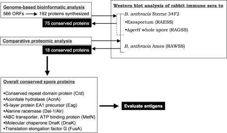
In this project we used two complementary approaches to select spore antigens that could produce antibodies cross-reactive against a broad range of B. cereus group spores. For the first method we used in silico filters to produce a list of 200 proteins by their conservation across five genomes. In the second approach we used a comparative proteomics analysis to detect cross-reacting spore antigens (Fig. 6). Both strategies have their drawbacks. Sequence-based analyses alone cannot predict the composition of the Bacillus spore especially considering the likely variability introduced by transcriptional and translational regulation of protein expression. However, any proteomics approach has inherent bias toward identification of abundant proteins. Combining both the approaches produced a relatively small list of potential conserved candidate antigens.
Fig. 6.
Schematic representation of the results obtained through the study design to obtain potential vaccine candidates.
The sera we used for challenging the proteins were taken from rabbits injected with B. anthracis exosporium (RAESS) and B. anthracis whole spore germination mutants (RAGSS) (44). Because B. anthracis has multiple genetic loci dedicated to triggering germination (49, 50) there is a possibility that a small portion of the spores may germinate in vivo and elicit responses to vegetative cell antigens. However, we found that results using RAGSS were mostly similar to those using sera from killed whole spores (RAWSS), indicating that germination in vivo is not a major issue for this ΔgerH mutant.
We identified 75 spore B. anthracis proteins from the bioinformatics-based screen that reacted against RAESS or RAGSS and 18 from the proteomics screen that reacted against RAESS, RAGSS, or RAWSS (Fig. 6). Only two protein candidates were found to be common to both approaches (Dal-1 and MetN). The activity of Dal-1 is higher in spores than in the vegetative cells (49, 51, 52), and the protein has been found to be a germination inhibitor in the exosporium (29, 51). MetN (methionine import ATP-binding protein) is responsible for energy coupling to the transmembrane transport system. MetN has not been reported as an immunogenic protein in any previous Bacillus studies, although immunogenic ABC transporters are being considered as potential vaccine candidates against several pathogens such as Helicobacter, Burkholderia, Streptococcus, Chlamydia, and Rickettsia species (53–58). B. anthracis spore antisera did not cross-react against the Dal-1 and MetN orthologs from the proteomes of the nine other strains tested. Our interpretation of these results is that the spore surface components proteins are in low abundance and/or elicit immune responses that in general cannot be detected by the relatively insensitive 2D gel electrophoresis. This is backed up by other studies (37, 59) that failed to detect several known spore proteins on the 2D protein gels. In some cases, the low abundance of the protein is the likely reason for the failure to detect in our studies, for instance, the Exs exosporium components (20, 36, 60, 61) and the recently discovered SoaA protein (62). In the case of the major exosporium component BclA (36, 63), a lack of trypsin cleavage sites precludes identification.
Four of the 18 cross-reacting proteins from the proteome-based screen (AcnA, Eag, DnaK, and FusA), were identified in B. anthracis spore lysates by Liu et al. (29). The remaining 18 proteins are not exclusively exosporium or endospore structural components but rather important core metabolic proteins presumably included to facilitate rapid growth upon germination. Translation elongation factors (Tuf and FusA) and chaperones (DnaK and GroEL) are examples of this class of spore proteins. Another class of the proteins listed in Table IV are those that are likely debris from the lysed mother cells adhered onto the surface of the spores. The Eag protein has been shown to be expressed only at the stationary phase of the vegetative cell (64). The RhsA protein probably also falls under the category of an incidental spore component. Another cross-reactive protein identified from this screen is the Crd protein. This large protein with 2,358 amino acids (molecular mass/pI, 244.9 kDa/4.17) contains 15 imperfect repeats of a 132-residue domain (PFAM (Protein Family database) accession number PF01345) that is found in three other B. anthracis proteins, GBAA1618 (molecular mass/pI, 523.3 kDa/4.06), GBAA3725 (molecular mass/pI, 219.8 kDa/4.2), and GBAA3721 (molecular mass/pI, 7.8 kDa/5.71). However, only Crd was detected in the protein gels. Genes encoding proteins with this motif are common in the B. cereus group but not found in the outgroup endospore-forming bacteria B. pseudomycoides DSMZ 12442 and B. subtilis. Proteins with multiple PF01345 domains appear in bacteria as diverse as Chlamydia trachomatis and the archaebacterium Methanobacterium. Crd lacks a classic type II signal sequence and LPXTG binding motif suggesting that it is neither cell wall- nor membrane-associated. The broad range of spores of B. cereus that cross-react to Crd suggests a promising candidate, and further investigation on the localization of this protein is necessary.
Several other researchers have demonstrated the power of proteomics analysis in a high throughput mode, including combining genome-based bioinformatics approaches (29, 37, 59). Of the proteins discussed in this report, Dal-1, Eag, Tuf, and FusA have been previously identified as immunogenic proteins from members of the B. cereus s.l. group (37, 59). The remaining four immunogenic proteins, MetN, Crd, AcnA, and DnaK, have not been reported previously.
One confounding experimental variable in the proteomics studies was the unexpected lack of cross-reaction to endospore antisera raised against B. anthracis by B. cereus ATCC 10987. B. cereus ATCC 10987 is much more closely related to B. anthracis than clade 2 and clade 3 strains that did cross-react to the sera. From our genomics analysis of the data it is clear that highly conserved orthologs of almost all the targets (in some proteins >97% similarity) found by the study are present in the genome of B. cereus ATCC 10987. Other strains that encode proteins with sequence very similar or identical to proteins in B. anthracis that elicit an immune response also do not cross-react to the sera. Expression patterns of the potential cross-reacting proteins during sporulation may vary in different genetic backgrounds, resulting in low abundance of the proteins in the electrophoretic maps. The design of this study, where the proteomes of 10 phylogenetically diverse strains were profiled, to a certain extent mitigates against unusual results in one or two individual strains.
Despite a high degree of chromosomal gene conservation and synteny among B. cereus s.l. genomes (2, 7), there are significant strain-specific differences in spore coat and endospore proteins, which may contribute to the survival and adaptation in host invasion (65). It is unlikely that the mammalian adaptive immune system is the main agent for selection as most members of this group would appear to infect insects and other invertebrates primarily (66).
Comparative analysis of the spore proteomes of the 10 diverse strains of the B. cereus group has shown that it is possible to distinguish between all these strains based on their 2D profiles with spots unique to each strain and only 72% overall matched spots. Ultrastructural analysis of the spores also showed variations, particularly in the exosporium, core dimension, and interspace regions. With further cross-reaction studies against other Bacillus species it may be possible to select a mixture of antigens (or a multiepitope antigen) specific for B. cereus group spores. Fluorescent tagged antibodies raised against these proteins could have a role as an environmental or clinical detection tool for B. cereus s.l. strains.
Acknowledgments
We sincerely thank Lieutenant M. Weiner for providing the gerH mutant B. anthracis 34F2 Sterne strain; Drs. D. Roth, J. He, and A. Greener for performing the TAP analysis; Drs. N. Nguyen and W. Hicks for mass spectrometry analysis; L. Liang for facilitating the generation of rabbit immune sera; and Dr. L. Baillie and S. Hibbs for providing the B. anthracis 34F2 Sterne spore suspension to inject rabbits. We appreciate the generosity of Dr. A. M. Freidlander for providing the RAWSS.
Footnotes
Published, MCP Papers in Press, February 9, 2009, DOI 10.1074/mcp.M800403-MCP200
The abbreviations used are: s.l., sensu lato (translation, “in the broad sense”); ATCC, American Type Culture Collection; BGSC, Bacillus Genetic Stock Collection; DSMZ, Deutsche Sammlung von Mikroorganismen und Zellkulturen; IS, interspace (distance between endospore and exosporium); NCBI, National Center for Biotechnology Information; RAESS, rabbit anti-exosporium sera; RAGSS, rabbit anti-GerH whole spore sera; RANS, rabbit anti-naïve sera; RAWSS, rabbit anti-whole spore (ungerminated) sera; TAP, Transcriptionally Active PCR; TEM, transmission electron microscopy; bis-Tris, 2-[bis(2-hydroxyethyl)amino]-2-(hydroxymethyl)propane-1,3-diol; BLAST, basic local alignment search tool; RTS, Rapid Translational System; 2D, two-dimensional; ABC, ATP-binding cassette; AMU, atomic mass unit; PA, protective antigen.
This work was supported by Joint Science and Technology Office-Chemical and Biological Defense/Defense Threat Reduction Agency Grant 9Y0010_05_NM_B (to T. D. R.).
The on-line version of this article (available at http://www.mcponline.org) contains supplemental material.
The whole genome shotgun data reported in this paper have been submitted to GenBank™ with NCBI Genome Project IDs 29709, 29713, 29671, 29651, 29657, 29649, 29701, and 29707.
REFERENCES
- 1.Helgason, E., Okstad, O. A., Caugant, D. A., Johansen, H. A., Fouet, A., Mock, M., Hegna, I., and Kolstø, A. B. (2000. ) Bacillus anthracis, Bacillus cereus, and Bacillus thuringiensis—one species on the basis of genetic evidence. Appl. Environ. Microbiol. 66, 2627 –2630 [DOI] [PMC free article] [PubMed] [Google Scholar]
- 2.Read, T. D., Peterson, S. N., Tourasse, N., Baillie, L. W., Paulsen, I. T., Nelson, K. E., Tettelin, H., Fouts, D. E., Eisen, J. A., Gill, S. R., Holtzapple, E. K., Okstad, O. A., Helgason, E., Rilstone, J., Wu, M., Kolonay, J. F., Beanan, M. J., Dodson, R. J., Brinkac, L. M., Gwinn, M., DeBoy, R. T., Madpu, R., Daugherty, S. C., Durkin, A. S., Haft, D. H., Nelson, W. C., Peterson, J. D., Pop, M., Khouri, H. M., Radune, D., Benton, J. L., Mahamoud, Y., Jiang, L., Hance, I. R., Weidman, J. F., Berry, K. J., Plaut, R. D., Wolf, A. M., Watkins, K. L., Nierman, W. C., Hazen, A., Cline, R., Redmond, C., Thwaite, J. E., White, O., Salzberg, S. L., Thomason, B., Friedlander, A. M., Koehler, T. M., Hanna, P. C., Kolstø, A. B., and Fraser, C. M. (2003. ) The genome sequence of Bacillus anthracis Ames and comparison to closely related bacteria. Nature 423, 81 –86 [DOI] [PubMed] [Google Scholar]
- 3.Ivanova, N., Sorokin, A., Anderson, I., Galleron, N., Candelon, B., Kapatral, V., Bhattacharyya, A., Reznik, G., Mikhailova, N., Lapidus, A., Chu, L., Mazur, M., Goltsman, E., Larsen, N., D'souza, M., Walunas, T., Grechkin, Y., Pusch, G., Haselkorn, R., Fonstein, M., Ehrlich, S. D., Overbeek, R., and Kyrpides, N. (2003. ) Genome sequence of Bacillus cereus and comparative analysis with. Bacillus anthracis. Nature 423, 87 –91 [DOI] [PubMed] [Google Scholar]
- 4.Rasko, D. A., Ravel, J., Økstad, O. A., Helgason, E., Cer, R. Z., Jiang, L., Shores, K. A., Fouts, D. E., Tourasse, N. J., Angiuoli, S. V., Kolonay, J., Nelson, W. C., Kolstø, A. B., Fraser, C. M., and Read, T. D. (2004. ) The genome sequence of Bacillus cereus ATCC 10987 reveals metabolic adaptations and a large plasmid related to Bacillus anthracis pXO1. Nucleic Acids Res. 32, 977 –988 [DOI] [PMC free article] [PubMed] [Google Scholar]
- 5.Han, C. S., Xie, G., Challacombe, J. F., Altherr, M. R., Bhotika, S. S., Brown, N., Bruce, D., Campbell, C. S., Campbell, M. L., Chen, J., Chertkov, O., Cleland, C., Dimitrijevic, M., Doggett, N. A., Fawcett, J. J., Glavina, T., Goodwin, L. A., Green, L. D., Hill, K. K., Hitchcock, P., Jackson, P. J., Keim, P., Kewalramani, A. R., Longmire, J., Lucas, S., Malfatti, S., McMurry, K., Meincke, L. J., Misra, M., Moseman, B. L., Mundt, M., Munk, A. C., Okinaka, R. T., Parson-Quintana, B., Reilly, L. P., Richardson, P., Robinson, D. L., Rubin, E., Saunders, E., Tapia, R., Tesmer, J. G., Thayer, N., Thompson, L. S., Tice, H., Ticknor, L. O., Wills, P. L., Brettin, T. S., and Gilna, P. (2006. ) Pathogenomic sequence analysis of Bacillus cereus and Bacillus thuringiensis isolates closely related to. Bacillus anthracis. J. Bacteriol. 188, 3382 –3390 [DOI] [PMC free article] [PubMed] [Google Scholar]
- 6.Hoffmaster, A. R., Ravel, J., Rasko, D. A., Chapman, G. D., Chute, M. D., Marston, C. K., De, B. K., Sacchi, C. T., Fitzgerald, C., Mayer, L. W., Maiden, M. C., Priest, F. G., Barker, M., Jiang, L., Cer, R. Z., Rilstone, J., Peterson, S. N., Weyant, R. S., Galloway, D. R., Read, T. D., Popovic, T., and Fraser, C. M. (2004. ) Identification of anthrax toxin genes in a Bacillus cereus associated with an illness resembling inhalation anthrax. Proc. Natl. Acad. Sci. U. S. A. 101, 8449 –8454 [DOI] [PMC free article] [PubMed] [Google Scholar]
- 7.Rasko, D. A., Altherr, M. R., Han, C. S., and Ravel, J. (2005. ) Genomics of the Bacillus cereus group of organisms. FEMS Microbiol. Rev. 29, 303 –329 [DOI] [PubMed] [Google Scholar]
- 8.Hofte, H., and Whiteley, H. R. (1989. ) Insecticidal crystal proteins of Bacillus thuringiensis. Microbiol. Rev. 53, 242 –255 [DOI] [PMC free article] [PubMed] [Google Scholar]
- 9.Rasko, D. A., Rosovitz, M. J., Økstad, O. A., Fouts, D. E., Jiang, L., Cer, R. Z., Kolstø, A. B., Gill, S. R., and Ravel, J. (2007. ) Complete sequence analysis of novel plasmids from emetic and periodontal Bacillus cereus isolates reveals a common evolutionary history among the B. cereus-group plasmids, including Bacillus anthracis pXO1. J. Bacteriol. 189, 52 –64 [DOI] [PMC free article] [PubMed] [Google Scholar]
- 10.Dixon, T. C., Meselson, M., Guillemin, J., and Hanna, P. C. (1999. ) Anthrax. N. Engl. J. Med. 341, 815 –826 [DOI] [PubMed] [Google Scholar]
- 11.Battisti, L., Green, B. D., and Thorne, C. B. (1985. ) Mating system for transfer of plasmids among Bacillus anthracis, Bacillus cereus, and Bacillus thuringiensis. J. Bacteriol. 162, 543 –550 [DOI] [PMC free article] [PubMed] [Google Scholar]
- 12.Van der Auwera, G. A., Timmery, S., Hoton, F., and Mahillon, J. (2007. ) Plasmid exchanges among members of the Bacillus cereus group in foodstuffs. Int. J. Food Microbiol. 113, 164 –172 [DOI] [PubMed] [Google Scholar]
- 13.Yuan, Y. M., Hu, X. M., Liu, H. Z., Hansen, B. M., Yan, J. P., and Yuan, Z. M. (2007. ) Kinetics of plasmid transfer among Bacillus cereus group strains within lepidopteran larvae. Arch. Microbiol. 187, 425 –431 [DOI] [PubMed] [Google Scholar]
- 14.Klee, S. R., Ozel, M., Appel, B., Boesch, C., Ellerbrok, H., Jacob, D., Holland, G., Leendertz, F. H., Pauli, G., Grunow, R., and Nattermann, H. (2006. ) Characterization of Bacillus anthracis-like bacteria isolated from wild great apes from Cote d'Ivoire and Cameroon. J. Bacteriol. 188, 5333 –5344 [DOI] [PMC free article] [PubMed] [Google Scholar]
- 15.Kudva, I. T., Griffin, R. W., Garren, J. M., Calderwood, S. B., and John, M. (2005. ) Identification of a protein subset of the anthrax spore immunome in humans immunized with the anthrax vaccine adsorbed preparation. Infect. Immun. 73, 5685 –5696 [DOI] [PMC free article] [PubMed] [Google Scholar]
- 16.Chitlaru, T., Gat, O., Grosfeld, H., Inbar, I., Gozlan, Y., and Shafferman, A. (2007. ) Identification of in vivo-expressed immunogenic proteins by serological proteome analysis of the Bacillus anthracis secretome. Infect. Immun. 75, 2841 –2852 [DOI] [PMC free article] [PubMed] [Google Scholar]
- 17.Warth, A. D., Ohye, D. F., and Murrell, W. G. (1963. ) The composition and structure of bacterial spores. J. Cell Biol. 16, 579 –592 [DOI] [PMC free article] [PubMed] [Google Scholar]
- 18.Aronson, A. I., and Fitz-James, P. (1976. ) Structure and morphogenesis of the bacterial spore coat. Bacteriol. Rev. 40, 360 –402 [DOI] [PMC free article] [PubMed] [Google Scholar]
- 19.Henriques, A. O., and Moran, C. P., Jr. (2000. ) Structure and assembly of the bacterial endospore coat. Methods 20, 95 –110 [DOI] [PubMed] [Google Scholar]
- 20.Henriques, A. O., and Moran, C. P., Jr. (2007. ) Structure, assembly, and function of the spore surface layers. Annu. Rev. Microbiol. 61, 555 –588 [DOI] [PubMed] [Google Scholar]
- 21.Takamatsu, H., and Watabe, K. (2002. ) Assembly and genetics of spore protective structures. CMLS Cell. Mol. Life Sci. 59, 434 –444 [DOI] [PMC free article] [PubMed] [Google Scholar]
- 22.Nicholson, W. L., Munakata, N., Horneck, G., Melosh, H. J., and Setlow, P. (2000. ) Resistance of Bacillus endospore to extreme terrestrial and extraterrestrial environments. Microbiol. Mol. Biol. Rev. 64, 548 –572 [DOI] [PMC free article] [PubMed] [Google Scholar]
- 23.Henriques, A. O., Costa, T., Martins, L. O., and Zilhao, R. (2004. ) Functional architecture and assembly of the spore coat, in Bacterial Spore Formers: Probiotics and Emerging Applications (Ricca, E., Henriques, A. O., Cutting, S. M., eds) pp. 34–52, Horizon Science Press, London
- 24.Gerhardt, P., Pankratz, H. S., and Scherrer, R. (1976. ) Fine structure of Bacillus thuringiensis spore. Appl. Environ. Microbiol. 32, 438 –440 [DOI] [PMC free article] [PubMed] [Google Scholar]
- 25.Chada, V. G., Sanstad, E. A., Wang, R., and Driks, A. (2003. ) Morphogenesis of Bacillus spore surfaces. J. Bacteriol. 185, 6255 –6261 [DOI] [PMC free article] [PubMed] [Google Scholar]
- 26.Barbosa, T. M., Serra, C. R., La Ragione, R. M., Woodward, M. J., and Henriques, A. O. (2005. ) Screening for Bacillus isolates in the broiler gastrointestinal tract. Appl. Environ. Microbiol. 71, 968 –978 [DOI] [PMC free article] [PubMed] [Google Scholar]
- 27.Giorno, R., Bozue, J., Cote, C., Wenzel, T., Moody, K. S., Mallozzi, M., Ryan, M., Wang, R., Zielke, R., Maddock, J. R., Friedlander, A., Welkos, S., and Driks, A. (2007. ) Morphogenesis of the Bacillus anthracis spore. J. Bacteriol. 189, 691 –705 [DOI] [PMC free article] [PubMed] [Google Scholar]
- 28.Driks, A. (1999. ) Bacillus subtilis spore coat. Microbiol. Mol. Biol. Rev. 63, 1 –20 [DOI] [PMC free article] [PubMed] [Google Scholar]
- 29.Liu, H., Bergman, N. H., Thomason, B., Shallom, S., Hazen, A., Crossno, J., Rasko, D. A., Ravel, J., Read, T. D., Peterson, S. N., Yates, J., and Hanna, P. C. (2004. ) Formation and composition of the Bacillus anthracis endospore. J. Bacteriol. 186, 164 –178 [DOI] [PMC free article] [PubMed] [Google Scholar]
- 30.Melly, E., Genest, P. C., Gilmore, M. E., Little, S., Popham, D. L., Driks, A., and Setlow, P. (2002. ) Analysis of the properties of spores of Bacillus subtilis prepared at different temperatures. J. Appl. Microbiol. 92, 1105 –1115 [DOI] [PubMed] [Google Scholar]
- 31.Zheng, L. B., and Losick, R. (1990. ) Cascade regulation of spore coat gene expression in. Bacillus subtilis. J. Mol. Biol. 212, 645 –660 [DOI] [PubMed] [Google Scholar]
- 32.Francis, A. W., Ruggiero, C. E., Koppisch, A. T., Dong, J., Song, J., Brettin, T., and Iyer, S. (2005. ) Proteomic analysis of Bacillus anthracis Sterne vegetative cells. Biochim. Biophys. Acta 1748, 191 –200 [DOI] [PubMed] [Google Scholar]
- 33.Margulies, M., Egholm, M., Altman, W. E., Attiya, S., Bader, J. S., Bemben, L. A., Berka, J., Braverman, M. S., Chen, Y. J., Chen, Z., Dewell, S. B., Du, L., Fierro, J. M., Gomes, X. V., Godwin, B. C., He, W., Helgesen, S., Ho, C. H., Irzyk, G. P., Jando, S. C., Alenquer, M. L., Jarvie, T. P., Jirage, K. B., Kim, J. B., Knight, J. R., Lanza, J. R., Leamon, J. H., Lefkowitz, S. M., Lei, M., Li, J., Lohman, K. L., Lu, H., Makhijani, V. B., McDade, K. E., McKenna, M. P., Myers, E. W., Nickerson, E., Nobile, J. R., Plant, R., Puc, B. P., Ronan, M. T., Roth, G. T., Sarkis, G. J., Simons, J. F., Simpson, J. W., Srinivasan, M., Tartaro, K. R., Tomasz, A., Vogt, K. A., Volkmer, G. A., Wang, S. H., Wang, Y., Weiner, M. P., Yu, P., Begley, R. F., and Rothberg, J. M. (2005. ) Genome sequencing in microfabricated high-density picolitre reactors. Nature 437, 376 –380 [DOI] [PMC free article] [PubMed] [Google Scholar]
- 34.Leighton, T. J., and Doi, R. H. (1971. ) The stability of messenger ribonucleic acid during sporulation in. Bacillus subtilis. J. Biol. Chem. 246, 3189 –3195 [PubMed] [Google Scholar]
- 35.Redmond, C., Baillie, L. W. J., Hibbs, S., Moir, A. J. G., and Moir, A. (2004. ) Identification of proteins in the exosporium of. Bacillus anthracis. Microbiology 150, 355 –363 [DOI] [PubMed] [Google Scholar]
- 36.Rode, L. J., Lewis, C. W., Jr., and Foster, J. W. (1962. ) Electron microscopy of spores of Bacillus megaterium with special reference to the effects of fixation and thin sectioning. J. Cell Biol. 13, 423 –435 [DOI] [PMC free article] [PubMed] [Google Scholar]
- 37.DelVecchio, V. G., Connolly, J. P., Alefantis, T. G., Walz, A., Quan, M. A., Patra, G., Ashton, J. M., Whittington, J. T., Chafin, R. D., Liang, X., Grewal, P., Khan, A. S., and Mujer, C. V. (2006. ) Proteomic profiling and identification of immunodominant spore antigens of Bacillus anthracis, Bacillus cereus and Bacillus thuringiensis. Appl. Environ. Microbiol. 72, 6355 –6363 [DOI] [PMC free article] [PubMed] [Google Scholar]
- 38.Mukhopadhyay, S., Miller, R. D., and Summersgill, J. T. (2004. ) Analysis of altered protein expression patterns of Chlamydia pneumoniae by an integrated Proteome-Works system. J. Proteome Res. 3, 878 –883 [DOI] [PubMed] [Google Scholar]
- 39.Mukhopadhyay, S., Good, D., Miller, R. D., Graham, J. E., Mathews, S. A., Timms, P., and Summersgill, J. T. (2006. ) Identification of Chlamydia pneumoniae proteins in the transition from reticulate to elementary body formation. Mol. Cell. Proteomics 5, 2311 –2318 [DOI] [PubMed] [Google Scholar]
- 40.Havlis, J., Thomas, H., Sebela, M., and Shevchenko, A. (2003. ) Fast-response proteomics by accelerated in-gel digestion of proteins. Anal. Chem. 75, 1300 –1306 [DOI] [PubMed] [Google Scholar]
- 41.Eng, J. K., McCormack, A. L., and Yates, J. R., III (1994. ) An approach to correlate mass spectral data of peptides with amino acid sequences in a protein databases. J. Am. Soc. Mass Spectrom. 5, 976 –989 [DOI] [PubMed] [Google Scholar]
- 42.Delcher, A. L., Bratke, K. A., Powers, E. C., and Salzberg, S. L. (2007. ) Identifying bacterial genes and endosymbiont DNA with Glimmer. Bioinformatics 23, 673 –679 [DOI] [PMC free article] [PubMed] [Google Scholar]
- 43.Liang, X., Teng, A., Braun, D. M., Felgner, J., Wang, Y., Baker, S. I., Chen, S., Zelphati, O., and Felgner, P. L. (2002. ) Transcriptionally active polymerase chain reaction (TAP) high throughput gene expression using genome sequence data. J. Biol. Chem. 277, 3593 –3598 [DOI] [PubMed] [Google Scholar]
- 44.Weiner, M. A., Read, T. D., and Hanna, P. C. (2003. ) Identification and characterization of the gerH operon of Bacillus anthracis endospores: a differential role for purine nucleosides in germination. J. Bacteriol. 185, 1462 –1464 [DOI] [PMC free article] [PubMed] [Google Scholar]
- 45.Bozue, J., Moody, K. L., Cote, C. K., Stiles, B. G., Friedlander, A. M., Welkos, S. L., and Hale, M. L. (2007. ) Bacillus anthracis spores of bclA mutant exhibit increased adherence to epithelial cells, fibroblasts, and endothelial cells but not to macrophages. Infect. Immun. 75, 4498 –4505 [DOI] [PMC free article] [PubMed] [Google Scholar]
- 46.Altschul, S. F., Gish, W., Miller, W., Myers, E. W., and Lipman, D. J. (1990. ) Basic local alignment search tool. J. Mol. Biol. 215, 403 –410 [DOI] [PubMed] [Google Scholar]
- 47.Altschul, S. F., and Koonin, E. V. (1998. ) Iterated profile searches with PSI-BLAST-a tool for discovery in protein databases. Trends Biochem. Sci. 23, 444 –447 [DOI] [PubMed] [Google Scholar]
- 48.Gardy, J. L., Laird, M. R., Chen, F., Rey, S., Walsh, C. J., Ester, M., and Brinkman, F. S. (2005. ) PSORTb v. 2.0: expanded prediction of bacterial protein subcellular localization and insights gained from comparative proteome analysis. Bioinformatics 21, 617 –623 [DOI] [PubMed] [Google Scholar]
- 49.Kanda-Nambu, K., Yasuda, Y., and Tochikubo, L. (2000. ) Isozymic nature of spore coat-associated alanine racemase of. Bacillus subtilis. Amino Acids 18, 375 –387 [DOI] [PubMed] [Google Scholar]
- 50.Behravan, J., Chirakkal, H., Masson, A., and Moir, A. (2000. ) Mutations in gerP locus of Bacillus subtilis and Bacillus anthracis affect access of germinants to their targets in spores. J. Bacteriol. 182, 1987 –1994 [DOI] [PMC free article] [PubMed] [Google Scholar]
- 51.Huang, C. M., Elmets, C. A., Tang, D. C., Li, F., and Yusuf, N. (2004. ) Proteomics reveals that proteins expressed during the early stage of Bacillus anthracis infection are potential targets for the development of vaccines and drugs. Genomics Proteomics Bioinformatics 2, 143 –151 [DOI] [PMC free article] [PubMed] [Google Scholar]
- 52.Steichen, C. T., Kearney, J. F., and Turnbough, C. L., Jr. (2007. ) Non-uniform assembly of the Bacillus anthracis exosporium and a bottle cap model for spore germination and outgrowth. Mol. Microbiol. 64, 359 –367 [DOI] [PubMed] [Google Scholar]
- 53.Weinberg, M. V., and Maier, R. J. (2007. ) Peptide transport in Helicobacter pylori: roles of dpp and opp systems and evidence for additional peptide transporters. J. Bacteriol. 189, 3392 –3402 [DOI] [PMC free article] [PubMed] [Google Scholar]
- 54.Harland, D. N., Dassa, E., Titball, R. W., Brown, K. A., and Atkins, H. S. (2007. ) ATP-binding cassette systems in Burkholderia pseudomallei and. Burkholderia mallei. BMC Genomics 28, 8 –83 [DOI] [PMC free article] [PubMed] [Google Scholar]
- 55.Harding, S. V., Sarkar-Tyson, M., Smither, S. J., Atkins, T. P., Oyston, P. C., Brown, K. A., Liu, Y., Wait, R., and Titball, R. W. (2007. ) The identification of surface proteins of. Burkholderia pseudomallei. Vaccine 25, 2664 –2672 [DOI] [PubMed] [Google Scholar]
- 56.Rodríguez-Ortega, M. J., Norais, N., Bensi, G., Liberatori, S., Capo, S., Mora, M., Scarselli, M., Doro, F., Ferrari, G., Garaguso, I., Maggi, T., Neumann, A., Covre, A., Telford, J. L., and Grandi, G. (2006. ) Characterization and identification of vaccine candidate proteins through analysis of the group A Streptococcus surface proteome. Nat. Biotechnol. 24, 191 –197 [DOI] [PubMed] [Google Scholar]
- 57.Schmitz-Esser, S., Linka, N., Collingro, A., Beier, C. L., Neuhaus, H. E., Wagner, M., and Horn, M. (2004. ) ATP/ADP translocases: a common feature of obligate intracellular amoebal symbionts related to Chlamydiae and Rickettsiae. J. Bacteriol. 186, 683 –691 [DOI] [PMC free article] [PubMed] [Google Scholar]
- 58.Bannantine, J. P., and Rockey, D. D. (1999. ) Use of primate model system to identify Chlamydia trachomatis protein antigens recognized uniquely in the context of infection. Microbiology 145, 2077 –2085 [DOI] [PubMed] [Google Scholar]
- 59.Ariel, N., Zvi, A., Makarova, K. S., Chitlaru, T., Elhanany, E., Velan, B., Cohen, S., Friedlander, A. M., and Shafferman, A. (2003. ) Genome-based bioinformatic selection of chromosomal Bacillus anthracis putative vaccine candidates coupled with proteomic identification of surface-associated antigens. Infect. Immun. 71, 4563 –4579 [DOI] [PMC free article] [PubMed] [Google Scholar]
- 60.Steichen, C. T., Chen, P., Kearney, J. F., and Turnbough, C. L., Jr. (2003. ) Identification of immuno-dominant protein and other proteins of the Bacillus anthracis exosporium. J. Bacteriol. 185, 1903 –1910 [DOI] [PMC free article] [PubMed] [Google Scholar]
- 61.Sylvestre, P., Couture-Tosi, E., and Mock, M. (2005. ) Contribution of ExsFA and ExsFB proteins to the localization of BclA on the spore surface and to the stability of the Bacillus anthracis exosporium. J. Bacteriol. 187, 5122 –5128 [DOI] [PMC free article] [PubMed] [Google Scholar]
- 62.Cote, C. K., Bozue, J., Moody, K. L., DiMezzo, T. L., Chapman, C. E., and Welkos, S. L. (2008. ) Analysis of a novel spore antigen in Bacillus anthracis that contributes to spore opsonization. Microbiology 154, 619 –632 [DOI] [PubMed] [Google Scholar]
- 63.Brahmbhatt, T. N., Janes, B. K., Stibitz, E. S., Darnell, S. C., Sanz, P., Rasmussen, S. B., and O'Brien, A. D. (2007. ) Bacillus anthracis exosporium protein BclA affects spore germination, interaction with extracellular matrix proteins, and hydrophobicity. Infect. Immun. 75, 5233 –5239 [DOI] [PMC free article] [PubMed] [Google Scholar]
- 64.Fouet, A., and Mesnage, S. (2002. ) Bacillus anthracis cell envelope components. Curr. Top. Microbiol. Immunol. 271, 87 –113 [DOI] [PubMed] [Google Scholar]
- 65.Anderson, I., Sorokin, A., Kapatral, V., Reznik, G., Bhattacharya, A., Mikhailova, N., Burd, H., Joukov, V., Kaznadzey, D., Walunas, T., Markd'souza, Larsen, N., Pusch, G., Liolios, K., Grechkin, Y., Lapidus, A., Goltsman, E., Chu, L., Fonstein, M., Ehrlich, S. D., Overbeek, R., Kyrpides, N., and Ivanova, N. (2005. ) Comparative genome analysis of Bacillus cereus group genomes with Bacillus subtilis. FEMS Microbiol. Lett. 250, 175 –184 [DOI] [PubMed] [Google Scholar]
- 66.Jensen, G. B., Hansen, B. M., Eilenberg, J., and Mahillon, J. (2003. ) The hidden lifestyles of Bacillus cereus and relatives. Environ. Microbiol. 5, 631 –640 [DOI] [PubMed] [Google Scholar]
- 67.Stewart, A. C., Osborne, B., and Read, T. D. (2009. ) DIYA: a bacterial annotation pipeline for any genomics lab. Bioinformatics, in press [DOI] [PMC free article] [PubMed]


