Abstract
Nondeletional hereditary persistence of fetal hemoglobin (nd-HPFH), a rare hereditary condition resulting in elevated levels of fetal hemoglobin (Hb F) in adults, is associated with promoter mutations in the human fetal globin (HBG1 and HBG2) genes. In this paper, we report a novel type of nd-HPFH due to a HBG2 gene promoter mutation (HBG2:g.-109G>T). This mutation, located at the 3′ end of the HBG2 distal CCAAT box, was initially identified in an adult female subject of Central Greek origin and results in elevated Hb F levels (4.1%) and significantly increased Gγ-globin chain production (79.2%). Family studies and DNA analysis revealed that the HBG2:g.-109G>T mutation is also found in the family members in compound heterozygosity with the HBG2:g.-158C>T single nucleotide polymorphism or the silent HBB:g.-101C>T β-thalassemia mutation, resulting in the latter case in significantly elevated Hb F levels (14.3%). Electrophoretic mobility shift analysis revealed that the HBG2:g.-109G>T mutation abolishes a transcription factor binding site, consistent with previous observations using DNA footprinting analysis, suggesting that guanine at position HBG2/1:g.-109 is critical for NF-E3 binding. These data suggest that the HBG2:g-109G>T mutation has a functional role in increasing HBG2 transcription and is responsible for the HPFH phenotype observed in our index cases.
Keywords: Nondeletional hereditary persistence of fetal hemoglobin, β-thalassemia, Fetal globin genes, HBG2, Mutation, Regulatory element, Transcription
Introduction
Hereditary persistence of fetal hemoglobin (HPFH) is a genetic condition characterized by persistent elevated levels of fetal hemoglobin (Hb F) in the adult erythroid stage [1]. HPFH heterozygotes have a normal clinical phenotype and hematological indices, except for the lower levels of hemoglobin A2 (Hb A2) that constitutes the hallmark of HPFH. The reason for that is the increased frequency of interactions between the locus control region (LCR) and the mutated γ-globin gene promoter compared to the normal situation. Subsequently, this shifts the transcriptional balance in favor to the otherwise silent, fetal globin genes, resulting in lower HBD gene transcription and Hb A2 levels [2].
The various forms of HPFH are distinguished into two main categories, namely, deletional and nondeletional HPFH (nd-HPFH), due to large deletions 3′ to the fetal globin genes or nucleotide substitutions in the fetal globin gene promoters, respectively [3]. The current explanation for the functional role of the latter HPFH category is the destabilization of a yet unknown silencing protein complex that permits LCR–gene interactions and recruitment of the basal transcriptional machinery to the mutated γ-globin gene promoter, leading to reactivation of fetal globin gene transcription [4].
To date, there are few γ-globin promoter mutations, leading to nd-HPFH clustered on or in close proximity to γ-globin gene cis-regulatory elements [5]. The most important nd-HPFH is the Greek type (HBG1:g.-117G>A [6]), whose functional role is also demonstrated in transgenic mice [7]. This mutation is located at the 5′ end of the distal γ-globin gene CCAAT box where important erythroid-specific transcription factors bind. Other mutations cluster approximately 200 bp upstream to the γ-globin genes transcription initiation site where a GC-rich region is located. Although clinically silent, the mutations leading to nd-HPFH provide valuable insights into the transcriptional regulation of the human fetal globin genes, which in turn can enable design of novel strategies for β-thalassemia therapeutics.
In this communication, we report the Hellenic type of nd-HPFH due to a novel G>T transversion in the HBG2 gene promoter, located at the 3′ end of the distal HBG2 CCAAT box.
Materials and methods
Case selection
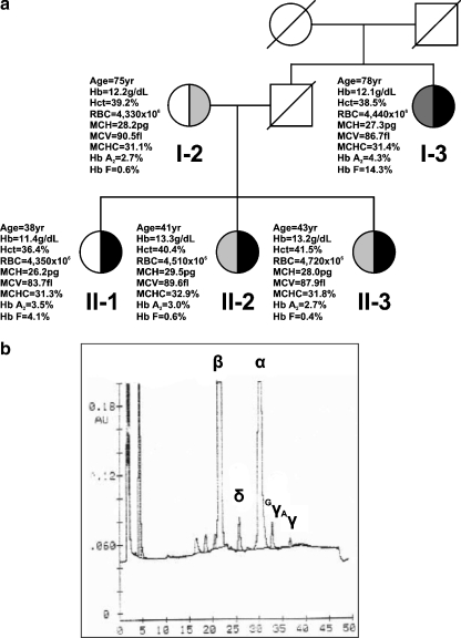
For the needs of this study, the mother and two sisters of the index case were also recruited. Unfortunately, the father of the index case was deceased, but, nevertheless, his sister was available for study (Fig. 1a). Also, 31 heterozygotes for the silent HBB:g.-101C>T β-thalassemia mutation have been included (Table 1), as well as 89 normal (nonthalassemic) individuals. The study was previously approved by the hospital’s ethics committee.
Fig. 1.
a Family tree showing the heterozygous and compound heterozygous cases for the Hellenic type of nd-HPFH, bearing the novel HBG2:g.-109G>T promoter mutation (depicted in black) and the HBG2:g.-158C>T polymorphism (depicted in light gray) or the silent HBB:g.-101C>T β-thalassemia nonsense mutation (depicted in dark gray), along with the corresponding hematological indices. b Chromatogram from RP-HPLC from the index case showing the significant increase in the Gγ/Aγ-globin chain ratio
Table 1.
Comparison of the hematological indices, Hb A2 and Hb F levels among the compound heterozygous for the novel HBG2:g.-109G>T mutation and the silent HBB:g.-101C>T β-thalassemia mutation (I-3; see also Fig. 1a), and heterozygotes for the silent HBB:g.-101C>T β-thalassemia mutation (note the markedly increased Hb F levels in case I-3)
| Hematological indices | HBB:g.-101C>T | ||
|---|---|---|---|
| HBG2:g.-109G>T (I-3) | Wild-type | Wild-type | |
| Females (n = 18) | Males (n = 13) | ||
| Age (years)/sex | 78/F | Adults | Adults |
| Hb (g/dL) | 12.1 | 12.4 ± 1.22 | 14.5 ± 0.84 |
| Hct (%) | 38.5 | 37.1 ± 2.76 | 43.1 ± 2.30 |
| RBC (×106) | 4,440 | 4,382 ± 446 | 5,144 ± 265 |
| MCH (pg) | 27.3 | 28.4 ± 1.54 | 28.5 ± 0.88 |
| MCV (fl) | 86.7 | 85.5 ± 4.03 | 84.3 ± 2.69 |
| MCHC (%) | 31.4 | 32.7 ± 1.04 | 33.9 ± 0.64 |
| Hb A2 (%) | 4.3 | 3.79 ± 0.24 | 3.92 ± 0.37 |
| Hb F (%) | 14.3 | 2.53 ± 1.8 | 1.60 ± 1.17 |
Hemoglobin studies
Blood samples were collected, with consent, in vacutainers with ethylenediaminetetraacetic acid as anticoagulant. Hematological indices were measured with an automated cell counter and Hb A2 and Hb F levels were quantitated, using cation exchange high-performance liquid chromatography (HPLC; VARIANT™, BioRad, Hercules, CA, USA). Globin chain quantitation was performed using reverse-phase HPLC, using the RP-18 column (Pharmacia, Uppsala, Sweden).
DNA analysis
Total genomic DNA isolation and γ-globin gene promoters’ amplification was done as previously described [8]. Mutation screening in the human γ-globin gene promoters was performed using denaturing gradient gel electrophoresis (DGGE) with a 40–70% linear gradient of denaturing agents (urea and formamide; 100% denaturant: 7 M urea, 40% formamide), as previously described [9]. HBB and HBD gene mutation screening was done as described in Losekoot and coworkers [10] and Papadakis and coworkers [11], respectively. Automated DNA sequencing was performed directly on the polymerase chain reaction (PCR) products. Screening for HBA2/1 gene deletions was done according to the multiplex gap-PCR strategy of Tan and coworkers [12], while a PCR-restriction fragment length polymorphism (RFLP) approach was used to screen for the commonest point mutations and small indels in the HBA2/1 genes, leading to nondeletional α-thalassemia in the Hellenic population [13].
Electrophoretic mobility shift assays
Electrophoretic mobility shift assays (EMSA) analysis was performed using total nuclear protein extracts from mouse erythroleukemia (MEL) and human K562 cells [14]. In brief, 5 μg nuclear extracts were used per reaction and competitions were done using 100-fold molar excess of the indicated double-stranded oligonucleotides before addition of the nuclear extract. Nucleotide sequence of the oligonucleotides used in these studies is available upon request. Supershift analysis was performed using a home-brewed GATA-1 antibody [15, 16].
Results
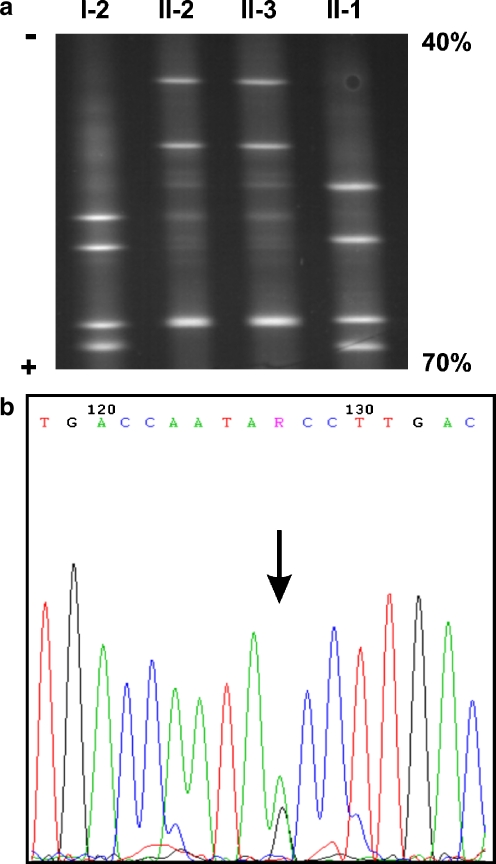
During routine carrier screening for β-thalassemia, an adult female subject was identified with moderately elevated Hb F levels (4.1%) and significantly increased Gγ/Aγ-globin chain ratio (Fig. 1b). An aberrant electrophoretic pattern was identified during mutation screening in the HBG2 promoter by DGGE analysis (Fig. 2a). DNA sequencing of the proximal HBG2 promoter region revealed a novel sequence variation, namely, a G>T transversion at position −109 relative to the gene’s transcription initiation site (HBG2:g.-109G>T; Fig. 2b). DNA sequencing did not reveal any other variant nucleotide in either one of the γ-globin genes promoters and distal 5′ regulatory regions between positions −672 and +25. Neither the HBG2:g.-158C>T (XmnI) nor the HBG1:g.-225-222(AGCA)del polymorphisms, which are often correlated with moderately increased HbF levels, were found in the index case.
Fig. 2.
a DGGE analysis of the promoter region of the HBG2 gene. Numbering correlates with the family tree in Fig. 1a. Note the mutant HBG2:g.-158 T (lower band) and HBG2:g.-109 T homoduplexes (upper band) that comigrate in lanes II-2 and II-3, compared to lanes I-2 and II-1, respectively. The 40–70% denaturing gradient corresponds to the top and bottom of the gel, respectively. b DNA sequencing analysis, performed in the forward and reverse (not shown) orientation, revealing a G>T transition at position −109 of the HBG2 gene promoter (arrow)
Hemoglobin studies and DNA analysis of the available family members indicated that the propositus mother (I-2) was a carrier of the HBG2:g.-158C>T (XmnI) polymorphism, while both sisters (II-2, II-3) were compound heterozygotes for the HBG2:g.-158C>T polymorphism and the novel HBG2:g.-109G>T mutation. Surprisingly, in the latter cases, Hb F levels were normal. Although the propositus father was deceased, his sister, who was available for study, presented with significantly increased Hb F levels (14.3%; Table 1, Fig. 1a). DNA analysis revealed that the propositus aunt (I-3) was also compound heterozygous for the HBG2:g.-109G>T promoter mutation and the silent HBB:g.-101C>T β-thalassemia mutation. The HBG2:g.-109G>T base substitution was neither identified in 31 β-thalassemic chromosomes, bearing the silent HBB:g.-101C>T mutation nor in 209 normal (nonthalassemic) chromosomes, suggesting that the novel HBG2:g.-109G>T variation is not a frequent polymorphism. Mutation screening in the HBD gene in the index case and her family members, using DGGE analysis [11], failed to identify any HBD mutations (data not shown). In addition, all family members were not carriers either of known HBA2/1 gene deletions or the commonest point mutations and small indels in the HBA2/1 genes, determined by gap-PCR and PCR-RFLP analysis, respectively.
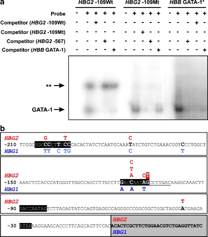
In order to verify the functional significance of the novel HBG2:g.-109G>T mutation, we measured Gγ-globin chain levels, using reverse-phase HPLC. Our results showed that the Gγ/Αγ-globin chain ratio was significantly increased, compared to normal (nonthalassemic) individuals and carriers for the HBB:g.-101C>T mutation (79.2/20.8 versus 40/60, respectively) [17]. Also, we performed EMSA analysis, using nuclear extracts from both MEL cells and human K562 cells, expressing the adult and fetal erythroid program, respectively, in order to assess whether the HBG2:g.-109G>T mutation affects protein binding in vitro. Indeed, EMSA analysis using a 40-bp oligonucleotide bearing the wild-type sequence (G) at position HBG2:g.-109 showed a double-band electrophoretic pattern, suggesting that two proteins and/or protein complexes bind to the oligonucleotide used. Addition of nonradioactive GATA-1 oligonucleotides, both from HBG2 and HBB promoters [15], resulted only in the lower band being efficiently competed, contrary to the upper band that remained visible (Fig. 3a), indicating that the lower band most likely represents GATA-1 binding. This finding was confirmed using GATA-1 antibody, resulting in a supershift of the lower (GATA-1) band (data not shown). EMSA analysis using a 40-bp oligonucleotide bearing the mutant sequence (T) at position HBG2:g.-109 revealed that binding of the protein (or protein complex) in the upper band was abolished. These data indicate that the upper band shift represent protein (or protein complex) binding only to the wild-type sequence at position HBG2:g.-109 (G) and that the HBG2:g.-109G>T transversion inhibits protein binding in vitro. Consistent with previous footprinting experiments [18], these data suggest that the HBG2:g.-109G>T mutation is responsible for the resulting nd-HPFH phenotype.
Fig. 3.
a Electrophoretic mobility shift assays of the oligonucleotides containing either the wild-type (Wt; HBG2:g-109 G) or mutant (Mt; HBG2:g-109 T) sequence using nuclear protein extracts prepared from dimethyl sulfoxide-induced MEL cells. The HBG2 −109Mt oligonucleotide most likely abolishes a NF-E3 binding site in vitro (depicted as two asterisks). Competition with nonlabeled HBG2 −109Wt oligonucleotide results in the disappearance of GATA-1 and NF-E3 band shifts, suggesting that both proteins bind to the HBG2 −109Wt oligonucleotide. Competition with nonlabeled HBG2 −567 or HBB GATA-1 oligonucleotides [15] results in the disappearance only of the GATA-1 (lower) band. Competition with nonlabeled HBG2 −567 or HBB GATA-1 oligonucleotides suggests that the affinity of GATA-1 is stronger to the former oligonucleotide. Use of nuclear protein extracts prepared from K562 cells yielded identical electrophoretic mobility shifts (not shown). The asterisk indicates the oligonucleotide sequence located in the HBB gene promoter region. b Schematic representation of the various nd-HPFH mutations reported to date for the HBG2 (in red) and HBG1 (in blue) globin genes (underlined sequences depict the two small deletions also leading to nd-HPFH). The novel nd-HPFH mutation reported in this study is highlighted in red. Gray box represents the sequences downstream to the transcription initiation site and sequences in solid black boxes represent phylogenetically conserved fetal globin genes cis-regulatory elements
Discussion
In this paper, we report the Hellenic type of nd-HPFH, resulting from a novel G>T transversion in the promoter region of the HBG2 gene. In the Hellenic population, the dominant nd-HPFH is the Greek type (HBG1:g.-117G>A [6]), accounting for almost 90% of the HPFH chromosomes [9]. The Cretan nd-HPFH (HBG1:g.-158C>T [9]) is the second most frequent type, followed by the HBG1:g.-201C>T and the HBG2:g.-196C>T mutations [19]. No deletional HPFH has even been reported for the Hellenic population.
The HBG2:g.-109G>T mutation was found both in heterozygosity as well as in compound heterozygosity with the HBG2:g.-158C>T (XmnI) polymorphism and the silent HBB:g.-101C>T β-thalassemia mutation, the latter being one of the most common HBB mutation leading to β-thalassemia in the Hellenic population [20, 21]. Interestingly, however, the resulting Hb F levels varied in each case. First of all, heterozygosity for the HBG2:g.-109G>T mutation resulted in moderately increased Hb F levels in the index case (4.1%). Also, compound heterozygosity with the silent HBB:g.-101C>T β-thalassemia mutation resulted in substantially increased Hb F levels (14.3%), as expected for compound heterozygous cases for nd-HPFH and β-thalassemia [22]. There are very few examples of nd-HPFH/β-thalassemia compound heterozygous cases, which are extremely rare and only occasionally reported in the literature [22, 23]. On the contrary, compound heterozygosity for the HBG2:g.-109G>T mutation and the HBG2:g.-158C>T polymorphism has no effect in the observed Hb F levels (Fig. 1a). There are two possible explanations for this observation: (a) the HBG2:g.-158C>T polymorphism itself exerts a silencing effect on the HBG2:g.-109G>T mutation or (b) another yet unidentified cis-regulatory element suppresses Hb F production in the sisters of the index case. The latter assumption resembles our recent findings [16] and reaffirms that the regulation of γ-globin gene expression and Hb F production is multifactorial and likely combinatorial and that many genetic factors can putatively impact Hb F production in adults.
The HBG2:g.-109G>T mutation resides at the 3′ end of the distal HBG2 CCAAT box where the CP1 and NF-E3 transcription factors bind [18]. On the basis of in vitro binding studies, GATA-1 and NF-E3 have both been implicated as candidate suppressors of γ-globin gene transcription that would act by binding to the region [18, 24]. In particular, guanosine at position HBG2:g.-109 has been previously shown to be a strong contact point for NF-E3 [24], suggesting that the HBG2:g.-109G>T transversion activates Hb F production most likely by abolishing NF-E3 binding. This assumption is in accordance with our EMSA analysis data indicating that the HBG2:g.-109G>T transversion results in abolishing protein binding in vitro (Fig. 3a). Taking into consideration that the Gγ-globin chain levels were doubled in the index case (I-1; Fig. 1a), these data strongly suggest that the HBG2:g.-109G>T mutation is responsible for the resulting HPFH phenotype.
The novel nd-HPFH mutation described in this paper is located at the 3′ end of the HBG2 gene’s distal CCAAT box, a phylogenetically conserved cis-regulatory element that has been shown to harbor many ubiquitous and erythroid-specific transcription factor binding sites [1]. Nine out of 21 nd-HPFH mutations have been identified in this region (Fig. 3b), suggesting that this regulatory element is critical for γ-globin gene transcription. Contrary, however, to the majority of these nd-HPFH mutations that have a substantial effect in Hb F production, the HBG2:g.-109G>T nd-HPFH mutation per se results in only moderate increase in HbF levels. In addition, the neighboring HBG2:g.-110A>C mutation, leading to the Czech nd-HPFH, also has a negligible effect in HbF production [25]. This is unexpected since the HBG1:g.-117G>A nd-HPFH mutation, residing at the 5′ end of the distal CCAAT box, leads to a substantial increase in γ-globin gene transcription. This can be explained by the fact that (a) different mutations in the distal CCAAT box abolish different transcription factors binding [24], which would exert a differential effect over γ-globin gene reactivation in the adult stage, (b) the HBG1:g.-117G>A and HBG2:g.-109G>T reside on different genes, hence their relative distance from the LCR, being directly proportional to the frequency of LCR/globin gene promoter interactions [26], varies. It is noteworthy that not all nd-HPFH mutations give a reproducible HPFH phenotype in transgenic mice (unpublished data).
Altogether, these data emphasize that γ-globin gene silencing should be the result of a gradual change in the transcription factor environment affecting the transcriptional balance between the HBG1/HBG2 and HBB genes.
Acknowledgements
This work was partly supported by grants from the Greek Ministry of Education (Pythagoras 52211.08) to PK and the Research Promotion Foundation (Cyprus, ΠΔΕ046_02) and the European Commission [International Thalassemia Network (ITHANET) Coordination action 026539] to GPP.
Footnotes
Marios Phylactides and Farzin Pourfarzad contributed equally to this work.
Contributor Information
George P. Patrinos, Phone: +31-10-7043949, FAX: +31-10-7044743, Email: g.patrinos@erasmusmc.nl
Panagoula Kollia, Phone: +30-1-2107274401, FAX: +30-1-2107274318, Email: pankollia@biol.uoa.gr.
References
- 1.Stamatoyannopoulos G, Grosveld F (2001) Hemoglobin switching. In: Stamatoyannopoulos G, Majerus P, Perlmutter R, Varmus H (eds) Molecular basis of blood diseases, 3rd edn. Saunders, Philadelphia, pp 135–182
- 2.Wijgerde M, Grosveld F, Fraser P (1995) Transcription complex stability and chromatin dynamics in vivo. Nature 377:209–213, doi:10.1038/377209a0 [DOI] [PubMed]
- 3.Bollekens JA, Forget BG (1991) Deltabeta thalassemia and hereditary persistence of fetal hemoglobin. Hematol Oncol Clin North Am 5:399–422 [PubMed]
- 4.Swank RA, Stamatoyannopoulos G (1998) Fetal gene reactivation. Curr Opin Genet Dev 8:366–370, doi:10.1016/S0959-437X(98)80095-6 [DOI] [PubMed]
- 5.Hardison RC, Chui DH, Giardine B, Reimer C, Patrinos GP, Anagnou N, Miller W, Wajcman H (2002) HbVar: a relational database of human hemoglobin variants and thalassemia mutations at the globin gene server. Hum Mutat 19:225–233, doi:10.1002/humu.10044 [DOI] [PubMed]
- 6.Gelinas R, Endlich B, Pfeiffer C, Yagi M, Stamatoyannopoulos G (1985) G to A substitution in the distal CCAAT box of the A gamma-globin gene in Greek hereditary persistence of fetal haemoglobin. Nature 313:323–325, doi:10.1038/313323a0 [DOI] [PubMed]
- 7.Berry M, Grosveld F, Dillon N (1992) A single point mutation is the cause of the Greek form of hereditary persistence of fetal haemoglobin. Nature 358:499–502, doi:10.1038/358499a0 [DOI] [PubMed]
- 8.Patrinos GP, Loutradi-Anagnostou A, Papadakis MN (1995) A novel DNA polymorphism of the Agamma globin gene (Agamma-588 A>G) is linked with the XmnI polymorphism (Ggamma-158 C>T). Hemoglobin 19:419–423, doi:10.3109/03630269509005835 [DOI] [PubMed]
- 9.Patrinos GP, Kollia P, Loutradi-Anagnostou A, Loukopoulos D, Papadakis MN (1998) The Cretan type of non-deletional hereditary persistence of fetal hemoglobin [Agamma-158 C>T] results from two independent gene conversion events. Hum Genet 102:629–634, doi:10.1007/s004390050753 [DOI] [PubMed]
- 10.Losekoot M, Fodde R, Hartveld CL, van Heeren H, Giordano PC, Bernini LF (1990) Denaturing gradient gel electrophoresis and direct sequencing of PCR amplified genomic DNA: a rapid and reliable diagnostic approach to beta thalassemia. Br J Haematol 76:269–274, doi:10.1111/j.1365-2141.1990.tb07883.x [DOI] [PubMed]
- 11.Papadakis MN, Papapanagiotou E, Loutradi-Anagnostou A (1997) Scanning method to identify the molecular heterogeneity of the delta-globin gene, especially in delta-thalassemias: detection of three novel mutations in the promoter region of the gene. Hum Mutat 9:465–472, doi:10.1002/(SICI)1098-1004(1997)9:5<465::AID-HUMU14>3.0.CO;2-0 [DOI] [PubMed]
- 12.Tan AS, Quah TC, Low PS, Chong SS (2001) A rapid and reliable 7-deletion multiplex polymerase chain reaction assay for alpha-thalassemia. Blood 98:250–251, doi:10.1182/blood.V98.1.250 [DOI] [PubMed]
- 13.Patrinos GP, van Baal S, Petersen MB, Papadakis MN (2005) Hellenic National Mutation database: a prototype database for mutations leading to inherited disorders in the Hellenic population. Hum Mutat 25:327–333, doi:10.1002/humu.20157 [DOI] [PubMed]
- 14.Papachatzopoulou A, Kaimakis P, Pourfarzad F, Menounos PG, Evangelakou P, Kollia P, Grosveld FG, Patrinos GP (2007) Increased gamma-globin gene expression in beta-thalassemia intermedia patients correlates with a mutation in 3′HS1. Am J Hematol 82:1005–1009, doi:10.1002/ajh.20979 [DOI] [PubMed]
- 15.Luo HY, Mang D, Patrinos GP, Pourfarzad F, Wuc CJY, Eung SH, Rosenfield CG, Daoust PR, Braun A, Grosveld FG, Steinberg MH, Chui DHK (2004) A novel single nucleotide polymorphism (SNP), T>G, in the GATA site at nucleotide (nt) −567 5′ to the Ggamma-globin gene may be associated with elevated Hb F. Blood 104:145a–146a
- 16.Chen Z, Luo HY, Basran RK, Hsu TH, Mang DW, Nuntakarn L, Rosenfield CG, Patrinos GP, Hardison RC, Steinberg MH, Chui DH (2008) A T-to-G transversion at nucleotide −567 upstream of HBG2 in a GATA-1 binding motif is associated with elevated hemoglobin F. Mol Cell Biol 28:4386–4393, doi:10.1128/MCB.00071-08 [DOI] [PMC free article] [PubMed]
- 17.Huisman TH, Harris H, Gravely M, Schroeder WA, Shelton JR, Shelton JB, Evans L (1977) The chemical heterogeneity of fetal hemoglobin in normal newborn infants and in adults. Mol Cell Biochem 17:45–55, doi:10.1007/BF01732554 [DOI] [PubMed]
- 18.Ronchi AE, Bottardi S, Mazzucchelli C, Ottolenghi S, Santoro C (1995) Differential binding of the NFE3 and CP1/NFY transcription factors to the human gamma- and epsilon-globin CCAAT boxes. J Biol Chem 270:21934–21941, doi:10.1074/jbc.270.37.21934 [DOI] [PubMed]
- 19.Tasiopoulou M, Boussiou M, Sinopoulou K, Moraitis G, Loutradi-Anagnostou A, Karababa P (2008) (G)gamma-196 C->T, (A)gamma-201 C->T: two novel mutations in the promoter region of the gamma-globin genes associated with nondeletional hereditary persistence of fetal hemoglobin in Greece. Blood Cells Mol Dis 40:320–322, doi:10.1016/j.bcmd.2007.10.007 [DOI] [PubMed]
- 20.Patrinos GP, Giardine B, Riemer C, Miller W, Chui DH, Anagnou NP, Wajcman H, Hardison RC (2004) Improvements in the HbVar human hemoglobin variants and thalassemia mutations for population and sequence variation studies. Nucleic Acids Res 32:D537–D541, doi:10.1093/nar/gkh006 [DOI] [PMC free article] [PubMed]
- 21.van Baal S, Kaimakis P, Phommarinh M, Koumbi D, Cuppens H, Riccardino F, Macek M Jr, Scriver CR, Patrinos GP (2007) FINDbase: a relational database recording frequencies of genetic defects leading to inherited disorders worldwide. Nucleic Acids Res 35:D690–D695, doi:10.1093/nar/gkl934 [DOI] [PMC free article] [PubMed]
- 22.Papadakis MN, Patrinos GP, Tsaftaridis P, Loutradi-Anagnostou A (2002) A comparative study of Greek non-deletional hereditary persistence of fetal hemoglobin and beta-thalassemia compound heterozygotes. J Mol Med 80:243–247, doi:10.1007/s00109-001-0312-4 [DOI] [PubMed]
- 23.Kollia P, Kalamaras A, Chassanidis C, Samara M, Vamvakopoulos NK, Radmilovic M, Pavlovic S, Papadakis MN, Patrinos GP (2008) Compound heterozygosity for the Cretan type of non-deletional hereditary persistence of fetal hemoglobin and beta-thalassemia or Hb Sabine confirms the functional role of the Agamma-158 C>T mutation in gamma-globin gene transcription. Blood Cells Mol Dis 41:263–264 [DOI] [PubMed]
- 24.Ronchi A, Berry M, Raguz S, Imam A, Yannoutsos N, Ottolenghi S, Grosveld F, Dillon N (1996) Role of the duplicated CCAAT box region in gamma-globin gene regulation and hereditary persistence of fetal haemoglobin. EMBO J 15:143–149 [PMC free article] [PubMed]
- 25.Indrak K, Indrakova J, Kutlar F, Pospisilova D, Sulovska I, Baysal E, Huisman THJ (1991) Compound heterozygosity for a beta0-thalassemia (frameshift codons 38/39; −C) and a nondeletional Swiss type of HPFH (A>C) at NT −110, Ggamma) in a Czechoslovakian family. Ann Hematol 63:111–115, doi:10.1007/BF01707283 [DOI] [PubMed]
- 26.Patrinos GP, de Krom M, de Boer E, Langeveld A, Imam AM, Strouboulis J, de Laat W, Grosveld FG (2004) Multiple interactions between regulatory regions are required to stabilize an active chromatin hub. Genes Dev 18:1495–1509, doi:10.1101/gad.289704 [DOI] [PMC free article] [PubMed]


