Short abstract
Interplay between Rho and Rac controls the invasive behavior of melanoma cells.
Abstract
Genome-wide analysis of regulators of Rho-family small GTPases has identified critical elements that control the morphology and invasive behavior of melanoma cells.
It is important to identify the mechanisms by which tumor cells invade surrounding tissues, in hopes of being able to develop therapies that will improve patient survival. The degradation of the basement membrane and/or degradation of other extracellular matrix protein barriers that may be present in the neighboring connective tissue has been thought to be necessary for tumor cells to invade. Drugs to inhibit an important class of extracellular proteases, the matrix metalloproteases (MMPs), were therefore developed, but a major setback was the limited success of these drugs in prolonging patient survival.
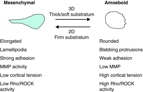
Among the various explanations proposed [1-3], a particularly intriguing one was provided by Wolf and Friedl in 2003 [4]. They found that for some fibrosarcoma (HT1080) and breast cancer (MDA-MB-231) cells, inhibition of MMP activity did not block invasion through three-dimensional tissue, but instead led to a conversion in cell morphology and type of migration. Before treatment the cells typically had an elongated, 'mesenchymal' morphology during invasion, but addition of MMP inhibitors resulted in a rounded, 'amoeboid' morphology - with little difference in the rate of invasion as the amoeboid cells could squeeze though gaps in the matrix. Mesenchymal migration is more fibroblast-like, with elongated cells that have stress fibers and can exert force to restructure the extracellular matrix, whereas amoeboid migration is characterized by round/ellipsoid cells with high cortical tension and low, but significant, adhesion to matrix [5,6]. Mesenchymal cells move through extension of lamellar structures that attach and pull the cell forwards, whereas amoeboid cells tend to produce blebbing protrusions and use cortical contraction to squeeze the cell through spaces in the extracellular matrix [7] (Figure 1).
Figure 1.
Characteristics of the mesenchymal and amoeboid cell phenotypes.
In the same year, Sahai and Marshall [8] extended these results to other tumor types, including melanoma and squamous cell carcinoma, and began the dissection of the signaling pathways that regulate the conversion. Amoeboid behavior was dependent on the presence of a three-dimensional tissue environment and was inhibited by C3 exoenzyme, a toxin that inhibits the A, B and C isoforms of the small GTPase Rho. The Rho family of GTPases, which also includes the Rac and Cdc42 proteins, is involved in controlling many cellular properties, including morphogenesis, cell motility and the organization of the cytoskeleton. The activity of these small GTPases is controlled by guanine-nucleotide exchange factors (GEFs) that promote the active GTP-bound form and by GTPase-activating proteins (GAPs) that favor the inactive GDP-bound form.
As RhoA, B and C are likely to perform distinct functions in migration and invasion [7,9], it will be important to elucidate which of these isoforms contribute to amoeboid-type invasion. Notably, a role for RhoC in amoeboid behavior has been proposed in an elegant study using intravital imaging of cells in zebrafish xenografts [10], with the caveat that overexpression may obscure isoform-specific functions. Further evidence has indicated a role for the serine/threonine kinase ROCK, a Rho effector that is a key regulator of myosin contractility, probably acting by stiffening the cell cortex [11,12]. In a paper published recently in Cell, Sanz-Moreno et al. [13] now provide insight into the roles of Rho and Rac and their interacting GEFs and GAPs in the interconversion of a melanoma cell line between mesenchymal and amoeboid modes.
There are more than 80 Rho GEFs and more than 70 Rho GAPs in the human genome [14-16], a vast excess over the number of different Rho-family GTPases. Although part of this discrepancy can be accounted for by tissue-specific expression of a number of these regulators, it is likely that a significant fraction of them is expressed in any given cell. The reigning hypothesis is that different GEFs mediate specific inputs from a small subset of receptors to the respective GTPases [17]. However, the precise connections between receptors and GEFs remains largely uncharted. To date, even less is known about how GAPs are regulated [16].
Rac and Rho control different morphologies
To identify Rho GEFs and GAPs that control interconvertibility, Sanz-Moreno et al. [13] used small interfering RNAs (siRNAs) to knockdown expression of 83 Rho GEFs in the melanoma cell line A375M2. This line displays a predominantly amoeboid phenotype with a minority of cells that migrate in a mesenchymal fashion. The authors found that depletion of DOCK3, a GEF specific for Rac, or of NEDD9, an adaptor protein of the p130Cas family that binds DOCK3, reduced the fraction of elongated (mesenchymal) cells. Moreover, siRNA-mediated depletion of Rac1 itself also reduced the fraction of elongated cells, while conversely, inhibitors of ROCK or myosin increased the levels of GTP-bound Rac (Rac-GTP) concomitantly with the fraction of elongated cells. Thus, these observations indicate that Rac1 signaling is important for maintaining the mesenchymal mode.
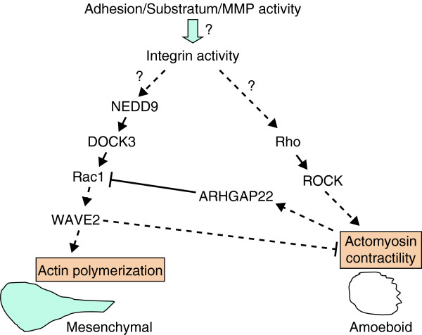
Sanz-Moreno et al. also identified WAVE2, a protein that promotes actin nucleation downstream of Rac, as a critical mediator of the elongated phenotype. Interestingly, depletion of either Rac1 or WAVE2 stimulated actomyosin contractility, evidenced by increased phosphorylation of the regulatory subunit of myosin II. This indicates that Rac, through WAVE2, could promote mesenchymal behavior in a dual fashion - by stimulating actin polymerization and cell protrusion and by restraining myosin contractility (Figure 2). Precisely how WAVE2 negatively regulates contractility remains to be defined. Activated Rac has also been shown to stimulate the activity of p190RhoGAP (which downregulates the activity of Rho isoforms), either by directly binding to it and relieving autoinhibition [18] or by promoting its phosphorylation by tyrosine kinases [19]. This suggests there may be additional mechanisms by which Rac can inhibit the Rho-mediated amoeboid phenotype.
Figure 2.
Signaling control of mesenchymal and amoeboid cell phenotypes. The reciprocal inhibitory relationship between Rac and Rho signaling cascades establishes a bistable switch that controls the mesenchymal and amoeboid phenotypes. Mesenchymal morphology is controlled by a pathway that activates Rac1 via the adaptor protein NEDD9 and the Rac-specific GEF DOCK3. Rac1 activation results in actin polymerization mediated by the actin-nucleation protein WAVE2, which promotes cell elongation. WAVE2 somehow also suppresses actomyosin contractility and, consequently, amoeboid behavior. On the other hand, Rho/ROCK activation stimulates actomysoin contractility, thereby promoting the amoeboid phenotype, and inhibits Rac by activating the Rac-specific GAP, ARHGAP22. Presumably both Rac1 and Rho activation are ultimately controlled by integrin activity, but precisely how the extracellular environment favors either Rac or Rho signaling remains to be resolved. Solid arrows, direct connections; dashed arrows, indirect connections.
To elucidate how ROCK signaling suppresses Rac activation, Sanz-Moreno et al. screened an siRNA library targeting 72 Rho-family GAPs. They identified one GAP, ARHGAP22, whose silencing led to increased numbers of elongated cells and increased the levels of Rac-GTP. If ARHGAP22 was depleted, the reduction in Rac-GTP levels by ROCK activation was blocked, indicating that ARHGAP22 did indeed mediate a Rho/ROCK-driven negative input to Rac. Thus, increased Rho signaling, via the Rho effector ROCK, might not only promote amoeboid behavior directly, but also actively restrain mesenchymal-type movement by activating ARHGAP22, in a manner yet to be elucidated. This reciprocal inhibitory relationship between the Rac and Rho GTPases could form a bistable switch that reinforces selection of one mode of migration over the other.
Metastasis is a multistep process that requires the adaptability of malignant cells to different microenvironments. Sanz-Moreno et al. also employed intravital imaging of GFP-expressing melanoma cells in subcutaneous xenografts, powerfully illustrating this adaptability and emphasizing the importance of examining migration mechanisms in live animals. Whereas in the core of the tumor, melanoma cells predominantly moved in an elongated/mesenchymal fashion, cells at the tumor edge migrated into the matrix in a rounded/amoeboid manner. Suppression of ARHGAP22 activity increased the proportion of elongated cells, consistent with the in vitro studies indicating that this should increase Rac activation. Treatment with the ROCK inhibitor Y27632 also reduced amoeboid movement and increased the number of cells moving in the mesenchymal mode within the tumor. Intriguingly, the increased mesenchymal movement did not occur out in the matrix, only in the tumor interior.
A confounding feature of cancer is heterogeneity among tumors (even within a specific type such as melanoma), and so an important question is the relevance of studies focusing on one, or even two, lines for the disease as a whole. To address this issue, the authors evaluated 11 melanoma cell lines in total, and found that silencing of ARHGAP22 expression led to increased proportions of elongated cells in most cases, whereas silencing of DOCK3 or NEDD9 reduced the proportions of elongated cells. This evaluation of a set of melanoma cell lines makes a strong case for the importance of the biaxial signaling network identified in the control of melanoma cell morphology and migration, with the caveat that cell shape rather than motility was used as a readout.
Driving forward
The study of Sanz-Moreno et al. [13] suggests a model in which Rac activity promotes mesenchymal migration and RhoA promotes amoeboid migration (Figure 2). Although mutual antagonism between the Rac and Rho GTPases has been observed in many other cellular settings [20,21], a critical question is precisely how this antagonistic relationship becomes integrated in coordinated cell behavior. One instance is the initiation of epithelial cell-cell adhesion, where rounds of activation and de-activation of Rac at the contacting membrane lead to expansion of the contact zone between cells [22]. Another example is the neuronal growth cone, where an appropriate level of Rac activity is required for persistent lamellar protrusion and neurite outgrowth [23]. For melanoma, Sanz-Moreno et al. identify mutual suppression mechanisms mediated by ARHGAP22 and WAVE2 function that can help maintain the cell in a single mode for effective movement. The details of how ARHGAP22 is activated and how WAVE2 leads to Rho suppression are areas for further study.
On an operational level, these studies indicate an approach to testing whether motility in vivo can be interpreted in terms of mesenchymal versus amoeboid modes of invasion. Treatment with ROCK inhibitors such as Y27632 can be used to inhibit amoeboid motility while maintaining or increasing the proportion of cells that are invading in the mesenchymal mode. Conversely, inhibitors of Rac or MMPs can be used to inhibit cells migrating in the mesenchymal mode and possibly increase migration in the amoeboid mode. Intravital time-lapse imaging will help distinguish between simply morphological alterations and changes in real invasive properties. Treatment of tumors with these or related inhibitors provides an opportunity to evaluate in vivo the contributions of mesenchymal and amoeboid motility to tumor cell invasion and metastasis.
An important consequence of the plasticity of the invasive behavior of tumor cells is that interfering with elements that control either migration mode would allow cells to switch to a different mode of invasion and consequently may have little effect on the extent of cell invasion. In line with previous observations [8], Sanz-Moreno et al. show that simultaneous blocking of both modes of migration diminishes this opportunistic behavior and significantly inhibits invasion. These findings have important therapeutic implications, indicating the potential clinical benefit of combinations of drugs targeting the respective modes of invasion.
An intriguing question that remains to be addressed is what are the upstream signals that lead to activation of Rac, thereby driving mesenchymal motility or activation of RhoA for amoeboid motility. There are three general conditions that we know of that contribute to the selection of the mesenchymal or amoeboid mode: MMP activity levels; rigidity of the substratum; and level of integrin activity. Inhibition of MMPs, reduced matrix rigidity, or reduced levels of integrin activity result in amoeboid migration. The challenge will be to construct a unifying model that integrates these features (Figure 2). Only then will it be possible to connect this intricate signaling network to the tumor microenvironment that is driving it.
Acknowledgments
Acknowledgements
MS and JES acknowledge financial support from the NIH (CA87567and NS060023 to MS and CA77522 and CA100324 to JES). JES is the Betty and Sheldon Feinberg Senior Faculty Scholar in Cancer Research. We thank P Friedl and R Ruggieri for critical reading of the manuscript and helpful comments.
Contributor Information
Marc Symons, Email: msymons@nshs.edu.
Jeffrey E Segall, Email: segall@aecom.yu.edu.
References
- Martin MD, Matrisian LM. The other side of MMPs: protective roles in tumor progression. Cancer Metastasis Rev. 2007;26:717–724. doi: 10.1007/s10555-007-9089-4. [DOI] [PubMed] [Google Scholar]
- Overall CM, Kleifeld O. Towards third generation matrix metalloproteinase inhibitors for cancer therapy. Br J Cancer. 2006;94:941–946. doi: 10.1038/sj.bjc.6603043. [DOI] [PMC free article] [PubMed] [Google Scholar]
- Pavlaki M, Zucker S. Matrix metalloproteinase inhibitors (MMPIs): the beginning of phase I or the termination of phase III clinical trials. Cancer Metastasis Rev. 2003;22:177–203. doi: 10.1023/A:1023047431869. [DOI] [PubMed] [Google Scholar]
- Wolf K, Mazo I, Leung H, Engelke K, von Andrian UH, Deryugina EI, Strongin AY, Brocker EB, Friedl P. Compensation mechanism in tumor cell migration: mesenchymalamoeboid transition after blocking of pericellular proteolysis. J Cell Biol. 2003;160:267–277. doi: 10.1083/jcb.200209006. [DOI] [PMC free article] [PubMed] [Google Scholar]
- Wolf K, Friedl P. Molecular mechanisms of cancer cell invasion and plasticity. Br J Dermatol. 2006;154(Suppl 1):11–15. doi: 10.1111/j.1365-2133.2006.07231.x. [DOI] [PubMed] [Google Scholar]
- Friedl P, Wolf K. Tumour-cell invasion and migration: diversity and escape mechanisms. Nat Rev Cancer. 2003;3:362–374. doi: 10.1038/nrc1075. [DOI] [PubMed] [Google Scholar]
- Simpson KJ, Dugan AS, Mercurio AM. Functional analysis of the contribution of RhoA and RhoC GTPases to invasive breast carcinoma. Cancer Res. 2004;64:8694–8701. doi: 10.1158/0008-5472.CAN-04-2247. [DOI] [PubMed] [Google Scholar]
- Sahai E, Marshall CJ. Differing modes of tumour cell invasion have distinct requirements for Rho/ROCK signalling and extracellular proteolysis. Nat Cell Biol. 2003;5:711–719. doi: 10.1038/ncb1019. [DOI] [PubMed] [Google Scholar]
- Wheeler AP, Ridley AJ. RhoB affects macrophage adhesion, integrin expression and migration. Exp Cell Res. 2007;313:3505–3516. doi: 10.1016/j.yexcr.2007.07.014. [DOI] [PubMed] [Google Scholar]
- Stoletov K, Montel V, Lester RD, Gonias SL, Klemke R. High-resolution imaging of the dynamic tumor cell vascular interface in transparent zebrafish. Proc Natl Acad Sci USA. 2007;104:17406–17411. doi: 10.1073/pnas.0703446104. [DOI] [PMC free article] [PubMed] [Google Scholar]
- Wilkinson S, Paterson HF, Marshall CJ. Cdc42-MRCK and Rho-ROCK signalling cooperate in myosin phosphorylation and cell invasion. Nat Cell Biol. 2005;7:255–261. doi: 10.1038/ncb1230. [DOI] [PubMed] [Google Scholar]
- Fackler OT, Grosse R. Cell motility through plasma membrane blebbing. J Cell Biol. 2008;181:879–884. doi: 10.1083/jcb.200802081. [DOI] [PMC free article] [PubMed] [Google Scholar]
- Sanz-Moreno V, Gadea G, Ahn J, Paterson H, Marra P, Pinner S, Sahai E, Marshal CJ. Rac activation and inactivation control plasticity of tumor cell movement. Cell. 2008;135:510–523. doi: 10.1016/j.cell.2008.09.043. [DOI] [PubMed] [Google Scholar]
- Rossman KL, Der CJ, Sondek J. GEF means go: turning on RHO GTPases with guanine nucleotide-exchange factors. Nat Rev Mol Cell Biol. 2005;6:167–180. doi: 10.1038/nrm1587. [DOI] [PubMed] [Google Scholar]
- Moon SY, Zheng Y. Rho GTPase-activating proteins in cell regulation. Trends Cell Biol. 2003;13:13–22. doi: 10.1016/S0962-8924(02)00004-1. [DOI] [PubMed] [Google Scholar]
- Bos JL, Rehmann H, Wittinghofer A. GEFs and GAPs: critical elements in the control of small G proteins. Cell. 2007;129:865–877. doi: 10.1016/j.cell.2007.05.018. [DOI] [PubMed] [Google Scholar]
- Schiller MR. Coupling receptor tyrosine kinases to Rho GTPases - GEFs what's the link. Cell Signal. 2006;18:1834–1843. doi: 10.1016/j.cellsig.2006.01.022. [DOI] [PubMed] [Google Scholar]
- Bustos RI, Forget MA, Settleman JE, Hansen SH. Coordination of Rho and Rac GTPase function via p190B RhoGAP. Curr Biol. 2008;18:1606–1611. doi: 10.1016/j.cub.2008.09.019. [DOI] [PMC free article] [PubMed] [Google Scholar]
- Nimnual AS, Taylor LJ, Bar-Sagi D. Redox-dependent downregulation of Rho by Rac. Nat Cell Biol. 2003;5:236–241. doi: 10.1038/ncb938. [DOI] [PubMed] [Google Scholar]
- Burridge K, Wennerberg K. Rho and Rac take center stage. Cell. 2004;116:167–179. doi: 10.1016/S0092-8674(04)00003-0. [DOI] [PubMed] [Google Scholar]
- Koh CG. Rho GTPases and their regulators in neuronal functions and development. Neurosignals. 2006;15:228–237. doi: 10.1159/000101527. [DOI] [PubMed] [Google Scholar]
- Yamada S, Nelson WJ. Localized zones of Rho and Rac activities drive initiation and expansion of epithelial cell-cell adhesion. J Cell Biol. 2007;178:517–527. doi: 10.1083/jcb.200701058. [DOI] [PMC free article] [PubMed] [Google Scholar]
- Woo S, Gomez TM. Rac1 and RhoA promote neurite outgrowth through formation and stabilization of growth cone point contacts. J Neurosci. 2006;26:1418–1428. doi: 10.1523/JNEUROSCI.4209-05.2006. [DOI] [PMC free article] [PubMed] [Google Scholar]


