

Abstract
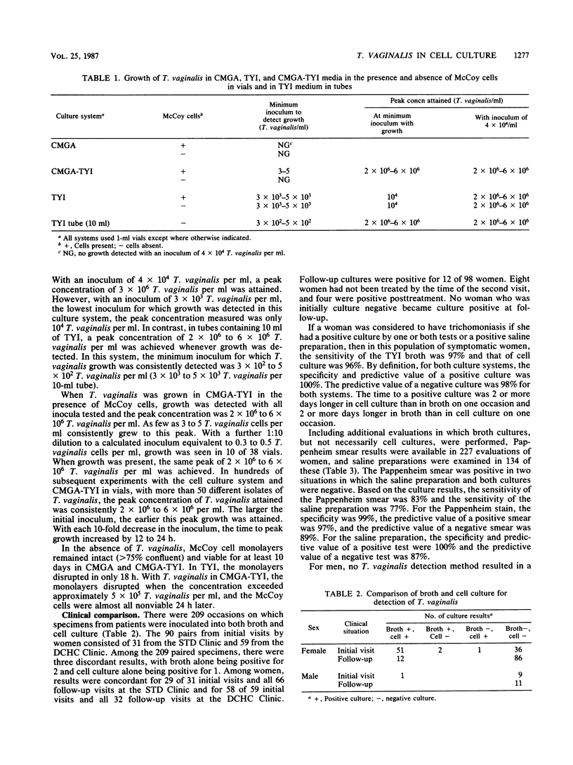
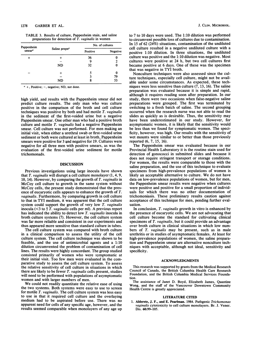
Trichomonas vaginalis can be grown in cell culture. We studied the growth kinetics of T. vaginalis in McCoy cell culture compared with that in a conventional broth medium (Diamond TYI-S-33 medium supplemented with 10% heat-inactivated bovine serum [TYI]). In the presence of McCoy cells and two parts cell culture medium to one part TYI, a peak concentration of 2 X 10(6) to 6 X 10(6) T. vaginalis per ml was consistently achieved with inocula as low as three T. vaginalis cells per ml. Without cells, this medium did not support growth of T. vaginalis. T. vaginalis in TYI in 1-ml vials with or without McCoy cells demonstrated poor growth. In tubes containing 10 ml of TYI, inocula grew to 2 X 10(6) to 6 X 10(6) T. vaginalis per ml, but at least 3 X 10(3) T. vaginalis per tube was required to initiate growth. Thus, in vitro, cell culture was more sensitive than TYI broth in detecting low numbers of T. vaginalis. In a subsequent clinical comparison of broth and cell culture for isolation of T. vaginalis from 188 vaginal specimens and 21 urethral specimens from men, the results were in agreement for 206 specimens (98.6%). There were no situations in which culture was negative and a saline preparation showed motile trichomonads. For women, using a positive culture as the indicator of true positivity, the sensitivity of detection of T. vaginalis was 83% with the Pappenheim stain and 77% with saline preparations. These studies show that cell culture can be used for isolation of T. vaginalis from clinical specimens; it gave results comparable to those of broth culture for the group of mainly symptomatic women. Further studies should be performed to determine its utility in clinical populations such as asymptomatic women and men with and without symptoms, in which T. vaginalis is more likely to be present in low numbers.
Full text
PDF

Selected References
These references are in PubMed. This may not be the complete list of references from this article.
- Alderete J. F., Pearlman E. Pathogenic Trichomonas vaginalis cytotoxicity to cell culture monolayers. Br J Vener Dis. 1984 Apr;60(2):99–105. doi: 10.1136/sti.60.2.99. [DOI] [PMC free article] [PubMed] [Google Scholar]
- Brown M. T. Trichomoniasis. Practitioner. 1972 Nov;209(253):639–644. [PubMed] [Google Scholar]
- CHRISTIAN R. T., MILLER N. F., LUDOVICI P. P., RILEY G. M. A study of Trichomonas vaginalis in human cell culture. Am J Obstet Gynecol. 1963 Apr 1;85:947–954. doi: 10.1016/s0002-9378(16)35599-5. [DOI] [PubMed] [Google Scholar]
- Catterall R. D. Trichomonal infections of the genital tract. Med Clin North Am. 1972 Sep;56(5):1203–1209. doi: 10.1016/s0025-7125(16)32345-8. [DOI] [PubMed] [Google Scholar]
- Diamond L. S., Harlow D. R., Cunnick C. C. A new medium for the axenic cultivation of Entamoeba histolytica and other Entamoeba. Trans R Soc Trop Med Hyg. 1978;72(4):431–432. doi: 10.1016/0035-9203(78)90144-x. [DOI] [PubMed] [Google Scholar]
- Fleury F. J. Diagnosis of Trichomonas vaginalis infection. JAMA. 1979 Dec 7;242(23):2556–2557. [PubMed] [Google Scholar]
- Fouts A. C., Kraus S. J. Trichomonas vaginalis: reevaluation of its clinical presentation and laboratory diagnosis. J Infect Dis. 1980 Feb;141(2):137–143. doi: 10.1093/infdis/141.2.137. [DOI] [PubMed] [Google Scholar]
- Garber G. E., Proctor E. M., Bowie W. R. Immunogenic proteins of Trichomonas vaginalis as demonstrated by the immunoblot technique. Infect Immun. 1986 Jan;51(1):250–253. doi: 10.1128/iai.51.1.250-253.1986. [DOI] [PMC free article] [PubMed] [Google Scholar]
- Gentry G. A., Lawrence N., Lushbaugh W. Isolation and differentiation of herpes simplex virus and Trichomonas vaginalis in cell culture. J Clin Microbiol. 1985 Aug;22(2):199–204. doi: 10.1128/jcm.22.2.199-204.1985. [DOI] [PMC free article] [PubMed] [Google Scholar]
- Heath J. P. Behaviour and pathogenicity of Trichomonas vaginalis in epithelial cell cultures: a study by light and scanning electron microscopy. Br J Vener Dis. 1981 Apr;57(2):106–117. doi: 10.1136/sti.57.2.106. [DOI] [PMC free article] [PubMed] [Google Scholar]
- Krieger J. N. Urologic aspects of trichomoniasis. Invest Urol. 1981 May;18(8):411–417. [PubMed] [Google Scholar]
- McCann J. S. Comparison of direct microscopy and culture in the diagnosis of trichomoniasis. Br J Vener Dis. 1974 Dec;50(6):450–452. doi: 10.1136/sti.50.6.450. [DOI] [PMC free article] [PubMed] [Google Scholar]
- Pindak F. F., Gardner W. A., Jr, Mora de Pindak M. Growth and cytopathogenicity of Trichomonas vaginalis in tissue cultures. J Clin Microbiol. 1986 Apr;23(4):672–678. doi: 10.1128/jcm.23.4.672-678.1986. [DOI] [PMC free article] [PubMed] [Google Scholar]
- Rothenberg R. B., Simon R., Chipperfield E., Catterall R. D. Efficacy of selected diagnostic tests for sexually transmitted diseases. JAMA. 1976 Jan 5;235(1):49–51. [PubMed] [Google Scholar]
- Spence M. R., Hollander D. H., Smith J., McCaig L., Sewell D., Brockman M. The clinical and laboratory diagnosis of Trichomonas vaginalis infection. Sex Transm Dis. 1980 Oct-Dec;7(4):168–171. doi: 10.1097/00007435-198010000-00004. [DOI] [PubMed] [Google Scholar]