Abstract
We present two rare cases of anomalous vertebral artery (VA) with retroesophageal right subclavian artery. One patient had a right VA arising from the right common carotid artery (CCA), and a left VA originating from the third branch off the aorta. Both VAs ascended anteriorly to the transverse foramen of C5 to C6 vertebra and entered the transverse foramen of C4. The other patient had a right VA arising from the right CCA and entering the transverse foramen of C5. The presence of anomalous variations of the origin and course of vertebral artery might have serious implications in angiographic and surgical procedures, and it is of great importance to be aware of such a possibility.
Keywords: Subclavian artery, Vertebral artery, Variations
INTRODUCTION
The advances in and popularity of the endovascular intervention in recent years has required more accurate knowledge and a greater understanding of the variation in great vessels5,6). Anomalous variation in the vertebral artery (VA) is regarded as an embryonic maldevelopment, and its incidence is low. We present two rare cases of anomalous origin and anomalous cervical course of VA with retroesophageal right subclavian artery (RERSA); one was bilateral VA variation, the other was unilateral. The literature on the variations of VAs is reviewed and the possible embryonic development of these variational patterns and their clinical importance is discussed briefly.
CASE REPORT
Case 1
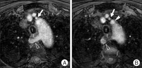
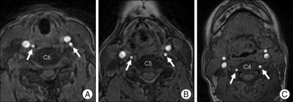
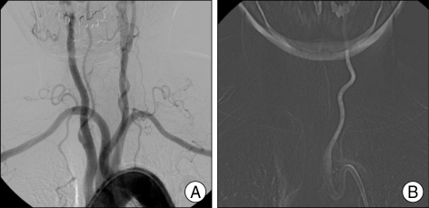
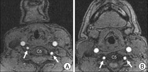
A 71-year-old woman presented for evaluation of her headache. She had been treated for hypertension for several years. She had no neurological deficits. Brain magnetic resonance (MR) imaging and angiography showed a cerebral aneurysm at the right paraclinoid portion. Source images of time of flight MR angiography showed the origin and course of the right SA running behind the esophagus upward and to the right toward the upper extremity, and the left VA branches off before the left SA originating from the aortic arch (Fig. 1). They also demonstrated that both VAs ascended anteriorly to the transverse foramen of C5 to C6 vertebra and entered the transverse foramen of C4 (Fig. 2). Diagnostic cerebral angiography and aortography were performed. In aortogram, the right common carotid artery (CCA) originated from the aorta as the first branch, and the right VA originated from the right CCA. The left VA originated from the aorta as the third branch, and the right SA originated on the left side of the aortic arch as the fifth branch (Fig. 3). Cerebral angiography confirmed an aneurysm of the right posterior communicating artery. The patient underwent coil embolization under general anesthesia and was discharged without complications.
Fig. 1.
A : Axial source images of time of flight magnetic resonance angiography showing that the right subclavian artery (SA) (open arrowhead) originates on the left side of the aortic arch and runs behind the esophagus. B : The left vertebral artery (arrow) branches off before the left SA (arrowhead) originating from the aortic arch. T : trachea, E : esophagus.
Fig. 2.
Axial source images of time of flight magnetic resonance angiography showing both vertebral arteries (VAs). Both VAs (arrows) ascend anteriorly to the transverse foramen of C6 (A) to C5 (B) vertebra and enter the transverse foramen of C4 (C).
Fig. 3.
A : Aortogram showing the right vertebral artery (VA) originating from the right common carotid artery and the right subclavian artery originating on the left side of the aortic arch. B : The left VA originates from the aortic arch as the third branch.
Case 2
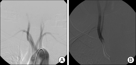
A-52-year-old women presented for dizziness evaluation. Brain MR imaging and angiography revealed hypervascular mass in the right cerebellopontine angle (CPA). Diagnostic cerebral angiography confirmed an arteriovenous malformation in the right CPA, with a feeding artery of the anterior inferior cerebellar artery and a draining vein of the superior petrosal sinus. Aortogram and cerebral angiography demonstrated that the right SA originated as the last branch of the aortic arch and the right VA originated from the right CCA (Fig. 4). Source images of MR angiography showed the right VA entering the transverse foramen of C5 (Fig. 5). But, the origin and course the left VA was not anomalous. The patient underwent gamma knife surgery and was discharged.
Fig. 4.
Aortogram showing the right subclavian artery originating on the left side of the aortic arch (A) and the right vertebral artery originating from the right commom carotid artery (B). The right subclavian artery runs behind the esophagus upward and to the right toward the upper extremity.
Fig. 5.
Axial source images of time of flight magnetic resonance angiography showing the right vertebral artery (VA) ascending anteriorly to the transverse foramen of C6 vertebra (A) and entering the transverse foramen of C5 (B). Arrows indicate both VAs.
DISCUSSION
A RERSA, also called an aberrant right SA, is a relatively common congenital anomaly of the aortic arch with an incidence of 0.5%. This variation is due to interruption of the fourth right aortic arch between the notches for the CCA and SA while the left fourth arch remains intact. A regression of the proximal portion of the right SA proximal portion occurs, and the retroesophageal aortic arch persists1).
In the case of RERSA, the right VA may originate from the right CCA4). Tsai et al.8) analyzed the pattern and prevalence of VA anomalies in 102 patients with RERSA. They reported that 13.7% of RERSAs had right VAs that originated from the right CCA, and 28.6% of them had a concomitant left VA anomaly, that is, the left VA originated as the third branch off the aortic arch. The incidence of anomalous origin of both VAs with RERSA, as seen in case 1, is extremely low.
Embryologically, the cervical segment of VA is formed by longitudinal anastomoses between the seven dorsal segmental arteries. The seventh segmental artery persists to form the VA and a part of SA, and the proximal part of the sixth dorsal segmental artery entering into the anastomosis disappears. When the right sixth dorsal segmental artery persists, the right VA will arise from the right CCA4). Furthermore, persistence of the first or second dorsal intersegmental artery to the fourth left aortic arch could result in the origination of the left VA from the aortic arch6).
In the cervical course of the VA, the VA generally enters the transverse foramen of the C6 vertebra and ascends through the transverse foramen. The other striking finding in our cases is that both VAs enter the transverse foramen of the C4 vertebra in case 1, and the right VA enters the C5 transverse foramen in case 2. Rieger and Huber3) reported that the VA enters the transverse foramen at C4 in 0.5% of cases and at C5 in 6.6%. Most anomalous courses of the VA are unilateral, and a bilateral anomaly, as seen in case 1, is very rare.
The anatomic variations of the VA are significant for diagnostic and surgical procedures in the head and neck regions2). Some authors suggested that anomalous origin of VA may lead to intracerebral disorders by altering the vascular hemodynamics, thereby placing patients at greater risk of thrombosis, aneurysm, occlusion, arterial dissection, and, potentially atherosclerosis2,6,7).
CONCLUSION
Two rare cases of anomalous origin and anomalous course of VA combined with RERSA were presented. In angiography, endovascular surgery, and head and neck surgery, including that of the cervical spine, it is important to know and to understand uncommon anomalous variation of the VAs, since this can allow physicians to avoid accidental damage to the VA during these procedures. Furthermore, it can aid their understanding of hemodynamics, save time, and conserve contrast media during angiographic catheterization2,8).
References
- 1.Fazan VPS, Ribeiro RA, Ribeiro JAS, Filho OAR. Right retroesophageal subclavian artery. Acta Cir Bras. 2003;18(Suppl 5):54–56. [Google Scholar]
- 2.Gluncic V, Ivikic G, Marin D, Percac S. Anomalous origin of both vertebral arteries. Clin Anat. 1999;12:281–284. doi: 10.1002/(SICI)1098-2353(1999)12:4<281::AID-CA8>3.0.CO;2-6. [DOI] [PubMed] [Google Scholar]
- 3.Rieger P, Huber G. Fenestration and duplicate origin of the left vertebral artery in angiography. Neuroradiology. 1983;25:45–50. doi: 10.1007/BF00327480. [DOI] [PubMed] [Google Scholar]
- 4.Roszel AJ, Kiely ML. A retroesophageal right subclavian artery with the right vertebral artery originating from the right common carotid artery. Clin Anat. 1991;4:373–379. [Google Scholar]
- 5.Sanudo J, Vazquez R, Puerta J. Meaning and clinical interest of the anatomical variations in the 21st century. Eur J Anat. 2003;7(Suppl 1):1–3. [Google Scholar]
- 6.Satti SR, Cerniglia CA, Koenigsberg RA. Cervical vertebral artery variations : an anatomic study. AJNR Am J Neuroradiol. 2007;28:976–980. [PMC free article] [PubMed] [Google Scholar]
- 7.Shoja MM, Tubbs RS, Khaki AA, Shokouhi G, Farahani RM, Moein A. A rare variation of the vertebral artery. Folia Morphol (Warsz) 2006;65:167–170. [PubMed] [Google Scholar]
- 8.Tsai IC, Tzeng WS, Lee T, Jan SL, Fu YC, Chen MC, et al. Vertebral and carotid artery anomalies in patients with aberrant right subclavian arteries. Pediatr Radiol. 2007;37:1007–1012. doi: 10.1007/s00247-007-0574-2. [DOI] [PubMed] [Google Scholar]


