Abstract
Pimecrolimus cream 1% has shown to be effective in patients with a variety of inflammatory cutaneous disorders. And it might be a useful modality in the treatment of seborrheic dermatitis. This prospective study was aimed at assessing the efficacy and tolerability of pimecrolimus cream 1% in the treatment of facial seborrheic dermatitis. Twenty patients were instructed to apply pimecrolimus cream 1% for 4 consecutive weeks. Assessment of the disease severity was performed at baseline and at week 1, 2, and 4. Clinical assessments of erythema, scaling, and pruritus were measured using a 4-point scale (0-3). Global assessments of the disease severity by patients and investigators were performed at each visit. Mean clinical scores of erythema, scaling, and pruritus significantly improved by 87.4%, 91.9%, and 91.5% respectively at week 4 (p<0.001). Improvements in the global assessment of disease severity determined by patients and investigators also showed excellent results. No specific adverse events other than transient burning and tingling sensations were noted. The relapse of facial seborrheic dermatitis was mostly observed between 3 to 8 weeks after the discontinuation of pimecrolimus. We suggest that the topical application of pimecrolimus cream 1% can be an effective and safe alternative for treatment of facial seborrheic dermatitis.
Keywords: Dermatitis, Seborrheic; Pimecrolimus
INTRODUCTION
Seborrheic dermatitis is a very common papulosquamous dermatoses, affecting over 5% of the general population with male predilections (1). This disease is characterized by mild to severe erythema, often accompanied by oily thick scales and mild inflammations. It typically affects areas containing sebaceous glands, particularly the face, ears, scalp, and upper part of the trunk (2).
Since seborrheic dermatitis commonly has a chronic and relapsing course with no permanent cure, various types of treatment have been introduced. The mainstays of treating facial seborrheic dermatitis rely on topical corticosteroids, antifungals, or a combination of both (3, 4). Although topical steroids rapidly reduce the severity of the disease by anti-inflammatory action, side effects associated with prolonged application and early relapse after discontinuation of treatment limit its wide usage (5). Topical antifungal agents are also frequently used in order to decrease the colonization of lipophylic yeasts, Malassezia (Pityrosporum ovale), but they show a relatively slow onset of the anti-inflammatory effect compared to topical steroids (4, 6).
Pimecrolimus cream 1% is a new topical macrolactam immunomodulator widely used for the treatment of atopic dermatitis via inhibition of T-cell activation and proinflammatory cytokine production (7). The cream has also shown to be effective in the treatment of other inflammatory skin disorders. Crutchfield (8) suggested that pimecrolimus cream 1% could be used as an effective treatment for facial seborrheic dermatitis. Rigopoulos et al. (9) conducted a randomized open-label clinical trial with pimecrolimus cream 1% and betamethasone 17-valerate 0.1% cream, and reported that pimecrolimus may be an excellent alternative modality for treating seborrheic dermatitis. We herein add our experience of rapid and sustained therapeutic response of pimecrolimus cream 1% in the treatment of facial seborrheic dermatitis.
MATERIALS AND METHODS
This study was a 4-week, open-label, clinical trial to investigate the efficacy of pimecrolimus cream 1% for the treatment of facial seborrheic dermatitis. It was conducted after the notice of purpose and obtaining signed informed consent from all subjects.
A total of 20 patients with a clinical diagnosis of facial seborrheic dermatitis were enrolled. They composed of 16 males and 4 females with a mean age of 47.9 yr (range, 22-79) and a mean duration of 3.8 yr (range, 1-13) of dermatitis. Patients who had taken topical or systemic treatment of corticosteroids, retinoids, or immune-modulating agents within 28 days prior to the study were excluded. Patients with any significant medical condition that could compromise immune responsiveness (lymphoma, acquired immunodefiuency syndrome, other immunodeficiency disorders, or a history of malignant diseases) were excluded. Exclusion criteria also included pregnancy, lactating women, and patients with other facial dermatoses. Enrolled patients were instructed to stop using systemic and topical antimicrobials and antifungal agents including tar, zinc pyrithione, or selenium sulfate for 14 days before starting the treatment, but the uas of non-medicated shampoo and facial cleansers were allowed the use of the study. Moreover, they were advised to minimize or avoid natural or artificial sunlight. Patients were informed to apply pimecrolimus cream 1% (Norvatis, Basel, Switzerland) to affected facial areas only in thin layers, twice daily for 4 consecutive weeks. If complete resolution of seborrheic dermatitis occurred early in the study, patients were instructed to use pimecrolimus cream for an additional week.
The evaluation of patients were performed at baseline and at week 1, 2, and 4 for the severity of seborrheic dermatitis. Four different sites (forehead/eyebrows, nasolabial folds, perioral areas/chin, and posterior aspect of the ears) were clinically assessed for erythema, scaling, and pruritus using a 4-point scale (0-3): 0, absent; 1, mild; 2, moderate; and 3, severe. Scores of each clinical symptom and sign at the baseline were compared to those at subsequent visits to assess the efficacy of pimecrolimus cream. In addition, patients themselves and physicians clinically performed the global assessments of the severity of seborrheic dermatitis at each visit as: completely clear (no evidence of seborrheic dermatitis); almost clear (>90% of clearance); marked improvement (75-90% of clearance); moderate improvement (50-75% of clearance); slight improvement (25-50% of clearance); no change compared with baseline; or worse compared with baseline. Local adverse reactions such as burning sensation, tingling, pain or soreness, or signs of viral or bacterial infections were recorded throughout the study as none, mild, moderate, or severe. A follow-up survey for recurrence of any erythema, scalings, or pruritus after the discontinuation of pimecrolimus was done by telephone. To assess the efficacy of pimecrolimus, data from scales of erythema, scaling, and pruritus were statistically analyzed by means of a repeated-measures analysis of variance (ANOVA) model.
RESULTS
The study patients had commonly been managed with topical corticosteroids, whereas only 1 patient had had a history of using a topical antifungal agent before visiting our clinic. Topical pimecrolimus was the first-line of therapy in 5 patients.
The 4-week duration of clinical study with pimecrolimus cream 1% was completed in 16 patients, whereas 4 patients discontinued treatment before the completion of the study by the reason of early resolution with their symptoms and signs.
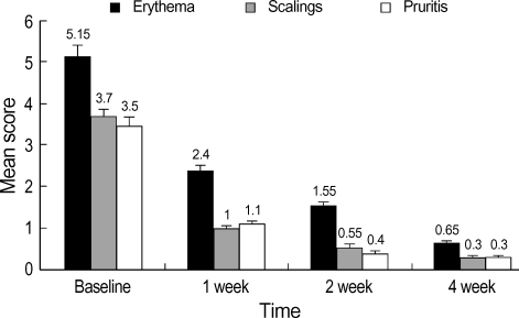
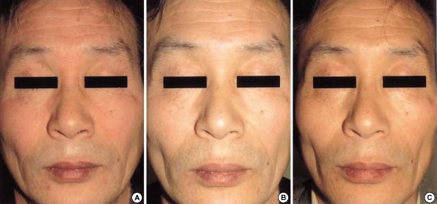
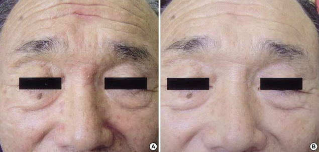
At the baseline, all patients had erythema, with a mean grade of 5.15; 19 (95%) patients had scaling, with a mean grade of 3.70; and 17 (85%) patients had pruritus, with a mean grade of 4.12. Fig. 1 shows the significant decrements in the mean severity scores of each clinical feature during the 4-week application of pimecrolimus cream 1%, and the most prominent reduction of the disease severity was observed at the end of the first week. At week 1, the mean score of scaling improved most rapidly at 73.0% compared to the mean baseline score. This improvement continued over the following week at 85.1%, and the overall scaling score showed a mean reduction of 91.9% at week 4 (p<0.001). Similar decrements of mean scores were also observed with erythema (p<0.001) and pruritus (p<0.001) at reductions of 53.4%, and 68.7% at week 1, 69.9% and 88.6% at week 2, and 87.4% and 91.5% at week 4 compared to the mean baseline scores, respectively. These changes were statistically significant (p<0.001) and clinically well correlated with photographs (Fig. 2, 3).
Fig. 1.
Improvements of mean scores of erythema, scaling, and pruritus in patients with seborrheic dermatitis (p<0.001).
Fig. 2.
Clinical features illustrating the significant improvement of seborrheic dermatitis at baseline (A), 1 week (B), and 4 weeks (C) after treatment with pimecrolimus cream 1%.
Fig. 3.
Clinical features illustrating the significant improvement of seborrheic dermatitis at baseline (A) and 2 weeks (B) after treatment with pimecrolimus cream 1%.
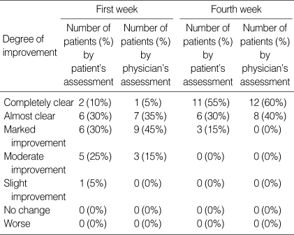
Global improvement of severity assessed by investigators and patients at the week 1 and 4 of treatment are shown in Table 1. Similar to the symptoms and signs, the patients' subjective response to pimecrolimus was mostly significant at the end of the first week. Investigators noticed above the moderate improvement of severity in all patients after the first week of treatment. At week 4, 3 patients (15%) reported marked improvement, 6 (30%) almost clear, and 11 (55%) complete clear of the dermatitis. From physician's point of view, 12 subjects (60%) including 4 prematurely discontinued patients experienced a complete clearance of clinical signs and symptoms within 4 weeks of pimecrolimus treatment.
Table 1.
Global improvement of severity assessed by patients and investigators
Pimecrolimus cream 1% was well tolerated, and adverse events were minimal. A total of 9 patients (45%) reported mild (1 patient) to moderate (8 patients) burning or tingling sensations lasting for approximately 20-30 min and mostly during the first 2 or 3 days of treatment. Only 1 patient experienced burning sensation for 7 days. These local adverse events did not require any further management nor did they lead to premature discontinuation of the therapy. No other local or serious adverse reactions associated with pimecrolimus were observed.
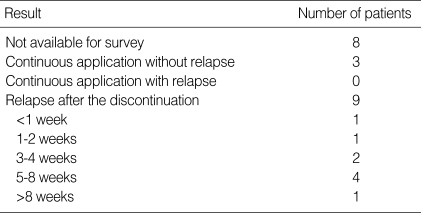
After the completion of the study, 12 patients were available for follow-up telephone surveys 4 to 8 weeks later. Three patients had continued to apply pimecrolimus cream 1% once daily and did not notice any relapse of symptoms or signs of the disease for 8 weeks. Nine patients not using any medications were aware of recurrence of the disease, mostly between 3 to 8 weeks after the discontinuation of pimecrolimus. Five out of 9 patients remained free of the disease for at least 4 weeks after the discontinuation of pimecrolimus, whereas 4 patients experienced relapse within 4 weeks (Table 2).
Table 2.
Results of follow-up telephone survey
DISCUSSION
Recent studies (8-11) proposed that pimecrolimus cream 1% is an effective and safe modality in controlling the symptoms and signs of seborrheic dermatitis (Table 3). In the study by Rigopoulos et al. (9) to determine the efficacy of pimecrolimus in comparison with potent topical corticosteroid in the treatment of seborrheic dermatitis, the complete resolution of symptoms was rapidly achieved within 9 days in both the pimecrolimus-treated and the topical corticosteroid-treated group. Although both agents were highly effective in the treatment of seborrheic dermatitis, the relapses were more common, rapid, and severe in the corticosteroid-treated group. Rallis et al. (10) treated 19 patients of seborrheic dermatitis with pimecrolimus, and clearance was observed after 1 to 3 weeks of treatment. In addition, patients who were previously unresponsive to topical corticosteroid and antifungals also showed clearance of seborrheic dermatitis with pimecrolimus treatment (8, 11). These results indicate that pimecrolimus cream 1% may be an effective nonsteroidal alternative for the treatment of seborrheic dermatitis. Similar results were reported in the use of topical tacrolimus for seborrheic dermatitis (12, 13). In our study, consistent with those previous reports, the mean severity scores of erythema, scaling, and pruritus showed statistically significant decrements after a 4-week application of pimecrolimus cream 1% relative to the baseline. The evident therapeutic responses mostly appeared within the first week of treatment and continued to improve until the end of the study. Improvements in global assessment of disease severity determined by patients and investigators also showed excellent results during the first week of the application, and they experienced continuous improvements throughout the duration of the study. According to the follow-up survey, improved states were maintained for at least 4 weeks in 5 out of 9 patients who did not use any medications after the completion of the study.
Table 3.
Review of the literature on seborrheic dermatitis successfully treated with pimecrolimus cream 1%
NM, not mentioned.
Previous studies have indicated that seborrheic dermatitis might develop as a result of inflammatory immune reactions to the lipophilic yeast Malassezia (14, 15). Humoral and cell-mediated immune response to Malassezia has also been advocated (16). Improvement of the disease by antifungal medications supports the possible role of Malassezia (14). Pimecrolimus selectively inhibits T cell activation via inhibition of the calcineurin-dependent nuclear transcription factor of activated T cells. It prevents the release of cytokines and proinflammatory mediators involved in the inflammatory response (7). Accordingly, the beneficial effect of pimecrolimus on the course of seborrheic dermatitis in our study is thought to be due to its anti-inflammatory and immunomodulatory properties. However, the possibility of direct antifungal activities of pimecrolimus cannot be excluded, since a recent report documented that tacrolimus, another topical immunomodulator, has antifungal activities against some of the Malassezia species (17).
The safety of pimecrolimus cream has been well documented, and in contrast to topical corticosteroids it does not induce skin atrophy or other corticosteroid-related adverse events, especially on the face (18, 19). Burning sensations and tingling are well-known local side effects of pimecrolimus, and 9 of 20 (45%) patients experienced these events in our study. However, most reactions were minimal and transient, beginning on day 1 and resolving within 3 days of treatment. No pain, soreness, or signs of viral or bacterial infections were detected.
In conclusion, our findings further support the previous reports that pimecrolimus cream 1% can be an effective and well-tolerated modality in the treatment of facial seborrheic dermatitis. However, since the most of previous encouraging reports had been studied in small numbers and in short-term, further clinical troals involving controlled, long-term follow-up protocols with a large scale of patients are warranted to assess the precise efficacy and preventive action of pimecrolimus in the management of seborrheic dermatitis.
Footnotes
This study was supported by Medical Research Institute Grant (2006-18) Pusan National Unirersity.
References
- 1.Fritsch PO, Reider N. Other Eczematous Eruption. In: Bolognia JL, Jorizzo JL, Rapini RP, editors. Dermatology. Philadelphia: Mosby; 2003. pp. 215–226. [Google Scholar]
- 2.Plewig G, Jansen T. Seborrheic Dermatitis. In: Freedberg IM, Eisen AZ, Wolff K, Austen KF, Goldsmith LA, Katz SI, editors. Fitzpatrick's Dermatology in General medicine. New York: McGraw-Hill; 2003. pp. 1198–1204. [Google Scholar]
- 3.Green CA, Farr PM, Shuster S. Treatment of seborrhoeic dermatitis with ketoconazole: II. Response of seborrhoeic dermatitis of the face, scalp and trunk to topical ketoconazole. Br J Dermatol. 1987;116:217–221. doi: 10.1111/j.1365-2133.1987.tb05815.x. [DOI] [PubMed] [Google Scholar]
- 4.Pierard-Franchimont C, Pierard GE. A double-blind placebo-controlled study of ketoconazole+desonide gel combination in the treatment of facial seborrheic dermatitis. Dermatology. 2002;204:344–347. doi: 10.1159/000063382. [DOI] [PubMed] [Google Scholar]
- 5.Faergemann J. Management of seborrheic dermatitis and pityriasis versicolor. Am J Clin Dermatol. 2000;1:75–80. doi: 10.2165/00128071-200001020-00001. [DOI] [PubMed] [Google Scholar]
- 6.Park EJ, Kim CW, Kim KH, Ahn KJ, Kim KJ. Therapeutic effect of itraconazole in seborrheic dermatitis. Korean J Med Mycol. 2004;9:222–229. [Google Scholar]
- 7.Gupta AK, Chow M. Pimecrolimus: a review. J Eur Acad Dermatol Venereol. 2003;17:493–503. doi: 10.1046/j.1468-3083.2003.00692.x. [DOI] [PubMed] [Google Scholar]
- 8.Crutchfield CE., 3rd Pimecrolimus: a new treatment for seborrheic dermatitis. Cutis. 2002;70:207–208. [PubMed] [Google Scholar]
- 9.Rigopoulos D, Ioannides D, Kalogeromitros D, Gregoriou S, Katsambas A. Pimecrolimus cream 1% vs. betamethasone 17-valerate 0.1% cream in the treatment of seborrhoeic dermatitis. A randomized open-label clinical trial. Br J Dermatol. 2004;151:1071–1075. doi: 10.1111/j.1365-2133.2004.06208.x. [DOI] [PubMed] [Google Scholar]
- 10.Rallis E, Nasiopoulou A, Kouskoukis C, Koumantaki E. Pimecrolimus cream 1% can be an effective treatment for seborrheic dermatitis of the face and trunk. Drugs Exp Clin Res. 2004;30:191–195. [PubMed] [Google Scholar]
- 11.Brownell I, Quan LT, Hsu S. Topical pimecrolimus in the treatment of seborrheic dermatitis. Dermatol Online J. 2003;9:13. [PubMed] [Google Scholar]
- 12.Braza TJ, DiCarlo JB, Soon SL, McCall CO. Tacrolimus 0.1% ointment for seborrhoeic dermatitis: an open-label pilot study. Br J Dermatol. 2003;148:1242–1244. doi: 10.1046/j.1365-2133.2003.05383.x. [DOI] [PubMed] [Google Scholar]
- 13.Meshkinpour A, Sun J, Weinstein G. An open pilot study using tacrolimus ointment in the treatment of seborrheic dermatitis. J Am Acad Dermatol. 2003;49:145–147. doi: 10.1067/mjd.2003.450. [DOI] [PubMed] [Google Scholar]
- 14.Faergemann J, Jones JC, Hettler O, Loria Y. Pityrosporum ovale (Malassezia furfur) as the causative agent of seborrheic dermatitis: new treatment options. Br J Dermatol. 1996;134(Suppl 46):12–15. doi: 10.1111/j.1365-2133.1996.tb15652.x. [DOI] [PubMed] [Google Scholar]
- 15.Neuber K, Kroger S, Gruseck E, Abeck D, Ring J. Effects of Pityrosporum ovale on proliferation, immunoglobulin (IgA, G, M) synthesis and cytokine (IL-2, IL-10, IFN gamma) production of peripheral blood mononuclear cells from patients with seborrhoeic dermatitis. Arch Dermatol Res. 1996;288:532–536. [PubMed] [Google Scholar]
- 16.Faergemann J. Pityrosporum species as a cause of allergy and infection. Allergy. 1999;54:413–419. doi: 10.1034/j.1398-9995.1999.00089.x. [DOI] [PubMed] [Google Scholar]
- 17.Sugita T, Tajima M, Ito T, Saito M, Tsuboi R, Nishikawa A. Anti-fungal activities of tacrolimus and azole agents against the eleven currently accepted Malassezia species. J Clin Microbiol. 2005;43:2824–2829. doi: 10.1128/JCM.43.6.2824-2829.2005. [DOI] [PMC free article] [PubMed] [Google Scholar]
- 18.Eichenfield LF, Lucky AW, Boguniewicz M, Langley RG, Cherill R, Marshall K, Bush C, Graeber M. Safety and efficacy of pimecrolimus (ASM 981) cream 1% in the treatment of mild and moderate atopic dermatitis in children and adolescents. J Am Acad Dermatol. 2002;46:495–504. doi: 10.1067/mjd.2002.122187. [DOI] [PubMed] [Google Scholar]
- 19.Queille-Roussel C, Paul C, Duteil L, Lefebvre MC, Rapatz G, Zagula M, Ortonne JP. The new topical ascomycin derivative SDZ ASM 981 does not induce skin atrophy when applied to normal skin for 4 weeks: a randomized, double-blind controlled study. Br J Dermatol. 2001;144:507–513. doi: 10.1046/j.1365-2133.2001.04076.x. [DOI] [PubMed] [Google Scholar]


