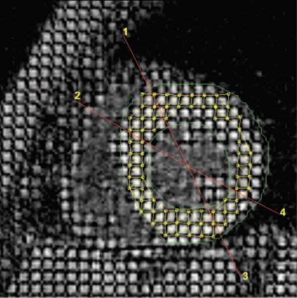
Figure 3.

Short-axis view depicting MRI interrogations similar to echocardiography in the parasternal-long axis. The anterior septum is between regions 1 and 2. The posterior wall is between 3 and 4. Note the midwall triangulation tiling deformation pattern used to determine strain between end diastole and end systole.
