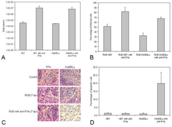
FIG. 4.
Growth of RUB in cells pretreated with anti-IFN serum. Cultures of Hs888Lu cells and HEF were incubated with anti-Type I IFN antiserum for 24 hours and then infected with RUB (MOI = 10 PFU/cell) in the presence of the anti-interferon serum. At 3 dpi, titer of virus present in the culture medium was quantitated by plaque assay (A) and the percentage of infected cells was determined (B). In both cases, the data presented are the average of 2 independent repetitions. Cells were stained with Giemsa for microscopy at 7 dpi (C) and the percentages of apoptotic cells were determined by TUNEL assay at 5 dpi (D; average of 2 independent repetitions).

