Abstract
BACKGROUND:
Abdominal surgery can lead to postoperative intra-abdominal adhesions (PIAAs) with significant morbidity and mortality. This study compares the use of honey with a standard bioresorbable membrane (Seprafilm™) to prevent the formation of PIAAs in rats.
METHODS:
Thirty rats underwent laparotomy, and PIAAs were induced by scraping the cecum. The animals were divided into three groups, each containing ten rats. Group 1 (control) represented the cecal abrasion group, with no intraperitoneal administration of any substance. Group 2 (honey group) underwent cecal abrasion and intraperitoneal administration of honey. Group 3 (Seprafilm™ group) underwent cecal abrasion and intraperitoneal Seprafilm™ application.
RESULTS:
Group 1 exhibited higher adhesion scores for adhesions between the abdominal wall and the organs. Groups 2 and 3 had decreased adhesive attachments to the intra-abdominal structures. Compared to group 1, the incidence of adhesion formation was lower in both group 2 (p=0.001) and group 3 (p=0.001). The incidence of fibrosis was also lower in group 2 (p=0.016) and group 3 (p=0.063) compared to group 1. There was no significant difference between the histopathological fibrosis scores for the rats in group 2 and those in group 3 (p= 0.688).
CONCLUSION:
This study suggests that both honey and Seprafilm™ decrease the incidence of PIAAs in the rat cecal abrasion model. Although the mechanism of action is not clear, intraperitoneal administration of honey reduced PIAAs. The outcome of this study demonstrates that honey is as effective as Seprafilm™ in preventing PIAAs.
Keywords: Postoperative intra-abdominal adhesions, Seprafilm, Honey, Rat, Cecal abrasion
INTRODUCTION
Postoperative intra-abdominal adhesion (PIAA) formation has long been considered an unavoidable consequence of laparotomy. Despite improvements in surgical techniques and instruments, PIAA formation is still a major source of morbidity and mortality. The incidence of adhesion formation has been reported to be 93 to 100% in patients who have undergone abdominal surgery.1 PIAAs after laparotomy can induce small-bowel obstruction, infarction, fistula formation, chronic pelvic pain, secondary female infertility and technical difficulty in cases of re-operation; they also generate a significant economic burden from the need for increased medical services.2, 3
The search for an effective anti-adhesion device has been continuing for decades. Many methods, drugs and materials have been evaluated to prevent PIAAs. The long list of failed materials includes corticosteroids, antihistamine, dextran, saline, anti-cytokine agents, recombinant tissue plasminogen activators, aprotinin, octreotide and heparin.2,4 Thus far, no ideal method has been found for the prevention of PIAA formation. Intra-abdominal administration of some anti-adhesive barriers, such as a bioresorbable membrane consisting of sodium hyaluronate and carboxymethylcellulose (Seprafilm™) (Genzyme Corporation, Cambridge, MA), may reduce postoperative adhesions, as demonstrated by some animal models and clinical studies.2,5,6
Honey has been used in burn victims and as a method of wound treatment for more than 2000 years. Honey is the foodstuff produced by honeybees from the nectar of flowers or secretions from other parts of the plant. It is rich in flavonoid components, such as luteolin, quercetin, apigenin, fisetin, kaempferol, isorhamnetin, acacetin, tamarixetin, chrysin and galangin, and, thus, exhibits antioxidant activity. Honey inhibits the growth of both gram-positive and gram-negative bacteria and provides anti-bacterial, anti-inflammatory, immuno-stimulatory, anti-ulcer and wound- and burn-healing effects.7 Certain physical properties of honey, such as its hygroscopicity, low pH and hypertonicity, are supposedly responsible for its wound-healing effects.7,8
The purpose of this study was to compare honey with a standard bioresorbable membrane (Seprafilm™) to prevent the formation of PIAAs in rats.
MATERIALS AND METHODS
This study was performed in the Animal Research Laboratory of Gazi University. Approval from the Research Committee of Gazi University was obtained before the study. A total of 30 female Wistar Albino rats, each weighing between 280–390 g, were used after overnight fasting.
The animals were divided into three groups, each containing ten rats. Group 1 (control group) represented the cecal abrasion group, with no intraperitoneal administration of any substance. Group 2 (honey group) underwent cecal abrasion and intraperitoneal honey administration (Mechihoney, Europa lmt., RICHLANDS QLD, AUSTRALIA). Group 3 (Seprafilm™ group) underwent cecal abrasion and intraperitoneal Seprafilm™ application. For anesthesia, the animals received 50 mg/kg of ketamine hydrochloride (Ketalar, 50 mg/mL; Parke-Davis, Istanbul) intramuscularly and 5 mg/kg of xylazine (Xylazine-20 Injection, Butler Company, Columbus, OH).
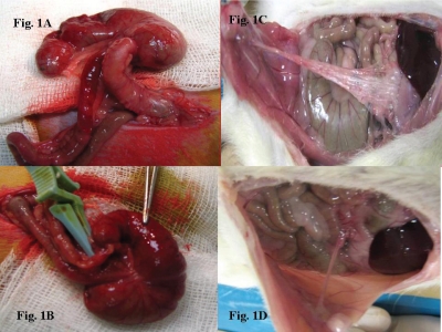
After shaving and disinfection of the skin, a 3-cm midline incision was made. The terminal ileum and cecum were mobilized and placed on wet gauze. Both sides of a 5- to 10-cm segment of terminal ileum and cecum were scraped until serosal petechiae appeared on the intestinal surfaces. The arteries of the scraped segments were then clamped for one minute to induce transient ischemia (scraping model, Figure 1A, 1B).8 In the control group, only this scraping model was performed, while in the honey group, the scraped areas were coated with 4 g of honey. In the Seprafilm™ group, a 20x30-mm sheet of material was applied directly to the abraded cecum before closure. The abdominal incision was closed in two layers with simple, continuous sutures of silk 3/0.
Figure 1 -.
A-B: Scraping model. C: Grade 3 adhesion in a control group animal, with extensive and thick adhesive bands and adhesions between the organs and the abdominal wall. D: Grade 1 adhesions in Seprafilm™ group and honey group animals with thin, easily removable adhesive bands between the organs and the abdominal wall.
All rats were allowed to resume their diets until the 10th postoperative day, at which point they were sacrificed with ether. The peritoneal cavities were entered via a reversed U-shaped incision in the anterior abdominal wall, which was retracted caudally to provide maximal exposure. Adhesions were examined macroscopically by two independent investigators (MA, HB) and graded blindly according to the Blauer and Collins scale (Table 1).9 Tissues containing adhesions were excised en bloc and the samples were fixed in a 10% formaldehyde solution. Samples were routinely processed by dehydration and paraffin embedding, and 5-μm cross-sections were prepared using a microtome. The samples were examined under a light microscope after hemotoxylin-eosin staining and were evaluated blindly by the same pathologist (IIG) to determine the general structure and the amount of fibroblastic activity and fibrosis present (Table 2). Thus, the fibrotic score of each rat was calculated according to the criteria mentioned.1
Table 1 -.
Blauer and Collins scale for macroscopic assessment of adhesion formation9
| Grade | Description of adhesions |
|---|---|
| 0 | No adhesion |
| 1 | Thin adhesive bands, easily removable |
| 2 | Thick adhesive bands limited to one area |
| 3 | Extensive and thick adhesive bands |
| 4 | Extensive and thick adhesive bands and adhesions between viscera and/or abdominal wall |
Table 2 -.
Scale for microscopic assessment of fibrosis1
| Grade | Histopathological signs |
|---|---|
| 0 | No fibrosis |
| 1 | Thin bunches of a cellular fibrosis |
| 2 | Wide areas of fibrosis with reduced vascularization |
| 3 | Areas of fibrosis formed by thick bunch of collagen |
Statistics
The Kruskal-Wallis test was used to test for differences in the grades of adhesions observed in the three groups. A Mann-Whitney U statistic analysis was used as a non-parametric test to determine differences in adhesion grading. A P-value ≤0.05 was considered significant, and a P-value <0.001 was considered highly significant.
RESULTS
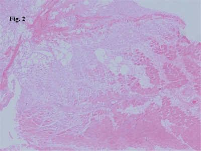
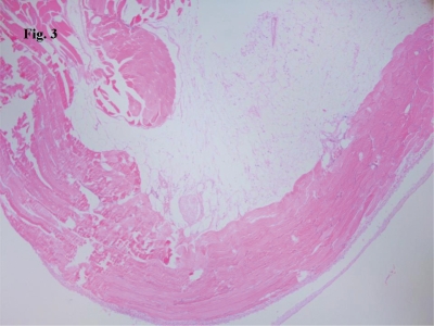
A total of 30 rats were randomly assigned to the control group (n = 10), the honey group (n = 10) or the Seprafilm™ group (n = 10). Five rats (one in the control group, two in the honey group and two in the Seprafilm™ group) died before re-laparotomy. These rats were replaced and the adhesion operation was redone for corresponding groups and included in the study. The intra-abdominal adhesions and histopathological fibrosis scores are summarized in table 3. Of the ten rats in the control group, five developed grade 4 adhesions, three developed grade 3 adhesions (Figure 1C), and two developed grade 2 adhesions. In the Seprafilm™ group, six developed grade 1 adhesions, and four developed grade 0 adhesions. In the honey group, five developed grade 1 adhesions (Figure 1D), and five developed grade 2 adhesions. Upon histological examination, tissue from rats in the control group demonstrated lymphocytes, plasma cells, polymorphonuclear leucocytes and areas of fibrosis containing fibroblasts (Figure 2). The rats in the honey and Seprafilm™ groups demonstrated decreased infiltration of inflammatory cells, fibroblasts and areas of fibrosis (Figure 3).
Table 3 -.
Intra-abdominal adhesions and histopathological fibrosis scores
| Adhesion score | Fibrosis score | |
|---|---|---|
| Control group (SD ±) | 3.3 (2–4) | 1.9 (0–3) |
| Honey group | 1.5 a (1–2) | 1.1 d (0–3) |
| Seprafilm group | 0.6 b, c (0–1) | 0.9 e, f (0–2) |
SD: standard deviation.
p=0.001; control group vs honey group;
p=0.001; control group vs seprafilm group;
p=0.003; seprafilm group vs honey group;
p=0.063; control group vs honey group;
p=0.016; control group vs seprafilm group;
p=0.068; seprafilm group vs honey group
Figure 2 -.
In the control group, the infiltration of inflammatory cells and areas of fibrosis consisting of fibroblasts are seen in the histopathological examination (H/E staining)
Figure 3 -.
In the Seprafilm™ group and the honey group fewer inflammatory cells and decreased fibrosis are seen (H/E staining)
The control group had higher adhesion scores between the abdominal wall and the organs (Figure 1C). The honey and Seprafilm™ groups had fewer adhesive attachments to the intra-abdominal structures (Figure 1D). Compared to the control group, both the honey (p=0.001) and Seprafilm™ (p=0.001) groups exhibited a lower incidence of adhesion formation. Comparison of the Seprafilm™ and honey groups showed that adhesion formation was less severe in the Seprafilm™ group (p=0.003). The Seprafilm™ group exhibited decreased fibrosis scores compared to the control group (p=0.016). The fibrosis scores in the honey group were also lower than the control group, but this difference was not significant (p=0.063). Importantly, there was no significant difference between the fibrosis scores of the honey and Seprafilm™ groups (p= 0.688).
DISCUSSION
PIAAs account for 40% of all cases of intestinal obstruction, with 60–70% of those involving the small bowel. Of the patients who require abdominal re-operation, 30–40% have postoperative adhesion-related intestinal obstruction. Enterocutaneous fistulas, intra-abdominal abscesses and ureteral obstructions can also develop as a result of PIAAs. In addition to causing substantial abdominal and pelvic pain, PIAAs are the leading cause of secondary infertility in women.10, 11 Therefore, PIAA formations not only represent a significant expenditure for the healthcare system, but they can also result in loss of work force capacity and impaired quality of life. In addition, PIAAs are a major source of morbidity following laparotomy and are the most common cause of high financial burdens. Total costs related to adhesions have been estimated at 1.2 billion U.S. dollars per year.12,13 Moreover, complications from adhesions after gynecological surgery were estimated to result in 226.8 million U.S. dollars in health care costs in 2006.14
Adhesion formation is a complicated process. Although the pathophysiology of adhesion formation is widely understood, an absolute solution to this problem does not yet exist. Adhesions can result from mechanical peritoneal damage, tissue ischemia or the presence of foreign materials. Additionally, two areas of injury must be in contact with each other. Both fibrinogenesis and fibrinolysis are activated, and a distortion of the dynamic balance between these two processes leads to adhesion formation.13,15
There are many experimental models for provoking peritoneal adhesions. The scraping model is very effective in engendering peritoneal adhesions because it involves two stages of damage, direct mechanical intestinal wall damage from gauze scraping and ischemic damage from vascular clamping (Figure 1A, 1B).8 As this model mimics abdominal surgery, we chose to use it in this study.
PIAAs at the peritoneum most commonly form within seven to ten days and become persistent after fourteen days. Therefore, this stage is critical in preventing adhesion formation, and efforts to combat adhesion formation concentrate mostly on this phase. We chose to wait ten days before performing re-laparotomy because other studies have shown the adhesion scores peak at this time.1,16
During the past decade, a variety of commercially available substances and materials have been used in attempts to reduce PIAAs.14 All of these materials have unique compositions and characteristics, with limitations and advantages regarding their use in the clinical setting. Ideally, such a barrier should be anti-adhesive, highly biocompatible, resorbable, adherent to the traumatized surface, effective on an oozing surface, applicable through the laparoscope and relatively inexpensive. As yet, such an ideal barrier does not exist.14
The mechanical separation of peritoneal surfaces represents a strategy for blocking peritoneal adhesion formation. This approach is particularly attractive, especially if the device can be easily applied by the surgeon and if the material is biodegradable and does not cause systemic effects. A bioresorbable membrane consisting of sodium hyaluronate and carboxymethylcellulose (Seprafilm™) is one such material used to prevent surgical adhesions. Within 24 hours of application, the Seprafilm™ turns into a gel, which remains in place to separate adhesiogenic tissues during the first few days after surgery when adhesions are most likely to develop. The material is cleared from the body within four weeks.2,4,12
Prospective, randomized trials have demonstrated that Seprafilm™ significantly reduces the incidence, severity and extent of adhesions following two-stage restorative proctocolectomy and uterine myomectomy. It has also been demonstrated to decrease the incidence of small-bowel obstruction after intestinal resection.10,12,17,18 This benefit was confirmed in our study, where Seprafilm™ was shown to significantly reduce postoperative adhesion formation compared to the control group. However, the therapeutic and physiological effects of Seprafilm™ may be limited to the site of application. The fact that the membrane is thin, crisp and filmy may make it difficult to use. This may be particularly true during difficult abdominal wound closures or cases involving a small incision.2
Natural honey has been used to treat burns and decubitis wounds since ancient times. Honey is becoming increasingly popular as a modern wound-dressing material, and studies have been published demonstrating its effectiveness. When honey is applied to wounds, it has been found to reduce inflammation, swelling and pain, eliminating the need for surgical removal, and induces a rapid clearance of infections. In the literature, it has been reported that honey carries no toxic effects. Honey is also considered sterile and inhibits the growth of both gram-positive and gram-negative bacteria. It has antifungal, cytostatic, anti-inflammatory, antitumoral and antimetastatic effects and promotes wound healing.8,19 However, studies regarding its effects on preventing PIAAs are limited.
The mechanisms underlying the effects of honey on wound healing are not entirely clear. However, several different characteristics of honey may have effects on various steps of the wound-healing process. Aysan, et al. suggested that one or more of the ingredients of honey, including cafeic acid, benzoic acid, phenolic acid, flavanoid glycons, inhibin and catalase, may be responsible for its effect. Inhibin and catalase have also been shown to promote the proliferation of epithelial cells. Another possible mechanism by which honey can promote wound healing is by increasing fibroblastic activity. Honey is hygroscopic, hypertonic and has a low pH. Hygroscopic substances decrease edema and constitute a fluid barrier to inhibit deepening of the wound. Hypertonicity contributes to antibacterial and antifungal properties. Thus, a hypertonic environment with a low pH and low moisture may promote the wound-healing process by degrading native collagen within the wound.8, 20
In the present study, we utilized a rat model to compare two types of anti-adhesive devices: an anti-adhesive sheet barrier membrane and honey. We found both materials, if applied just prior to laparotomy closure, significantly reduced the formation of PIAAs. Although the mechanism of action is not clear, intraperitoneal administration of honey can reduce PIAAs. Our study indicates that honey may be used as an anti-adhesive barrier and can produce results similar to those of various types of commercially available materials.
REFERENCES
- 1.Yilmaz HG, Tacyildiz IH, Keles C, Gedik E, Kilinc N. Micronized purified flavonoid fraction may prevent formation of intraperitoneal adhesions in rats. Fertil Steril. 2005;84(suppl 2):1083–88. doi: 10.1016/j.fertnstert.2005.03.076. [DOI] [PubMed] [Google Scholar]
- 2.Oncel M, Remzi FH, Senegore AJ, Connor JT, Fazio VW. Comparison of novel liquid (Adcon-P) sodium hyaluronate and carboxymethylcellulose membrane (Seprafilm) postsurgical adhesion formation in a murine model. Dis Colon Rectum. 2003;46:187–91. doi: 10.1007/s10350-004-6523-3. [DOI] [PubMed] [Google Scholar]
- 3.Van Der Krabben AA, Dijkstra FR, Nieuwenhuijzen M, Reijnen MM, Schaapveld M, van Goor H. Morbidity and mortality of inadvertent enterotomy during adhesiotomy. Br J Surg. 2000;87:467–71. doi: 10.1046/j.1365-2168.2000.01394.x. [DOI] [PubMed] [Google Scholar]
- 4.Hellebrekers BW, Trimbos-Kemper GC, van Blitterswijk CA, Bakkum EA, Trimbos JB. Effects of five different barrier materials on postsurgical adhesion formation in the rat. Hum Repro. 2000;15:1358–63. doi: 10.1093/humrep/15.6.1358. [DOI] [PubMed] [Google Scholar]
- 5.Beck DE. The role of Seprafilm bioresorbable membrane in adhesion prevention. Eur J Surg Suppl. 1997:49–55. [PubMed] [Google Scholar]
- 6.Altuntas I, Tahran O, Delibas N. Seprafilm reduces adhesions to polypropylene mesh and increases peritoneal hydroxyproline. Am Surg. 2002;68:759–61. [PubMed] [Google Scholar]
- 7.Erguder BI, Kilicoglu SS, Namuslu M, Kilicoglu B, Devrim E, Kismet K, et al. Honey prevent hepatic damage induced by obstruction of the common bile duct. World J Gastroenterol. 2008;14:3729–32. doi: 10.3748/wjg.14.3729. [DOI] [PMC free article] [PubMed] [Google Scholar]
- 8.Aysan E, Ayar E, Aren A, Cifter C. The role of intra-peritoneal honey administration in preventing post-operative peritoneal adhesions. Eur J Obstet and Gynecol and Reprod Biol. 2002;104:152–5. doi: 10.1016/s0301-2115(02)00070-2. [DOI] [PubMed] [Google Scholar]
- 9.Blauer KL, Collins RL. The effect of intraperitoneal progesterone on postoperative adhesion formation in rabbit. Fertil Steril. 1998;49:144–9. [PubMed] [Google Scholar]
- 10.Cohen Z, Senagore AJ, Dayton MT, Koruda MJ, Beck DE, Wolff BG, et al. Prevention of postoperative abdominal adhesions by a novel, glycerol/sodium hyaluronate/carboxymethylcellulose-based bioresorbable membrane: a prospective, randomized, evaluator-blinded multicenter study. Dis Colon Rectum. 2005;48:1130–9. doi: 10.1007/s10350-004-0954-8. [DOI] [PubMed] [Google Scholar]
- 11.Johnson P, Richard C, Ravid A, Spencer L, Pinto E, Hanna M, et al. Female infertility following ileal pouch anal anastomosis for ulcerative colitis. Dis Colon Rectum. 2004;47:1119–26. doi: 10.1007/s10350-004-0570-7. [DOI] [PubMed] [Google Scholar]
- 12.Becker JM, Dayton MT, Fazio VW, Beck DE, Stryker SJ, Wexner SD, et al. Prevention postoperative abdominal adhesion by asodium hyaluronate-based bioresorbable membrane: A prospective, randomized double-blind multicenter study. J Am Coll Surg. 1996;183:297–306. [PubMed] [Google Scholar]
- 13.Arikan S, Adas G, Barut G, Toklu AS, Kocakusak A, Uzun H, et al. An evaluation of low molecular weight heparin and hyperbaric oxygen treatment in the prevention of intra-abdominal adhesions and wound healing. Am J Surg. 2005;189:155–60. doi: 10.1016/j.amjsurg.2004.11.002. [DOI] [PubMed] [Google Scholar]
- 14.Bristow RE, Santillan A, Diaz-Montes TP, Gardner GJ, Giuntoli RL, 2nd, Peeler ST. Prevention of adhesion formation after radical hysterectomy using a sodium hyaluronate/carboxymethylcellulose (HA-CMC) barrier: A cost-effectiveness analysis. Gynecol Oncol. 2007;104:739–46. doi: 10.1016/j.ygyno.2006.09.029. [DOI] [PubMed] [Google Scholar]
- 15.Menger MD, Vollmar B. Surgical trauma: hyperinflammation vs immunosuppression? Langenbecks Arch Surg. 2004;389:475–84. doi: 10.1007/s00423-004-0472-0. [DOI] [PubMed] [Google Scholar]
- 16.Gomel V, Urman B, Gurgan T. Pathophysiology of adhesion formation and strategies for prevention. J Reprod Med. 1996;41:35–41. [PubMed] [Google Scholar]
- 17.Alponat A, Lakshminarasappa SR, Yavuz N, Goh PM. Prevention of adhesions by Seprafilm, an absorbable barrier. An incisional hernia model in rats. Am Surg. 1997;63:818–9. [PubMed] [Google Scholar]
- 18.Vrijland WW, Tseng LN, Eijkman HJ, Hop WC, Jakimowicz JJ, Leguit P, et al. Fewer intraperitoneal adhesions with use of hyaluronic acid-carboxymethylcellulose membrane. A randomized clinical trial. Ann Surg. 2002;235:193–9. doi: 10.1097/00000658-200202000-00006. [DOI] [PMC free article] [PubMed] [Google Scholar]
- 19.Hamzaoglu I, Saribeyoglu K, Durak H, Karahasanoglu T, Bayrak I, Altug T, et al. Protective covering of surgical wounds with honey impedes tumor implantation. Arch Surg. 2000;135:1414–7. doi: 10.1001/archsurg.135.12.1414. [DOI] [PubMed] [Google Scholar]
- 20.Bergmann A, Yanai J, Weiss J, Bell D, David MP. Acceleration of wound healing by topical application of honey. An animal model. Am J Surg. 1983;145:374–6. doi: 10.1016/0002-9610(83)90204-0. [DOI] [PubMed] [Google Scholar]


