Summary
Gadd45 genes have been implicated in stress signaling responses to various physiological or environmental stressors, resulting in cell cycle arrest, DNA repair, cell survival and senescence, or apoptosis. Evidence accumulated up to date suggests that Gadd45 proteins function as stress sensors, mediating their activity through a complex interplay of physical interactions with other cellular proteins that are implicated in cell cycle regulation and the response of cells to stress. These include PCNA, p21, cdc2/cyclinB1, and the p38 and JNK stress response kinases. Disregulated expression of Gadd45 has been observed in multiple types of solid tumors as well as in hematopoietic malignancies. Also, evidence has accumulated that Gadd45 proteins are intrinsically associated with the response of tumor cells to a variety of cancer therapeutic agents. Thus, Gadd45 proteins may represent a novel class of targets for therapeutic intervention in cancer. Additional research is needed to better understand which of the Gadd45 stress response functions may be targeted for chemotherapeutic drug design in cancer therapy.
Keywords: Gadd45, cellular stress, Cell cycle arrest, DNA repair, gene activation, Demethylation, Apoptosis, Survival, Senescence, PCNA, p21, sCdc2/cyclinB1, MAPK stress kinases
I. Gadd45 in cellular stress responses
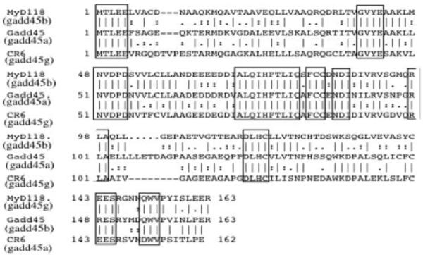
Gadd45 genes were cloned in this laboratory (Abdollahi et al, 1990; Zhang et al, 1999), in the laboratory of Dr. Smith (Beadling et al, 1993) and in the Fornace laboratoy at the NIH (Fornace 1992). Gadd45, MyD118, and CR6 (currently termed Gadd45a, Gadd45b, & Gadd45g, respectively) are referred to as Gadd45 proteins, gadd45 genes or Gadd45 family members. These small (18kd), evolutionarily conserved proteins are highly homologous to each other (55%-57% overall identity at the amino acid level), highly acidic (pI=4.0-4.2) (Figure 1), and are localized primarily within the cell nucleus (Abdollahi et al, 1990; Zhang et al, 1999; Vairapandi et al, 2002).
Figure 1. Homology of Gadd45a family of proteins.
Alignment of amino acid sequence of the various gadd45a family members including murine MyD118 (Gadd45b), murine Gadd45 (Gadd45a) and murine CR6 (Gadd45g). Boxes reveal conserved amino acid residues.
Gadd45 family members are rapidly induced by genotoxic stress agents (Papathanasiou et al, 1989; Fornace et al, 1991; Vairapandi et al, 2002), as well as by terminal differentiation and apoptotic cytokines (Abdollahi et al, 1990; Zhan et al, 1994; Zhang et al, 1999; Zhang 2000). Emerging evidence indicates that the proteins encoded by these genes play pivotal roles as stress sensors that modulate and integrate the response of mammalian cells to a variety of environmental and physiological stressors (Fornace, 1992; Fornace et al, 1992; Liebermann and and Hoffman 1998; Zhang et al, 1999) either dependent or independent of p53 (Carrier et al, 1995; Liebermann and Hoffman 1995; Zhan et al, 1996). They also function to modulate tumor formation in response to oncogenic stress (Tront et al, 2006). Gadd45 proteins appear to serve similar, but not identical, functions depending upon the particular stress response pathway activated. For example, only Gadd45b is induced by TGFb (Selvakumaran et al, 1994a; Yoo et al, 2003), whereas only Gadd45a is a p53 target (Kastan et al, 1992; Selvakumaran et al, 1994b; Guillouf et al, 1995). All three genes are induced with different expression kinetics during terminal hematopoietic differentiation, associated with growth arrest and apoptosis (Zhang et al, 1999). Distinct expression patterns for these genes were also observed in a variety of murine tissues (Zhang et al, 1999; Zhang 2000).
Importantly, individual members of the Gadd45 family are differentially induced by a variety of genotoxic and environmental stress agents (Takekawa et al, 1998; Shaulian and Karin 1999; Wang et al, 1999; Zhang et al, 1999, 2001; Zhang 2000), indicating that each gene is induced by a distinct subset of environmental stresses. To what extent the function of each of the Gadd45 proteins is unique or overlaps with the function of the remaining family member proteins remains to be determined.
As detailed below, Gadd45 genes have been implicated in the control of cell cycle arrest (Beadling et al, 1993; Liebermann and Hoffman 1998; Wang et al, 1999; Zhang et al, 1999; Vairapandi et al, 2002), DNA repair (Vairapandi et al, 1992; Smith et al, 1994, 2000), cell survival (Smith et al, 1996, 2000; De Smaele et al, 2001; Amanullah et al, 2003; Gupta et al, 2005, 2006), apoptosis (Selvakumaran et al, 1994a; Takekawa et al, 1998; Vairapandi et al, 2000; Azam et al, 2001; Zhang et al, 2001; Yoo et al, 2003), senescence (Tront et al, 2006), and susceptibility of cells for transformation in vitro and in tumor development in vivo (Hollander et al, 1999; Tront et al, 2006).
II. Mode of action of Gadd45 proteins
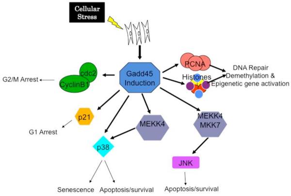
Evidence accumulated in recent years implies that Gadd45 proteins function as stress sensors, mediating their activity via a complex interplay of physical interactions with other cellular proteins. These interactions have been shown to regulate cell cycle control, DNA repair, epigenetic changes, apoptosis, survival and senescence (Figure 2).
Figure 2. Gadd45 function in stress signaling.
Summary of the various functions of Gadd45 family members affecting cellular processes such as cell cycle arrest, DNA repair, survival, apoptosis, senescence as well as epigenetic gene activation.
A. Cell cycle control
Inhibiting endogenous expression of Gadd45a, Gadd45b, or Gadd45g in human cells by antisense Gadd45 constructs was found to impair the G2/M checkpoint following exposure to UV radiation or MMS (Liebermann and Hoffman 1998; Wang et al, 1999; Vairapandi et al, 2002). In addition, microinjecting a Gadd45a expression vector into primary human fibroblasts arrested the cells at the G2/M boundary of the cell cycle (Wang et al, 1999). In another study (Fan et al, 1999), deregulated ectopic expression of CR6 (Gadd45g) in HeLa cells had little effect on HeLa cell growth under normal culture conditions. However, following serum withdrawal Gadd45g blocked HeLa cell G2/M transition and caused endoreduplication. In contrast, under normal culture conditions in USO2 cells, ectopic expression of any one of the Gadd45 proteins resulted in blocking either G1/S or G2/M transitions (Fan et al, 1999). In this laboratory it was observed that IPTG-induced ectopic expression of Gadd45a, Gadd45b, or Gadd45g in both H1299 and M1 cells, in the absence of genotoxic stress, retarded cell growth and increased accumulation of cells in the G1 phase of the cell cycle (Zhang et al, 2001).
G2/M cell cycle arrest mediated by Gadd45 proteins was shown to be due to their ability to interact with and inhibit the kinase activity of the cdc2/cyclinB1 complex (Zhan et al 1999; Vairapandi et al. 2002). Association of either Gadd45a or Gadd45b proteins with cdc2/cyclinB1 results in dissociation of the cdc2/cyclinB1 complex that, in turn, inhibits cdc2 kinase activity (Vairapandi et al, 2002). Their ability to arrest cells in G1 is less well understood. It is possible that interaction of Gadd45 proteins with p21 plays a role in G1 cell cycle arrest. As such, all 3 family members have been observed to interact with p21 (Zhang et al, 1993; Xiong etal, 1993; El-Deiry Et al, 1993), but the role for this interaction remains to be further elucidated.
B. DNA repair
Current evidence suggests that Gadd45a and Gadd45b function in DNA excision repair through their interactions with PCNA (Vairapandi et al. 1992; Smith et al, 1994, 2000). As such, experimental data obtained shows that interaction of either Gadd45a or Gadd45b with PCNA participates in nucleotide excision DNA repair (Vairapandi et al. 1992; Smith et al, 1994, 2000). Whether Gadd45g plays a role in DNA repair has not been established.
C. Demethylation dependent epigenetic gene activation
A recent study documented that Gadd45a promotes epigenetic gene activation by repair-mediated DNA demethylation (Barreto et al, 2007). Additionally, it has been shown that TAF12 recruits Gadd45a and the nucleotide excision repair complex to the promoter of rRNA genes, leading to active DNA demethylation (Schmitz et al, 2009). However, we have recently shown conserved DNA methylation in Gadd45a −/− mice, suggesting that Gadd45a promotion of epigenetic gene activation by repair-mediated DNA demethylation is not global (Engel et al, 2009). Other investigators have recently demonstrated that DNA demethylation in zebrafish involves the coupling of a deaminase, a glycosylase, and Gadd45b (Rai et al, 2008). Furthermore, transient activation of mature dentate granule cells has been observed to induce Gadd45b through an NMDAR-Ca2+-CaM kinase pathway. Gadd45b was shown to be a key factor in an epigenetic regulation path that actively demethylate specific CpG sites in regulatory regions of genes, resulting in increased late-phase expression in mature neurons. Release of these extrinsic factors from mature neurons in turn promotes several key aspects of adult neurogenesis (Ma et al, 2009). Gadd45 interactions with PCNA and/or histones (Carrier et al, 1999) may play a role in this function.
D. Apoptosis
Ample evidence exists that Gadd45 proteins have a pro-apoptotic function. For example, it was observed that blocking MyD118 (Gadd45b) by antisense expression in M1 myeloblastic leukemia cells impaired TGFb-induced cell death, thereby implicating Gadd45b as a positive modulator of TGFb-induced apoptosis (Selvakumaran et al, 1994a). Consistent with this notion, IPTG-inducible ectopic expression of Gadd45b accelerated TGFb-induced apoptosis in M1 cells (Zhang, 2000). More recently, it was demonstrated that TGFb-induced apoptosis is mediated by Gadd45b via p38 activation in primary hepatocytes from wild type mice, and was blocked in hepatocytes from Gadd45b−/− mice (Yoo et al, 2003). Furthermore, ectopic expression of all three Gadd45 proteins was shown to induce apoptosis in HeLa cells (Takekawa et al, 1998). This induction of apoptosis was shown to be dependent upon the interaction of Gadd45 proteins with MEKK4, an upstream activator of the stress induced p38/JNK kinases (Takekawa et al, 1998). That Gadd45 proteins can directly interact with and activate p38 kinase was observed as well (Bulavin et al, 2003). In addition, ectopic expression of all three Gadd45 proteins was shown to induce apoptosis in HeLa cells (Takekawa et al, 1998) as well as enhance stress mediated apoptosis in both M1 leukemia and H1299 lung carcinoma cells (Vairapandi et al, 2000; Azam et al, 2001; Zhang et al, 2001). Also, BRCA-1-mediated induction of gadd45a has been implicated in apoptosis of breast cancer cells (Harkin et al, 1999), whereas Gadd45g expression was shown to have a role in neuronal cell death (Kojima et al, 1999). Lastly, gadd45a has also been implicated in apoptosis of UV-irradiated keratinocytes (Hildesheim et al, 2002).
E. Survival
Intriguingly, in apparent contradiction to the role Gadd45 proteins play in cell death, many observations are consistent with a role in cell survival. Clonogenic survival assays with gadd45a−/− MEFs (Smith et al, 2000) and RKO cells expressing antisense Gadd45a RNA (Smith et al, 1996) showed that deficiency in Gadd45a increases the sensitivity of cells to killing by UV irradiation or cisplatin. It has been suggested that Gadd45b plays a role in TNFα NFϰB mediated cell survival of mouse embryo fibroblasts (De Smaele et al, 2001), although additiona data has challenged this view (Amanullah, et al, 2003). In addition, it was recently documented that Gadd45a and Gadd45b deficiency each sensitized hematopoietic cells to genotoxic stress induced apoptosis (Gupta et al, 2005). It was shown that in hematopoietic cells exposed to UV radiation, Gaddd45a and Gadd45b cooperate to promote cell survival by two distinct signaling pathways involving activation of a novel Gadd45a mediated p38-NF-ϰB-mediated survival pathway and Gadd4545b-mediated inhibition of the stress response MKK4-JNK pathway (Gupta et al, 2006). The role Gadd45 proteins play in DNA repair and cell cycle arrest is compatible with a survival function. Consistent with the idea that interaction of Gadd45 proteins with PCNA may promote cell survival by enhancing DNA repair, it was observed that Gadd45/PCNA interaction impedes their apoptotic function (Vairapandi et al, 2000; Azam et al, 2001).
F. Senescence
Senescence represents a physiological stress response associated with cellular aging. When primary mammalian cells are cultured in vitro they undergo a limited number of cell divisions and then arrest in a state known as replicative senescence. Such cells are irreversibly arrested in the G1 phase of the cell cycle and are no longer sensitive to growth factor stimulation (Drayton et al, 2002; Lundberg et al, 2000 & references therein). Senescence is a barrier that cells must overcome in order to become immortal and proliferate indefinitely, and, therefore, functions as a tumor-suppressing mechanism that limits the proliferative capacity of cells in vivo (Drayton et al, 2002; Lundberg et al, 2000). Recent data has shown that cellular senescence is in fact a physiological mechanism that constrains tumor development in vivo (Lundberg et al, 2000; Drayton et al, 2002). The signaling pathways that mediate replicative or oncogene induced senescence are not fully understood. In Mouse Embryo Fibroblasts (MEFs), p19ARF, which is encoded by a partially overlapping alternative reading frame at the p16INK4A locus, has been implicated as a major mediator of replicative senescence. p19ARF binds directly to and sequesters MDM2, thereby inhibiting the ability of MDM2 to induce degradation of the p53 tumor suppressor protein (Lundberg et al, 2000). This loss of MDM2 function, in turn, results in the stabilization of p53 and activation of p53-mediated growth arrest, believed to play a major role in the irreversible growth arrest associated with the senescent phenotype. The integrity of both the p16INK4A/pRB and p19ARF/p53 pathways is essential for oncogene-induced senescence (Drayton et al, 2002). Studies with MEFs deficient for one or more Gadd45 genes, notably Gadd45a, have provided evidence that Gadd45 genes play important roles in both replicative and oncogene mediated senescence (Hollander et al, 1999; Bulavin et al, 2003). The molecular nature of stress response pathways involving Gadd45 and its interacting proteins in MEF senescence, and whether there is crosstalk with the p16INK4A/pRB and p19ARF/p53 pathways remains to be determined.
III. Gadd45 as sensors of oncogenic stress which modulate tumor development
The complex role of stress sensors in monitoring oncogenic stress ultimately leading to tumor development is not fully understood. The best and most studied example of oncogenic stress in tumorigenesis is p53 and its varied cellular functions. Recent observations have implicated Gadd45 proteins as important sensors of oncogenic stress, both in vitro and in vivo.
It is known that whereas primary mouse cells require introduction of two activated oncogenes for transformation, disruption of certain key growth control genes allows single oncogene transformation (Lundberg et al, 2000; Drayton et al, 2002). It was shown that for MEFs obtained from Gadd45a−/− mice, H-ras was sufficient for transformation (Hollander et al, 1999; Bulavin et al, 2003). The role Gadd45b and/or Gadd45g play in susceptibility of MEFs to single oncogene transformation remains to be assessed.
Evidence was obtained that Gadd45 proteins also play a role in modulation of tumor development in vivo. Gadd45a−/− and Gadd45b−/− mice were observed to display increased mutation frequency, and susceptibility to ionizing radiation (IR) and chemical carcinogenesis (Hollander et al, 1999). Also, it has been documented that NF-ϰB-mediated repression of Gadd45a and Gadd45g is essential for cancer cell survival (Zerbini and Liebermann, 2005). More recently, evidence was obtained that loss of Gadd45a accelerates ras-driven mammary tumor formation. Ras-driven tumor formation in the absence of Gadd45a resulted in both a decrease in apoptosis, linked to a decrease in JNK activation, and a decrease in senescence, correlated with a decrease in p38 kinase activation (Tront et al, 2006). Altogether, these results provide a novel model for the tumor suppressive function of Gadd45a in the context of ras-driven breast carcinogenesis; Gadd45a elicits its function through activation of the stress induced JNK & p38 kinases, which contribute to increased apoptosis and ras-induced senescence. How loss of Gadd45a may effect breast carcinogensis driven by other oncogenes, and how loss of other Gadd45 genes may contribute to breast carcinogenesis are interesting questions to be addressed in future studies.
V. GADD45 disregulation in cancer
Although members of the Gadd45 family seem infrequently mutated in cancer based on current knowledge, reduced expression of the three Gadd45 family members due to promoter methylation has been observed in several types of human cancers.
The Gadd45a promoter is methylated in the majority of breast cancers, resulting in reduced expression when compared with normal breast epithelium (Zerbini and Lbermann, 2005). In pituitary adenomas, silencing of the Gadd45c gene is seen in 67% of patients. This downregulation is primarily associated with methylation of the Gadd45g gene and reversal of this epigenetic change results in re-expression of the protein (Bahar et al, 2004). Gadd45g is also down- regulated in anaplastic thyroid cancer and in 65% of hepatocellular carcinomas due to hypermethylation of the Gadd45g promoter (Sun et al, 2003). Interestingly, the Gadd45b gene is methylated and silenced in hepatocellular carcinoma as well, indicating a strong linkage between at least two Gadd45 genes and liver cancer.
Ying and colleagues analyzed the methylation status of two regions in the Gadd45g promoter in a total of 75 cell lines as well as primary tissues and tumors (Ying et al, 2005). They show that promoter hypermethylation is frequently detected in tumors cell lines, including 85% of non-Hodgkin, 50% of Hodgkin lymphoma, 73% of nasopharyngeal, 50% of cervical, 29% of esophageal, and 40% of lung carcinoma but not in immortalized normal epithelial cell lines, normal tissues, or peripheral blood mononuclear cells. To gain more insight into the Gadd45g methylation, they also did high-resolution bisulfite genomic sequencing. They found that densely methylated CpG sites were detected in all silenced cell lines, indicating that epigenetic silencing of Gadd45g could be involved in the pathogenesis of tumors.
Other observations showing that activated NF-ϰB leads to repression of GADD45a and GADD45g in various types of cancer (Zerbini and Libermann, 2005) together with the frequent constitutive activation of NF-ϰB in cancers suggest that there are at least two mechanisms whereby Gadd45 genes become repressed in cancer. Thus, repression of Gadd45 gene expression in various types of cancer via methylation or NF-kB activation may be two different methods through which Gadd45 deregulated expression may lead to tumorigenesis. Because activation of NF-ϰB is a critical step for many cells in escaping programmed cell death and is dependent on Gadd45a and Gadd45g repression, methylation of the Gadd45g gene as reported by Ying et al, may result in a similar phenotype.
Moreover, very recently, a methylation-mediated repression of Gadd45a was observed in prostate cancer. The role of Gadd45a as a potential therapeutic target has been highlighted by the fact that it is up-regulated on docetaxel treatment and may contribute to docetaxel-mediated cytotoxicity of prostate cancer cells (Ramachandran et al, 2009).
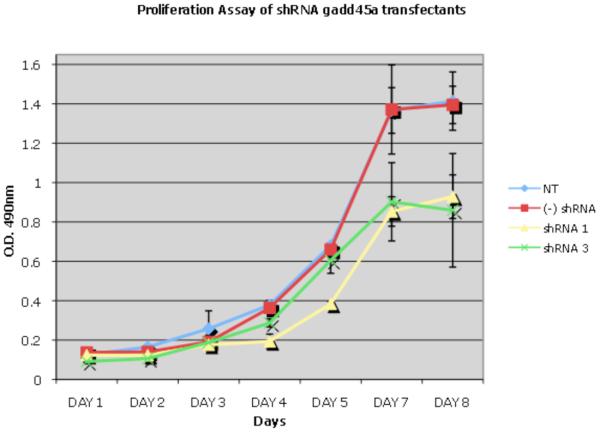
Interestingly, Gadd45a mutations have been documented in pancreatic cancer. One study has shown that gadd45a expression is elevated in several pancreatic ductal adenocarinoma cell lines, and loss of Gadd45a expression limits growth and survival of one cell line in culture (Schneider et al, 2006). In another study, it was observed that ectopic expression of Gadd45a in the PANC1 pancreatic cancer cell line resulted in apoptosis and cell cycle arrest (Li et al, 2009). These two studies suggest contradictory roles of gadd45a in pancreatic tumor cell growth and survival. In our lab, we have observed that inhibition of endogenous gadd45a expression in the PANC1 cell line by shRNA limits cell number, due to cell cycle arrest &/or apoptosis (Figure 3). As such, further investigation is needed to better define a role for Gadd45a and other family members in pancreatic cancer development.
Figure 3. Gadd45a knock-down decreases tumor cell proliferation.
PANC1 pancreatic adenocarcinoma tumor cells were transfected with shRNA vectors specific for Gadd45a (Open Biosystems). shRNA vectors 1 and 3 significantly decreased Gadd45a protein expression levels (data not shown). Proliferation was significantly decreased in Gadd45a knock-down PANC1 tumor cells as compared to no treatment or scrambed shRNA (−) controls. MTS assays were performed according to manufacturer's instructions (Promega). Bars-SD.
A small study in Japan attempted to correlate expression of Gadd45a and p53 inactivation in human pancreatic cancer (Yamasawa et al, 2002). This was important since gadd45a is a p53 target gene, although it has been shown by this lab as well as by others to also be expressed independently of p53. Interestingly, elevated Gadd45a expression levels were reported in 54% of human pancreatic ductal carcinomas and the frequency of point mutations was found to be almost 14% (Yamaguchi et al, 2002). Moreover, over-expression of Gadd45a protein, along with possible p53 loss of function, significantly contributed to poor prognosis, compared with patients with undetectable Gadd45a expression levels (Yamasawa et al, 2002). In resectable invasive pancreatic ductal carcinomas, Gadd45a is frequently mutated, and this mutation combined with the p53 status affects the survival of these patients (Yamasawa et al, 2002).
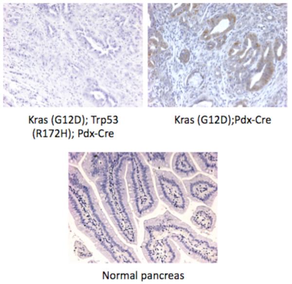
In order to further understand pancreatic cancer initiation and progression in vivo, several mouse models of this disease have been established. Hingorani et al developed the mouse model KrasG12D;Pdx1-cre, where endogenous expression of KrasG12D was directed to progenitor cells of the mouse pancreas (Hingorani et al, 2003). In addition, a second mouse model (KrasG12D;P53R172HPdx1-cre) in which both k-ras is activated and p53 inactivated recapitulates human disease in vivo. Immunohistochemistry of tumors was performed from each of the two mouse pancreatic cancer models. Gadd45a expression is upregulated in the tumor microenvironment when compared to normal pancreatic tissue (Figure 4). This upregulation, however, is p53 dependent, as tumors with inactive p53 do not express this protein (Figure 4).
Figure 4. Gadd45a immunohistochemistry of normal and diseased pancreas.
Paraffin-embedded tissue sections were stained according to manufacturer's instructions (Santa Cruz Biotechnology). Anti-Gadd45a antibody was utilized at a 1:50 dilution followed by appropriate biotinylated secondary antibody (1:200). Top left panel: Tissue section obtained from pancreatic tumor mouse model initiated by loss of p53 as well as activation of k-ras. Top right panel: Tissue section obtained from pancreatic tumor mouse model initiated by activation of k-ras. Bottom panel Normal pancreatic tissue. Tissue sections were kindly provided by Dr. Sunil Hingorani, Fred Hutchinson Cancer Research Center.
These observations on primary mouse tumors appear contradictory to results obtained in cell lines, where elevated Gadd45a can promote tumor survival and growth and yet loss of Gadd45a expression in the mouse model is associated with the more aggressive tumor state (KrasG12D;P53R172HPdx1-cre). However, it is known that the biological effects of Gadd45a are dependent upon the cellular context within which it is expressed. Since the genetic background of the pancreatic tumor cell lines is not known, varies from one line to another, as well as from one laboratory to the next, our studies will focus on the complementary approaches of animal models and human tumors to elucidate the role of Gadd45a in pancreatic ductal adenocarcinoma (PDA).
Finally, a recent study has documented Gadd45 downregulation in AML (Perugini et al, 2009). Furthermore we have observed that Gadd45a and Gadd45b function as suppressor of BCR-ABL driven leukemogenesis in a mouse model, using adaptive transplantation of BCR-ABL transduced wt and Gadd45 knockout hematopoietic cells into irradiated mice (Xiojen Sha, unpublished). In combination, these observations suggest a role for Gadd45 disregulation in hematopoietic malignancies.
VI. Prospects of targeting Gadd45 proteins in cancer therapy
To conclude, Gadd45 proteins play important roles in modulating diverse molecular pathways of stress signaling in response to a wide variety of extrinsic and physiological stress agents. Disregulated expression of Gadd45 expression has been observed in multiple types of solid tumors as well as in hematopoietic malignancies. These observations, in conjunction with the fact that Gadd45 proteins are intrinsically associated with tumor cell response to a variety of cancer therapeutic agents imply that Gadd45 proteins may serve as a novel class of targets for therapeutic intervention of cancer. Additional research is needed to pinpoint which of the Gadd45 stress response functions may be best suited for the target of cancer therapeutics.
Acknowledgments
Supported By NIH grants 1R01HL084114-01 (DAL), 1R01CA122376-01 (DAL), NIH, RO1 CA081168-06 (BH). Tissue sections were kindly provided by Dr. Sunil Hingorani, Fred Hutchinson Cancer Research Center.
Abbreviations
- GGR
global genomic repair
- MEFs
Mouse Embryo Fibroblasts
- NER
nucleotide excision repair
Biographies
 Alexandra Cretu
 Xiojen Sha
 Jennifer Tront
 Barbara Hoffman
 Dan A Liebermann
References
- Abdollahi A, Hoffman-Liebermann B, Liebermann D. Sequence and expression of a cDNA encoding MyD118: A novel myeloid differentiation primary response gene induced by multiple cytokines. Oncogene. 1990;6:165–167. [PubMed] [Google Scholar]
- Amanullah A, Azam N, Balliet A, Hollander C, Hoffman B, Fornace A, Liebermann D. Cell signalling: cell survival and a Gadd45-factor deficiency. Nature. 2003;424:741–42. doi: 10.1038/424741b. [DOI] [PubMed] [Google Scholar]
- Azam N, Vairapandi M, Zhang W, Hoffman B, Liebermann DA, Azam N, Vairapandi M, Zhang W, Hoffman B, Liebermann DA. Interaction of CR6 (GADD45g) with proliferating cell nuclear antigen impedes negative growth control. J Biol Chem. 2001;276:2766–74. doi: 10.1074/jbc.M005626200. [DOI] [PubMed] [Google Scholar]
- Bahar A, Bicknell JE, Simpson DJ, Clayton RN, Farrell WE. Loss of expression of the growth inhibitory gene GADD45gamma, in human pituitary adenomas, is associated with CpG island methylation. Oncogene. 2004;23:93–44. doi: 10.1038/sj.onc.1207193. [DOI] [PubMed] [Google Scholar]
- Barreto G, Schäfer A, Marhold J, Stach D, Swaminathan SK, Handa V, Döderlein G, Maltry N, Wu W, Lyko F, Niehrs C. Gadd45a promotes epigenetic gene activation by repair-mediated DNA demethylation. Nature. 2007;445:671–5. doi: 10.1038/nature05515. [DOI] [PubMed] [Google Scholar]
- Beadling C, Johnson KW, Smith KA. Isolation of interleukin 2-induced immediate-early genes. Proc Natl Acad Sci USA. 1993;90:2719–23. doi: 10.1073/pnas.90.7.2719. [DOI] [PMC free article] [PubMed] [Google Scholar]
- Bulavin DV, Kovalsky O, Hollander MC, Fornace AJ., Jr Loss of oncogenic H-ras-induced cell cycle arrest and p38 mitogen-activated protein kinase activation by disruption of Gadd45a. Mol Cell Biol. 2003;23:3859–71. doi: 10.1128/MCB.23.11.3859-3871.2003. [DOI] [PMC free article] [PubMed] [Google Scholar]
- Carrier F, Georgel PT, Pourquier P, Blake M, Kontny HU, Antinore MJ, Gariboldi M, Myers TG, Weinstein JN, Pommier Y, Fornace AJ., Jr Gadd45, a p53-responsive stress protein, modifies DNA accessibility on damaged chromatin. Mol Cell Biol. 1999;19(3):1673–85. doi: 10.1128/mcb.19.3.1673. [DOI] [PMC free article] [PubMed] [Google Scholar]
- Carrier F, Smith ML, Bae I, Kilpatrick KE, Lansing TJ, Chen CY, Engelstein M, Friend SH, Henner WD, Gilmer TM, Fornace AJ., Jr Characterization of human Gadd45, a p53-regulated protein. J Biol Chem. 1995;269:32672–7. [PubMed] [Google Scholar]
- Chan TA, Hwang PM, Hermeking H, Kinzler KW, Vogelstein B. Cooperative effects of genes controlling the G(2)/M checkpoint. Genes Dev. 2000;14:1584–8. [PMC free article] [PubMed] [Google Scholar]
- Engel N, Tront JS, Erinle T, Nguyen N, Latham KE, Sapienza C, Hoffman B, Liebermann DA. Conserved DNA methylation in Gadd45a(−/−) mice. Epigenetics. 2009;4 doi: 10.4161/epi.4.2.7858. Epub ahead of print. [DOI] [PMC free article] [PubMed] [Google Scholar]
- De Smaele E, Zazzeroni F, Papa S, Nguyen DU, Jin R, Jones J, Cong R, Franzoso G. Induction of Gadd45b by NF-ϰB downregulates pro-apoptotic JNK signalling. Nature. 2001;414:308–13. doi: 10.1038/35104560. [DOI] [PubMed] [Google Scholar]
- Deng C, Zhang P, Harper JW, Elledge SJ, Leder P. Mice lacking p21CIP1/WAF1 undergo normal development, but are defective in G1 checkpoint control. Cell. 1995;82:675–84. doi: 10.1016/0092-8674(95)90039-x. [DOI] [PubMed] [Google Scholar]
- Drayton S, Peters G. Immortalisation and transformation revisited. Curr Opin Genet Dev. 2002;12:98–104. doi: 10.1016/s0959-437x(01)00271-4. [DOI] [PubMed] [Google Scholar]
- El-Deiry WS, Tokino T, Velculescu VE, Levy DB, Parsons R, Trent JM, Lin D, Mercer WE, Kinzler KW, Vogelstein B. WAF1, a potential mediator of p53 tumor suppression. Cell. 1993;75:817–25. doi: 10.1016/0092-8674(93)90500-p. [DOI] [PubMed] [Google Scholar]
- Elledge SJ. Cell cycle checkpoints: preventing an identity crisis. Science. 1996;274:1664–72. doi: 10.1126/science.274.5293.1664. [DOI] [PubMed] [Google Scholar]
- Fornace AJ, Jr, Nebert DW, Hollander MC, Luethy JD, Papathanasiou M, Fargnoli J, Holbrook NJ. Induction by ionizing radiation of the Gadd45 gene in cultured human cells: lack of mediation by protein kinase C. Mol Cell Biol. 1991;11:1009–16. doi: 10.1128/mcb.11.2.1009. [DOI] [PMC free article] [PubMed] [Google Scholar]
- Fornace AJ, Jackman J, Hollander MC, Hoffman-Liebermann B, Liebermann DA. Genotoxic-stress-response genes and growth-arrest genes: gadd, MyD, other genes induced by treatments eliciting growth arrest. Ann N Y Acad Sci. 1992;663:139–154. doi: 10.1111/j.1749-6632.1992.tb38657.x. [DOI] [PubMed] [Google Scholar]
- Fornace AJ., Jr Mammalian genes induced by radiation; activation of genes associated with growth control. Annu Rev Genet. 1992;26:507–526. doi: 10.1146/annurev.ge.26.120192.002451. [DOI] [PubMed] [Google Scholar]
- Guillouf C, Grana X, Selvakumaran M, Hoffman B, Giordano A, Liebermann DA. Dissection of the genetic programs of p53 G1 growth arrest and apoptosis: Blocking p53-induced apoptosis unmasks G1 arrest. Blood. 1995;85:2691–98. [PubMed] [Google Scholar]
- Gupta M, Gupta SK, Hoffman B, Liebermann DA. Gadd45a and Gadd45b Protect Hematopoietic Cells From UV Induced Apoptosis Via Distinct Signaling Pathways including p38 actiivation and JNK inhibition. J Biol Chem. 2006;281:17552–17558. doi: 10.1074/jbc.M600950200. [DOI] [PubMed] [Google Scholar]
- Gupta M, Gupta SK, Balliet AG, Hollander MC, Fornace AJ, Hoffman B, Liebermann DA. Hematopoietic Cells from Gadd45a and Gadd45b Deficient Mice are Sensitized to Genotoxic-stress Induced Apoptosis. Oncogene. 2005;24:7170–9. doi: 10.1038/sj.onc.1208847. [DOI] [PubMed] [Google Scholar]
- Harkin DP, Bean JM, Miklos D, Song YH, Truong VB, Englert C, Christians FC, Ellisen LW, Maheswaran S, Oliner JD, Haber DA. Induction of GADD45 and JNK/SAPK-dependent apoptosis following inducible expression of BRCA1. Cell. 1999;97:575–86. doi: 10.1016/s0092-8674(00)80769-2. [DOI] [PubMed] [Google Scholar]
- Harper JW, Adami GR, Wei N, Keyomarsi K, Elledge SJ. The p21 Cdk-interacting protein Cip1 is a potent inhibitor of G1. Cell. 1993;75:805–816. doi: 10.1016/0092-8674(93)90499-g. [DOI] [PubMed] [Google Scholar]
- Hildesheim J, Bulavin DV, Anver MR, Alvord WG, Hollander MC, Vardanian L, Fornace AJ., Jr Gadd45a protects against UV irradiation-induced skin tumors, promotes apoptosis and stress signaling via MAPK and p53. Cancer Res. 2002;62:7305–15. [PubMed] [Google Scholar]
- Hingorani SR, Petricoin EF, Maitra A, Rajapakse V, King C, et al. Preinvasive and invasive ductal pancreatic cancer and its early detection in the mouse. Cancer Cell. 2003;4:437–450. doi: 10.1016/s1535-6108(03)00309-x. [DOI] [PubMed] [Google Scholar]
- Hollander MC, Sheikh MS, Bulavin DV, Lundgren K, AugeriHenmueller L, Shehee R, Molinaro TA, Kim KE, Tolosa E, Ashwell JD, Rosenberg MP, Zhan Q, Fernandez- Salguero PM, Morgan WF, Deng CX, Fornace AJ., Jr Genomic instability in Gadd45a- deficient mice. Nat Genet. 1999;23:176–84. doi: 10.1038/13802. [DOI] [PubMed] [Google Scholar]
- Jin S, Antinore MJ, Lung FD, Dong X, Zhao H, Fan F, Colchagie AB, Blanck P, Roller PP, Fornace AJ, Jr, Zhan Q. The GADD45 inhibition of Cdc2 kinase correlates with GADD45- mediated growth suppression. J Biol Chem. 2000;275:16602–8. doi: 10.1074/jbc.M000284200. [DOI] [PubMed] [Google Scholar]
- Jonsson ZO, Hubsche U. Proliferating cell nuclear antigen: more than a clamp for DNA polymerases. Bio Essays. 1997;19:967–975. doi: 10.1002/bies.950191106. [DOI] [PubMed] [Google Scholar]
- Kastan MB, Zhan Q, El-Deiry WS, Carrier F, Jacks T, Walsh WV, Plunkett BS, Vogelstein B, Fornace AJ., Jr A mammalian cell cycle checkpoint utilizing p53 and Gadd45 is defective in Ataxia-telangiectasia. Cell. 1992;71:587–597. doi: 10.1016/0092-8674(92)90593-2. [DOI] [PubMed] [Google Scholar]
- Kelman Z, Hurwitz J. Protein-PCNA interactions: a DNA-scanning mechanism? Trends Biochem Sci. 1998;23:236–238. doi: 10.1016/s0968-0004(98)01223-7. [DOI] [PubMed] [Google Scholar]
- Kojima S, Mayumi-Matsuda K, Suzuki H, Sakata T. Molecular cloning of rat GADD45g, gene induction and its role during neuronal cell death. FEBS Lett. 1999;446:313–7. doi: 10.1016/s0014-5793(99)00234-3. [DOI] [PubMed] [Google Scholar]
- Li Y, Qian H, Li X, Wang H, Yu J, Liu Y, Zhang X, Liang X, Fu M, Zhan Q, Lin C. Adenoviral-mediated gene transfer of Gadd45a results in suppression by inducing apoptosis and cell cycle arrest in pancreatic cancer cell. J Gene Med. 2009;11:3–13. doi: 10.1002/jgm.1270. [DOI] [PubMed] [Google Scholar]
- Liebermann DA, Hoffman B. Waxman S, editor. MyD/GADD genes in terminal differentiation growth arrest and apoptosis in “Differentiation Therapy”. Ares-Serono Symposia-Series Challange of Modern Medicine. 1995;10:107–116. [Google Scholar]
- Liebermann DA, Hoffman B. MyD genes in negative growth control. Oncogene. 1998;17:3319–30. doi: 10.1038/sj.onc.1202574. [DOI] [PubMed] [Google Scholar]
- Lu B, Yu H, Chow C, Li B, Zheng W, Davis RJ, Flavell RA. GADD45g mediates the activation of the p38 and JNK MAP kinase pathways and cytokine production in effector TH1 cells. Immunity. 2001;14:583–90. doi: 10.1016/s1074-7613(01)00141-8. [DOI] [PubMed] [Google Scholar]
- Lundberg AS, Hahn WC, Gupta P, Weinberg RA. Genes involved in senescence and immortalization. Curr Opin Cell Biol. 2000;12:705–9. doi: 10.1016/s0955-0674(00)00155-1. [DOI] [PubMed] [Google Scholar]
- Ma DK, Jang MH, Guo JU, Kitabatake Y, Chang ML, Pow-Anpongkul N, Flavell RA, Lu B, Ming GL, Song H. Neuronal activity-induced Gadd45b promotes epigenetic DNA demethylation and adult neurogenesis. Science. 2009;323:1074–7. doi: 10.1126/science.1166859. [DOI] [PMC free article] [PubMed] [Google Scholar]
- Fan W, Richter G, Cereseto A, Beadling C, Smith KA. Cytokine response gene 6 induces p21 and regulates both cell growth and arrest. Oncogene. 1999;18:6573–82. doi: 10.1038/sj.onc.1203054. [DOI] [PubMed] [Google Scholar]
- O'Connor PM. Mammalian G1 and G2 phase checkpoints. Cancer Surv. 1997;29:151–82. [PubMed] [Google Scholar]
- Papa S, Zazzeroni F, Bubici C, Jayawardena S, Alvarez K, Matsuda S, Nguyen DU, Pham CG, Nelsbach AH, Melis T, De Smaele E, Tang WJ, D'Adamio L, Franzoso G. Gadd45 b mediates the NF-ϰ B suppression of JNK signalling by targeting MKK7/JNKK2. Nat Cell Biol. 2004;6:146–53. doi: 10.1038/ncb1093. [DOI] [PubMed] [Google Scholar]
- Papathanasiou MA, Kerr NC, Robbins JH, McBride OW, Alamo I, Jr, Barrett SF, Hickson ID, Fornace AJ., Jr Mammalian genes coordinately regulated by growth arrest signals and DNA-damaging agents. Mol Cell Biol. 1989;10:4196–203. doi: 10.1128/mcb.9.10.4196. [DOI] [PMC free article] [PubMed] [Google Scholar]
- Perugini M, Kok CH, Brown AL, Wilkinson CR, Salerno DG, Young SM, Diakiw SM, Lewis ID, Gonda TJ, D'Andrea RJ. Repression of Gadd45α by activated FLT3 and GM-CSF receptor mutants contributes to growth, survival and blocked differentiation. Leukemia. 2009 doi: 10.1038/leu.2008.349. in press. [DOI] [PubMed] [Google Scholar]
- Ramachandran K, Gopisetty G, Gordian E, Navarro L, Hader C, Reis IM, Schulz WA, Singal R. Methylation-mediated repression of GADD45α in prostate cancer and its role as a potential therapeutic target. Cancer Res. 2009;69:1527–35. doi: 10.1158/0008-5472.CAN-08-3609. [DOI] [PubMed] [Google Scholar]
- Rai K, Huggins IJ, James SR, Karpf AR, Jones DA, Cairns BR. DNA demethylation in zebrafish involves the coupling of a deaminase, a glycosylase, Gadd45. Cell. 2008;135:1201–12. doi: 10.1016/j.cell.2008.11.042. [DOI] [PMC free article] [PubMed] [Google Scholar]
- Sancar A. Mechanisms of DNA excision repair. Science. 1994;266:1954–1956. doi: 10.1126/science.7801120. [DOI] [PubMed] [Google Scholar]
- Schmitz KM, Schmitt N, Hoffmann-Rohrer U, Schäfer A, Grummt I, Mayer C. TAF12 recruits Gadd45a and the nucleotide excision repair complex to the promoter of rRNA genes leading to active DNA demethylation. Mol Cell. 2009;33:344–53. doi: 10.1016/j.molcel.2009.01.015. [DOI] [PubMed] [Google Scholar]
- Schneider G, Weber A, Zechner U, Oswald F, Friess HM, Schmid RM, Liptay S. Gadd45a is highly expressed in pancreatic ductal adenocarcinoma cells and required for tumor cell viability. Int J Cancer. 2006;11:2405–11. doi: 10.1002/ijc.21637. [DOI] [PubMed] [Google Scholar]
- Selvakumaran M, Lin HK, Miyashita T, Wang HG, Krajewski S, Reed JC, Hoffman B, Liebermann D. Immediate early up-regulation of bax expression by p53 but not TGFb1: A paradigm for distinct apoptotic pathways. Oncogene. 1994b;9:1791–1798. [PubMed] [Google Scholar]
- Selvakumaran M, Lin HK, Tjin Tham Sjin R, Reed J, Liebermann D, Hoffman B. The novel primary response gene MyD118 and the proto-oncogenes myb, myc and bcl-2 modulate transforming growth factoMol. Cell Biol. 1994a;14:2352–2360. doi: 10.1128/mcb.14.4.2352. [DOI] [PMC free article] [PubMed] [Google Scholar]
- Shaulian E, Karin M. Stress-induced JNK activation is independent of Gadd45 induction. J Biol Chem. 1999;274:29595–8. doi: 10.1074/jbc.274.42.29595. [DOI] [PubMed] [Google Scholar]
- Smith ML, Chen IT, Zhan Q, Bae I, Chen CY, Gilmer TM, Kastan MB, O'Connor PM, Fornace AJ., Jr Protein interaction of the p53-regulatproliferating cell nuclear antigen. Science. 1994;266:1376–80. doi: 10.1126/science.7973727. [DOI] [PubMed] [Google Scholar]
- Smith ML, Ford JM, Hollander MC, Bortnick RA, Amundson SA, Seo YR, Deng CX, Hanawalt PC, Fornace AJ., Jr p53-mediated DNA repair responses to UV radiation: studies of mouse cells lacking p53, p21, and/or Gadd45 genes. MCB. 2000;20:3705–14. doi: 10.1128/mcb.20.10.3705-3714.2000. [DOI] [PMC free article] [PubMed] [Google Scholar]
- Smith ML, Kontny HU, Zhan Q, Sreenath A, O'Connor PM, Fornace AJ., Jr Antisense GADD45 expression results in decreased DNA repair and sensitizes cells to u.v.-irradiation or cisplatin. Oncogene. 1996;13:2255–63. [PubMed] [Google Scholar]
- Sun L, Gong R, Wan B, Huang X, Wu C, Zhang X, Zhao S, Yu L. GADD45g, down- regulated in 65 % hepatocellular carcinoma (HCC) from 23 Chinese patients, inhibits cell growth and induces cell cycle G2/M arrest for hepatoma Hep-G2 cell lines. Mol Biol Rep. 2003;30:249–53. doi: 10.1023/a:1026370726763. [DOI] [PubMed] [Google Scholar]
- Takekawa M, Saito H. A family of stress-inducible GADD45-like proteins mediate activation of the stress-responsive MTK1/MEKK4/MAPKKK. Cell. 1998;95:521–30. doi: 10.1016/s0092-8674(00)81619-0. [DOI] [PubMed] [Google Scholar]
- Tront JS, Hoffman B, Liebermann DA. Gadd45a Suppresses Ras-driven Mammary Tumorigenesis by Activation of JNK and p38 Stress Signaling Resulting in Apoptosis and Senescence. Cancer Res. 2006;66:8448–54. doi: 10.1158/0008-5472.CAN-06-2013. [DOI] [PubMed] [Google Scholar]
- Vairapandi M, Azam N, Balliet AG, Hoffman B, Liebermann DA. Characterization of MyD118, Gadd45, PCNA interacting domains: PCNA impedes MyD/Gadd Mediated Negative Growth Control. J Biol Chem. 2000;275:16810–16819. doi: 10.1074/jbc.275.22.16810. [DOI] [PubMed] [Google Scholar]
- Vairapandi M, Balliet AG, Hoffman B, Liebermann DA. The differentiation primary response gene MyD118, related to GADD45, encodes for a nuclear protein which interacts with PCNA and p21WAF1/CIP1. Oncogene. 1996;12:2579–2594. [PubMed] [Google Scholar]
- Vairapandi M, Balliet AG, Hoffman B, Liebermann DA. GADD45b and GADD45g are cdc2/cyclinB1 kinase inhibitors with a role in S and G2/M cell cycle checkpoints induced by genotoxic stress. J Cell Physiol. 2002;192:327–38. doi: 10.1002/jcp.10140. [DOI] [PubMed] [Google Scholar]
- Wang X, Gorospe M, Holbrook NJ. Gadd45 is not required for activation of c-Jun N- terminal kinase or p38 during acute stress. J Biol Chem. 1999;274:29599–602. doi: 10.1074/jbc.274.42.29599. [DOI] [PubMed] [Google Scholar]
- Wang XW, Zhan Q, Coursen JD, Khan MA, Kontny HU, Yu L, Hollander MC, O'Connor PM, Fornace AJ, Jr, Harris CC. GADD45 induction of a G2/M cell cycle checkpoint. Proc Natl Acad Sci USA. 1999;96:3706–11. doi: 10.1073/pnas.96.7.3706. [DOI] [PMC free article] [PubMed] [Google Scholar]
- Xiong Y, Hannon GJ, Zhang H, Casso D, Kobayashi R, Beach D. p21 is a universal inhibitor of cyclin kinases. Nature. 1993;366:701–4. doi: 10.1038/366701a0. [DOI] [PubMed] [Google Scholar]
- Yamasawa K, Nio Y, Dong M, Yamaguchi K, Itakura M. Clinicopathological significance of abnormalities in Gadd45 expression and its relationship to p53 in human human pancreatic cancer. Clin Cancer Res. 2002;8:2563–9. [PubMed] [Google Scholar]
- Yang J, Zhu H, Murphy TL, Ouyang W, Murphy KM. IL-18-stimulated GADD45 b required in cytokine-induced, but not TCR-induced, IFN-g production. Nat Immunol. 2001;2:157–64. doi: 10.1038/84264. [DOI] [PubMed] [Google Scholar]
- Yang Q, Manicone A, Coursen JD, Linke SP, Nagashima M, Forgues M, Wang XW. Identification of a functional domain in a Gadd45-mediated G2/M checkpoint. J Biol Chem. 2000;275:36892–8. doi: 10.1074/jbc.M005319200. [DOI] [PubMed] [Google Scholar]
- Ying J, Srivastava G, Hsieh WS, Gao Z, Murray P, Liao SK, Ambinder R, Tao Q. The stress-responsive gene GADD45G is a functional tumor suppressor, with its response to environmental stresses frequently disrupted epigenetically in multiple tumors. Clin Cancer Res. 2005;11:6442–9. doi: 10.1158/1078-0432.CCR-05-0267. [DOI] [PubMed] [Google Scholar]
- Yoo J, Ghiassi M, Jirmanova L, Balliet AG, Hoffman B, Fornace AJ, Jr, Liebermann DA, Bottinger EP, Roberts AB. TGF-b-induced apoptosis is mediated by Smad-dependent expression of GADD45B through p38 activation. JBC. 2003;278:43001–7. doi: 10.1074/jbc.M307869200. [DOI] [PubMed] [Google Scholar]
- Zerbini LF, Libermann TA. Life and death in cancer. GADD45 and g are critical regulators of NF-ϰB mediated escape from programmed cell death. Cell Cycle. 2005;4:18–20. doi: 10.4161/cc.4.1.1363. [DOI] [PubMed] [Google Scholar]
- Zhan Q, Antinore MJ, Wang XW, Carrier F, Smith ML, Harris CC, Fornace AJ., Jr Association with Cdc2 and inhibition of Cdc2/Cyclin B1 kinase activity by the p53-regulated protein Gadd45. Oncogene. 1999;18:2892–900. doi: 10.1038/sj.onc.1202667. [DOI] [PubMed] [Google Scholar]
- Zhan Q, Fan S, Smith ML, Bae I, Yu K, Alamo I, Jr, O'Connor PM, Fornace AJ., Jr Abrogation of p53 function affects gadd gene responses to DNA base-damaging agents and starvation. DNA Cell Biol. 1996;15:805–15. doi: 10.1089/dna.1996.15.805. [DOI] [PubMed] [Google Scholar]
- Zhan Q, Lord KA, Alamo I, Jr, Hollander MC, Carrier F, Ron D, Kohn KW, Hoffman B, Liebermann DA, Fornace AJ., Jr The gadd and MyD genes define a novel set of mammalian genes encoding acidic proteins that synergistically suppress cell growth. Mol Cell Biol. 1994;14:2361–2371. doi: 10.1128/mcb.14.4.2361. [DOI] [PMC free article] [PubMed] [Google Scholar]
- Zhang H, Xiong Y, Beach D. Proliferating cell nuclear antigen and p21 are components of multiple cell cycle kinase complexes. Mol Biol Cell. 1993;4:897–906. doi: 10.1091/mbc.4.9.897. [DOI] [PMC free article] [PubMed] [Google Scholar]
- Zhang W, Bae I, Krishnaraju K, Azam N, Fan W, Smith K, Hoffman B, Liebermann DA. CR6: A third member in the MyD118 & Gadd 45 gene family which functions in negative growth control. Oncogene. 1999;18:4899–4907. doi: 10.1038/sj.onc.1202885. [DOI] [PubMed] [Google Scholar]
- Zhang W, Hoffman B, Liebermann DA. Ectopic expression of MyD118/Gadd45/CR6 (Gadd45b/α/g) sensitizes neoplastic cells to genotoxic stress-induced apoptosis. Int J Oncol. 2001;18:749–57. [PubMed] [Google Scholar]
- Zhang W. The MyD118/Gadd4/CR6 gene gamily in negative growth control. Temple University; 2000. Thesis. [Google Scholar]


