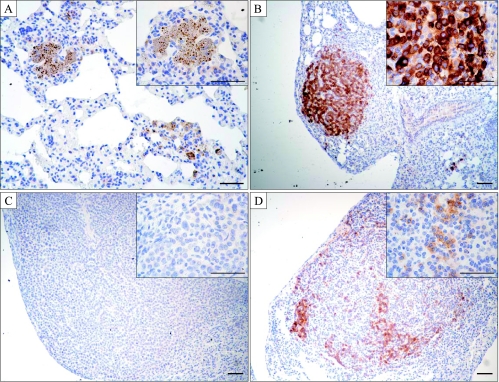
Figure 7.
Immunohistochemistry for IGF-IR in the lungs from mice treated with doxycycline for 8 months followed by a 1 month withdrawal of doxycycline. (A) Mouse with apparent regressed tumor lesions, (B) a tumor containing primarily IGF-IR-positive cells, (C) a tumor containing exclusively IGF-IR-negative cells, and (D) a number containing both IGF-IR-positive and IGF-IR-negative cells. The inset is a higher magnification of the stained slide. Scale bars, 50 µm; insets, 100 µm.

