Abstract
Little is known about osteonal bone mineral and matrix properties, although these properties are of major importance for the understanding of bone alterations related to age and bone diseases such as osteoporosis. During aging, bone undergoes modifications that compromise their structural integrity as shown clinically by the increase of fracture incidence with age. Based on Fourier transform infrared (FTIR) analysis from baboons between 0 and 32 yr of age, consistent systematic variations in bone properties as a function of tissue age are reported within osteons. The patterns observed were independent of animal age and positively correlated with bone tissue elastic behavior measured by nano-indentation. As long as tissue age is expressed as a percentage of the entire osteon radius, osteonal analyses can be used to characterize disease changes independent of the size of the osteon. These mineral and matrix analyses can be used to explain bone fragility. The mineral content (mineral-to-matrix ratio) was correlated with the animal age in both old (interstitial) and newly formed bone tissue, showing for the first time that age-related changes in BMC can be explain by an alteration in the mineralization process itself and not only by an imbalance in the remodeling process.
Key words: bone quality, aging, osteon, Fourier transform infrared microspectroscopy, Fourier transform infrared imaging, nonhuman primate model
INTRODUCTION
Aging is a natural process that negatively affects bone integrity in ways that are not completely understood but that manifest clinically in the increase of fracture incidence with age.(1,2) Because fracture risk cannot be explained simply by reduced “bone mass,”(1–6) the tissue and animal age modifications in bone properties need to be characterized and related to mechanical behavior to help understand the etiology of age-related skeletal fragility. Osteons, representing the newly remodeled cortical bone, are formed by resorption of existing bone coupled with circumferential apposition of mineralized collagen fiber lamellae surrounding a Haversian canal. These tree ring-like structures with the newest mineralized layers deposited nearest to the blood vessel canal (osteonal center) present a natural gradient in tissue age, from the center (youngest tissue) to the periphery (oldest tissue).
Infrared and Raman microspectroscopy and imaging have been used to describe chemical changes in human bone related to aging, diseases, and pharmacology of treatments.(7–11) These methods provide spatial information in addition to the geometry and density provided by techniques routinely applied in the clinic. Previous studies using spectroscopy reported that in normal cortical and trabecular bone, the mineral content, carbonate substitution, crystallinity, and collagen maturity increased with tissue age.(12,13) The same patterns with tissue age were described for mineral properties in osteonal bone.(14,15) Additional studies showed these specific variations linked with tissue age seem to be lost in bone of osteoporotic and osteopenic individuals.(16,17) Unfortunately, using human bone biopsies, the animal-age effects cannot be easily distinguished. Another limitation of these studies was the small amount of tissue available in the biopsies, raising questions about whether the sampled osteons and trabeculae are representative.
To address these questions and with the intention in future studies to characterize the changes related to bone diseases or drug therapies by the analysis of newly formed bone, this paper examines variations in the tissue age and animal age in multiple osteons from healthy baboon femurs by Fourier transform infrared microspectroscopy (FTIRM) and imaging (FTIRI). Additionally, mechanical properties of osteonal tissue obtained by atomic force microscopy (AFM) nano-indentation are correlated with compositional bone properties obtained by FTIRI.
Baboon bone, like human bone, is constantly remodeling and presents similar Haversian microstructures that result from secondary remodeling. The baboon, a well-established model for the study of human bone maintenance and turnover, provides an excellent opportunity for examining animal and tissue age-related variation in the chemical and compositional properties as well as the mechanical properties of bone within osteons. Baboons have a lifespan approximately equivalent to one third of the human(18) and develop similar skeletal fragility with aging.(19,20) Moreover, the female baboon lifespan is sufficiently long to provide evidence of reproductive senescence and natural menopause associated with changes in hormone levels that play an important role in bone metabolism and loss.(19,21)
MATERIALS AND METHODS
Animals
Twenty-seven femurs from 1 male and 26 female baboons (Papio hamadryas spp.), ranging in age from 0 to 31 yr, were used in this study. All animals were from the colony at the Southwest National Primate Research Center/Southwest Foundation for Biomedical Research (SNPRC/SFBR, San Antonio, TX, USA). This colony was established during the 1950s with wild-caught baboons from Kenya to develop the baboon model in medical research.(22) Our study was conducted in accordance with the Guide for the Care and Use of Laboratory Animals.(23) The Institutional Animal Care and Use Committee approved all procedures during the baboons' lives at SNPRC/SFBR in accordance with established guidelines. Inclusion criteria were death by natural causes and absence of reported metabolic bone disease. The population was divided into nine different average age groups (0, 1, 2.5, 6, 9, 13.5, 18.5, 27, and 31 yr). Each group consisted of three animals (Table 1). Our goal during this work was not to assess sex-specific differences. However, with the exception of one animal in the youngest group, we examined only females.
Table 1.
Baboon Population Used
| Age group (yr) | Sex | Age |
| 0 | F | Stillborn |
| M | Stillborn | |
| F | 0.2 | |
| 1 | F | 0.9 |
| F | 1.1 | |
| F | 1.2 | |
| 2.5 | F | 2.35* |
| F | 2.4* | |
| F | 2.8*† | |
| 6 | F | 6 |
| F | 6.1 | |
| F | 6.2 | |
| 9 | F | 8.9 |
| F | 9.1 | |
| F | 9.3 | |
| 13.5 | F | 13.2‡ |
| F | 13.5‡ | |
| F | 13.7‡ | |
| 18.5 | F | 17.5 |
| F | 18.8 | |
| F | 19.1 | |
| 27 | F | 26.4 |
| F | 27.2 | |
| F | 27.3 | |
| 31 | F | 30.5 |
| F | 32.5 | |
| F | 32 |
All samples were used for FTIR.
* Samples used for Raman.
† Samples used for EDX.
‡ Samples used for AFM nano-indentation.
Sample preparation
Bone samples were collected during routine necropsies, wrapped in saline-soaked gauze, sealed in plastic bags, and maintained in frozen storage at –20°C before specimen preparation. Specimens were initially fixed with 80% ethanol (EtOH). The segments were slowly dehydrated through a series of increasing concentrations of EtOH, cleared with xylene, and finally infiltrated and embedded in polymethylmethacrylate (PMMA) using the Erben method.(24) For the FTIRI analysis, 2-μm-thick sections were cut from the embedded undecalcified bone blocks with a Leica microtome (SM 2500; Leica). The sections were transferred onto BaF2 windows (SpectraTek, Hopewell Junction, NY, USA) and mounted onto a fixed stage on the FTIR microspectrometer. The residual bone blocks, from which the sections were cut, were used for the Raman spectroscopy, nano-indentation, and energy dispersive X-ray (EDX).
FTIR microspectroscopy and imaging analysis
Spectra were acquired with a Spectrum Spotlight 300 Imaging System (Perkin Elmer Instruments, Shelton, CT, USA), consisting of a step-scanning FT-IR spectrometer with an MCT (mercury-cadmium-telluride) focal plane array detector placed at the image focal plane of an IR microscope. Single spectra and images were collected in the transmission mode at a spectral resolution of 4 cm−1 in the frequency region between 2000 and 800 cm−1 with an IR detector pixel size of 6.25× 6.25 μm. Two different IR methods were used to analyze individual osteons: line mode, where sequential single spectra were recorded in 10-μm steps along three different lines starting from the center and moving to the periphery of osteons; and image mode, in which sequential FTIR images (12.5 × 12.5 μm) from the center toward the periphery were recorded along three different orientations in the same osteons. Data obtained with these two sampling methods were statistically indistinguishable for both spectral parameters and each age group. The linear regression calculated between the results obtained by the line and the image mode for each IR parameters showed very high levels of correlation and slopes close to unity verifying that the results obtained were the same for both IR sampling methods. Therefore, in this study, IR data represent average results from both methods. From each baboon sample (n = 27), four different osteons were randomly chosen from the cortical section and analyzed. The mineral-to-matrix ratio was also calculated with FTIR imaging on the overall cortical bone tissue (osteonal and primary interstitial) between the endocortical surface and the periosteal surface from each baboon sample. All spectra were baseline corrected, and the embedding medium was spectrally subtracted using ISYS software (Spectral Dimensions, Olney, MD, USA). The results are presented by age group with SD. Error bars were removed on some figures in this work for clarity.
The following FTIR parameters reviewed in detail elsewhere(25,26) were calculated using ISYS software. Mineral-to-matrix ratio, which is linearly related to mineral content,(27) was calculated by integrating the phosphate area peaks between 916 and 1180 cm−1 and the amide I mode from 1592 to 1712 cm−1. Carbonate-to-phosphate ratio, which reflects the level of carbonate substitution in the hydroxyapatite (HA) crystal,(28) was calculated through the integrated area of the ν2 carbonate peak (840–892 cm−1) and that of the phosphate. Crystallinity, related to the size and perfection of HA crystal,(29) was calculated as the ratio of relative peak height subbands at 1030 and 1020 cm−1 within the broad phosphate contour. The collagen cross-linking network maturity was estimated by the intensity ratio of amide I subbands at 1660 and 1690 cm−1.(30) Data for all parameters are presented as the mean of the results obtained from three baboon samples in the same age group and include four osteons in each sample and three lines per osteon.
Raman microspectroscopy analysis
Raman spectra were acquired with a Kaiser Optical Systems Raman Microprobe with a Leica microscope (Model DMLP). This instrument uses a 785-nm diode laser to generate ∼7–10 mW of single mode laser power at the sample with a spot size of ∼2 μm using a ×100 objective. The backscattered radiation illuminates a near-IR CCD (Model DU 401-BR-DD; ANDOR Technology). Spectra were recorded from 1800 to 400 cm−1, with a resolution of 4 cm−1. A line scan was generated in which sequential single spectra were recorded in 3-μm steps along three different lines starting from the osteonal center to the periphery in four different osteons chosen randomly for each sample analyzed. To compare Raman and FTIR data, the baboons of the 2.5-yr-old group were analyzed by Raman because these had shown the steepest gradients by FTIR. Detector integration time for each scan was 20-s exposure time. The final spectra were the average of two scan accumulations. The same embedded and polished bone blocks used to cut IR sections were placed directly on the stage of the Raman microscope, and the transverse cross-section was oriented perpendicular to the laser beam incident from the microscope ×100 objective.
The following Raman parameters were calculated as reviewed elsewhere.(8,31,32) The carbonate-to-phosphate ratio was calculated from the height of the carbonate ν1 band (∼1070 cm−1) and ν1 phosphate peak (960 cm−1), where the substitution of the PO4 −3 functional group by CO3 −2 (B-type carbonate) exhibits a distinct peak at 1070 cm−1.(33) Crystallinity (crystal size and perfection) of the HA crystal was approximated from the full width at half maximum (FWHM) of ν1 phosphate peak intensity(34) such that crystallinity = 1/bandwidth. A sharper peak (decreasing FWHM) indicates greater crystallinity. The mineral-to-matrix ratio, generally calculated from the intensity of the phosphate peak to the intensity of the amide I band (∼1665 cm−1), was determined using the phenylalanine ring-breathing mode at 1004 cm−1 in place of amide I as the matrix index because the spectra recorded were too noisy around the amide I and amide III peaks.
Nano-indentation
To correlate mineral and matrix properties calculated by FTIR and Raman with local tissue mechanical properties, nano-indentation was performed along radial lines of the baboon osteons. The three samples of the 13-yr-group were selected for nano-indentation. Transverse sections 3 mm thick were cut from the embedded, undecalcified bone blocks. The specimens were polished anhydrously to avoid demineralization and reprecipitation.(35,36) The RMS roughness of the sample surfaces was assessed by atomic force microscopy (AFM) and was <15 nm over an area of 5 × 5 μm.
Nano-indentation uses a depth-sensing indenter that is pressed into the material with a specified load at a constant rate and withdrawn, producing an output curve of load versus displacement. The indentation modulus, Ei, was calculated from the unloading portion of the curve(35) as:
The hardness, H, is the average pressure under load, and was calculated as:
where Ac and S are the contact area and stiffness, respectively, at maximum load, P max. E is the Young's modulus, and υ is the Poisson's ratio, where the subscripts s and t refer to the sample and tip materials, respectively.
Indentation was performed on three osteons per sample, in regions that had been previously imaged with FTIR. Each osteon was indented with three radial lines, with indents placed at the center of each lamella. The scanning nano-indenter (TriboIndentor; Hysitron, Minneapolis, MN, USA) used for these experiments consisted of a nano-indentation transducer with a Berkovich diamond indenter tip and a scanning probe microscope. The shape of the indenter tip was characterized using the method proposed by Oliver and Pharr.(37) Before indentation, a 40 × 40-μm surface topography scan of the sample was performed to locate the area of interest within an osteon. Then, before each indent, a 20 × 20-μm scan was made to accurately place an indent on the center of each lamella. The tip was loaded into the sample at a rate of 50 μN/s, held at a maximum load of 700 μN for 10 s, and unloaded at a rate of 50 μN/s. The load-unload rates and the hold time, as well as the maximum load, were chosen based on prior experiments and for reasons described previously.(35,36)
EDX analysis
Quantitative EDX analysis and additional FTIR analysis were conducted on the same four osteons from one baboon of the 2.5-yr age group. Electron micrographs of the osteons were obtained at ×1000 magnification with a scanning electron microscope (Quanta 600; FEI). Information about the chemical elements along osteons was obtained from the EDX spectra at 20 KV (model Phoenix; EDAX). For each osteon, EDX line analysis was performed along three different directions from the center to the periphery of the osteon. Each sequential single EDX point in ∼0.3-μm steps along the lines was recorded at 20 kV for 200 ms.
Statistical analysis
The mean ± SD for each osteon and each animal was calculated for all FTIR, Raman, and nano-indentation data. These were checked by ANOVA in each age range to ensure that the groups could be combined. All statistical analyses were performed with SigmaStat (Systat Software, San Jose, CA, USA). Linear or polynomial regression analyses were performed to detect correlations between tissue and animal age, physicochemical parameters from the FTIR and Raman data, the mechanical parameters from nano-indentation, or chemical content given by EDX. Regressions used the values obtained from each baboon age group as indicated. Correlations are reported as the square of the Pearson's correlation coefficient (r 2). The level of significance was set at p < 0.01 for all statistical analyses.
RESULTS
At all ages examined, the cortical baboon samples presented well-defined osteons (Figs. 1A and 1C), with the exception of the youngest group for which visualizing osteons was extremely difficult. Typical IR and Raman spectra (Figs. 1B and 1D) of osteonal tissue from dehydrated bones show the spectral contribution of major molecular species from both the mineral and matrix components.
FIG. 1.
Typical photomicrographs of the osteons obtained by the optical attachments of the FTIR (A) and Raman (C) spectrometer. The dotted circles represent the outline of the Haversian canal. The other line defines the peripheries of the osteon used for analyses. Typical FTIR (B) and Raman (D) spectrum recorded from osteonal tissue in baboon cortical bone. The bands of interest for this study were CO3 −2 (840–890 cm−1), PO4 −3 (916–1180 cm−1), and amide I (1592–1712 cm−1) for IR analyses and PO4 −3 (960 cm−1), Ring mode of phenylalanine (1004 cm−1), and CO3 −2 (1065–1071 cm−1) for Raman analyses. Spectra were baselined, and PMMA (embedding media) spectra were subtracted.
Some osteonal FTIR parameters were correlated with tissue and animal age
Independent of the baboon age group considered, the osteons observed in the same sections showed significant variability in their diameters, which ranged between 200 and 500 μm. To compare the results, the spectroscopic parameters were plotted as a function of the radial distance across the osteons and were expressed as percentage from the osteonal center (0%) to the periphery (100%).
The mineral content increased linearly as a function of increasing distance from the osteonal center to the periphery with a strong correlation coefficient (r 2 = ∼0.93, p < 0.001) as shown for the analysis of the 2.5-yr-old animals (Fig. 2A). This behavior held across all age groups examined (Fig. 2B). The slopes and the correlation coefficients for each individual age group were similar and independent of the age of the baboon (Table 2). However, the absolute values of these mineral-to-matrix ratios seemed to depend on animal age. The variation of the mineral-to-matrix ratio was nonlinearly correlated (r 2 = ∼0.9, p < 0.01) with the baboon age, regardless of the section analyzed along osteons (Fig. 2C).This ratio increased rapidly with baboon age until 15–20 yr and gradually decreased until 31 yr, the oldest age tested. Compared with the total cortical bone area (osteonal plus interstitial lamellae) from the same baboon sections (Fig. 2D), the average mineral-to-matrix ratio calculated over the entire osteon, regardless of animal age, was smaller than that calculated in the cortical bone. A similar relationship with animal age was observed as that within osteons.
FIG. 2.
FTIR analysis of the mineral-to-matrix ratio. (A) Typical data obtained for this ratio as a function of the distance along osteons from one baboon age group (2.5 yr). (B) Average mineral-to-matrix ratio calculated along the osteons for the nine baboon age groups. (C) Mineral-to-matrix ratio plotted as a function of the baboon age for each osteonal sections (0.83 < r 2 < 0.89). (D) Comparison of the mineral-to-matrix ratio plotted as a function of the baboon age recorded in the total cortical bone (triangles) and in individual osteons (diamonds) from the same baboon section. Values are mean ± SD (n = 36).
Table 2.
FTIRI Properties Correlated With Tissue Age for the Nine Age Groups
| Age group (yr) |
Min/matrix ratio |
CO3/PO4 ratio |
1030/1020 ratio |
1660/1690 ratio |
||||
| Slope | r2 | Slope | r2 | Slope | r2 | Slope | r2 | |
| 0 | 0.61 | 0.90* | −0.01 | 0.93† | 0.16 | 0.93† | 1.71 | 0.96† |
| 1 | 0.68 | 0.93* | −0.006 | 0.99† | 0.16 | 0.96* | 1.32 | 0.93† |
| 2.5 | 0.97 | 0.91* | −0.006 | 0.94† | 0.16 | 0.95† | 0.84 | 0.97† |
| 6 | 0.74 | 0.99* | −0.007 | 0.93† | 0.23 | 0.97* | 1.71 | 0.96† |
| 9 | 0.63 | 0.96* | −0.007 | 0.92† | 0.19 | 0.93† | 0.97 | 0.95† |
| 13.5 | 0.89 | 0.98* | −0.005 | 0.96† | 0.16 | 0.94† | 0.89 | 0.96† |
| 18.5 | 0.91 | 0.96* | −0.006 | 0.98† | 0.22 | 0.95† | 0.99 | 0.95† |
| 27 | 0.77 | 0.99* | −0.004 | 0.96† | 0.14 | 0.93† | 0.89 | 0.94† |
| 31 | 0.92 | 0.94* | −0.007 | 0.98† | 0.22 | 0.95† | 1.32 | 0.93† |
Slope and correlation reported as the square of the Pearson's correlation coefficient (r 2) for each analyzed IR parameter plotted as a function of the anatomical location inside the osteons calculated for each baboon age groups shown in Figs. 2B, 3B, 4B, and 4D. Each age group consisted of three animals.
* p < 0.001.
† p < 0.01.
The carbonate-to-phosphate ratio decreased with tissue age (Figs. 3A and 3B) but increased with animal age (Fig. 3C). The variation of the ratio with distance from the osteon center exhibited two-phase behavior in all age groups (Figs. 3A and 3B). In the first phase, the carbonate-to-phosphate ratio decreased linearly until a minimum value was reached, typically at ∼40% from the osteon center. In the second phase, the ratio was essentially constant. The slopes and correlation coefficients of the first linear phase were similar and independent of animal age (Table 2). The decrease in carbonate-to-phosphate ratio was not caused by a decrease in carbonate content as shown by the increase in the carbonate-to-matrix ratio along the osteons (Fig. 3D) but by an increase in the phosphate content close to Haversian canal. The carbonate substitution in the osteonal tissue seemed dependent on the animal age. The ratio was linearly correlated with the baboon age for each section along osteons (Fig. 3C).
FIG. 3.
FTIR analysis of carbonate substitution. (A) Typical data obtained for the carbonate-to-phosphate (CO3/PO4) ratio as a function of the distance along osteons from one baboon age group (2.5 yr). (B) CO3/PO4 ratio along the osteons for the nine baboon age groups. (C) CO3/PO4 ratio plotted in function of the baboon age for each section along the line from the osteon center (0.6 < r 2). (D) Carbonate-to-matrix (CO3/amide I) ratio plotted as a function of the distance along osteons from 2.5-yr-old baboon group. Values are mean ± SD (n = 36).
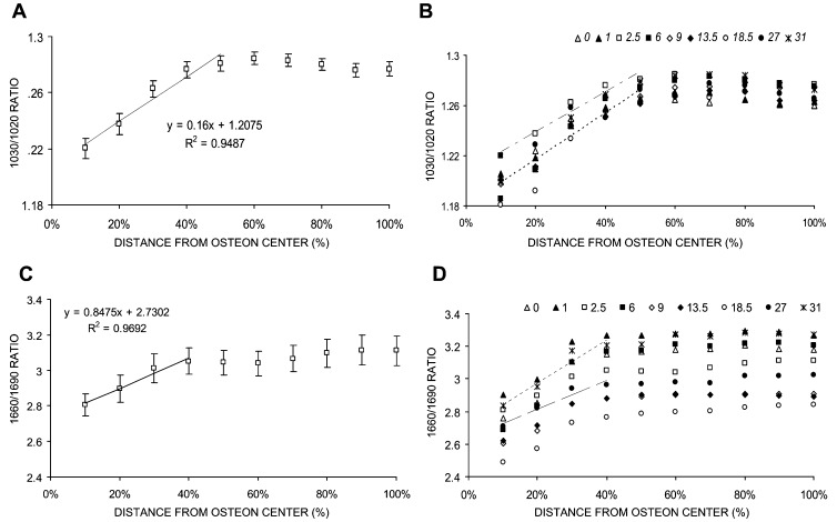
The crystallinity parameter (1030/1020 cm−1 peak ratio) increased with tissue age independently of the animal age (Figs. 4A and 4B). The relationship was not constant along the osteon and again was biphasic. In the first one half of the osteon starting at the center, the ratio increased linearly and reached a plateau. The slopes and the correlation coefficients were comparable for all the samples regardless of animal age (Table 2). In contrast to the mineral-to-matrix ratio and carbonate-to-phosphate ratio, the crystallinity parameter was independent of animal age.
FIG. 4.
Crystallinity (1030/1020 cm−1) and cross-link (1660/1690 cm−1) ratio in baboon osteonal bone. (A) Typical data for the crystallinity ratio plotted as a function of the distance along osteons from the 2.5-yr-old baboon group and (B) crystallinity along the osteons for the nine baboon age groups. (C) Typical data for the cross-link ratio plotted as a function of the distance along osteons from one baboon age group (2.5 yr) and (D) cross-link ratio along the osteons for the nine baboon age groups. Value are mean ± SD (n = 36).
The collagen cross-link ratio increased as a function of the tissue age across the osteon regardless of animal age (Figs. 4C and 4D), with a pattern similar to that recorded for the crystallinity parameter. The relationship with tissue age also was biphasic. During the first phase, close to the osteon center, the ratio increased linearly and reached a constant value. The slopes and the correlation coefficients were comparable for all baboon age groups (Table 2).
Raman spectroscopy was used to explore changes close to the osteonal center
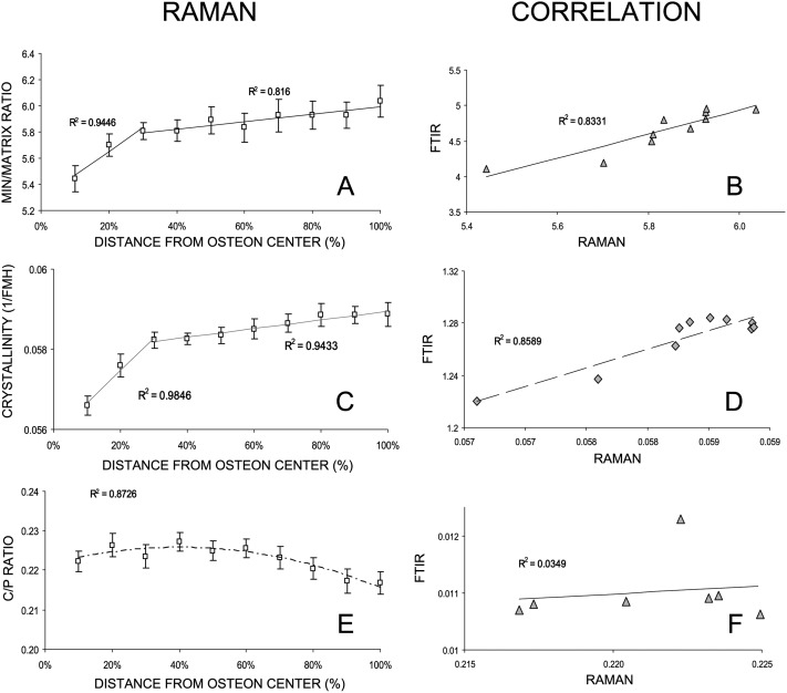
The Raman analysis of the 2.5-yr-old baboon group (Fig. 5) showed similar mineral properties with tissue age along the osteons as the FTIR data; however, unlike the FTIR data, the Raman mineral-to-matrix ratio was biphasic (Fig. 5A). In the first phase, nearest the center of the osteon, the mineral-to-matrix ratio increased rapidly, followed by a more gradual but still linearly increasing second phase. A strong correlation was found between the Raman and FTIR ratios taken from the same samples (Fig. 5B). Mineral crystallinity parameter, as determined by Raman, increased along the osteons (Fig. 5C). The crystallinity increased strongly and quickly around the osteonal center and more slowly toward the periphery. The FTIR and Raman crystallinity data were highly correlated (Fig. 5D). The Raman carbonate-to-phosphate ratio decreased slightly, but significantly toward the periphery of the osteon (Fig. 5E). In comparison with the FTIR data for the same animal group, the trend observed by Raman was different. The correlation between FTIR and Raman for this parameter was not significant (Fig. 5F).
FIG. 5.
Raman data and correlation between Raman and FTIR parameters. All parameters were plotted as a function of the distance along individual osteons from the 2.5-yr baboon age group. Where shown, error bars are ±SD (n = 36). (A) Typical Raman data for mineral-to-matrix ratio and (B) the linear correlation found between the Raman ratio and the FTIR mineral-to-matrix ratio. (C) Typical Raman data for the crystallinity (=1/bandwidth [960 cm−1]) and (D) the linear correlation between the crystallinity parameter calculated by Raman and by FTIR. (E) Raman carbonate-to-phosphate ratio along osteons and (F) the linear regression calculated between Raman and FTIR parameters.
Specific mechanical properties along osteons
When the mechanical properties were examined in the 13.5-yr-old baboon group, the indentation modulus and hardness increased with tissue age (Fig. 6). The correlation with tissue age was significant (r 2 = ∼0.93, p < 0.001) for the indentation modulus (Fig. 6A), whereas no correlation existed for hardness with tissue age (Fig. 6B). The correlation between indentation modulus and hardness with the FTIR parameters described previously are summarized in Table 3.
FIG. 6.
Mechanical properties evaluated by nano-indentation from the 13.5-yr-old baboon age group. Mean ± SD (n = 27). Indentation modulus (A) and hardness (B) data plotted as a function of the distance along osteons expressed in percentage from the osteonal center (0%) to the periphery (100%).
Table 3.
Mechanical and Chemical Relationship in Osteonal Bone
| Min/matrix ratio | CO3/PO4 ratio | 1030/1020 ratio | 1660/1690 ratio | |
| Indentation modulus (GPa) | 0.86* | −0.95* | 0.96* | 0.93* |
| Hardness (GPa) | 0.52 | −0.55 | 0.58 | 0.44 |
Correlation between mechanical parameters obtained by nano-indentation (Fig. 6) and those calculated by FTIR microspectroscopy reported as the square of the Pearson's correlation coefficient (r 2) from the 13-yr-old group.
* p < 0.001.
Mineral content changes with tissue age
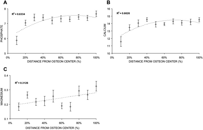
Both calcium (Ca) and phosphate (P) content increased nonlinearly with tissue age as shown by the data from a baboon in the 2.5-yr-old group (Figs. 7A and 7B). For magnesium (Mg), the increase observed was not significant (Fig. 7C). The correlation coefficients between the EDX data for calcium and phosphate and the FTIR data along osteons were highly significant (Table 4). The correlations between FTIR parameters and magnesium content were not significant.
FIG. 7.
EDX analyses of osteons from one baboon in the 2.5-yr group. (A) Relative phosphate, (B) calcium, and (C) magnesium content calculated by EDX and plotted as a function of the distance along osteons. The units shown are arbitrary. The same osteons used here were also used to calculate the FTIR parameters described previously. Mean ± SD (n = 12).
Table 4.
Mineral Content
| P | Ca | Mg | Min/matrix ratio | CO3/PO4 ratio | 1030/1020 ratio | 1660/1690 ratio | |
| P | 1 | — | — | 0.61* | −0.88† | 0.84† | 0.94† |
| Ca | 0.98 | 1 | — | 0.59* | −0.85† | 0.86† | 0.96† |
| Mg | 0.51 | 0.42 | 1 | 0.64* | −0.19 | 0.28 | 0.38 |
Correlation between mineral content calculated by EDX analyses (Fig. 7) and the FTIRI parameters reported as the square of the Pearson's correlation coefficient (r 2) calculated from the 2.5-yr-old baboon age group.
* p < 0.05.
† p < 0.001.
DISCUSSION
This study reports the detailed description and comparisons of the spatially resolved physicochemical, compositional, and mechanical properties within individual baboon osteons. Correlations are reported as a function of tissue age within osteons and as a function of animal age. Linear and biphasic relationships were observed with tissue age for FTIR and Raman compositional bone parameters. Significant correlations with Raman, EDX, and nano-indentation measured existed for the FTIR compositional data.
Previous FTIR microspectroscopy and imaging studies showed reproducible patterns for bone mineral properties across cortical osteons and along forming trabecular surfaces from normal human iliac crest biopsies.(14,15) The patterns observed for normal bone within osteons and in trabeculae were altered in osteoporotic human bone.(16,17) However, because bone is a very heterogeneous tissue(38,39) that is strongly modified by aging, these patterns would be difficult to interpret or to compare with biopsies from patients of different ages. Using a nonhuman primate model, we observed reproducible variations, independent of the animal age, in mineral and matrix properties at specific distances normalized to the width of the osteon. This normalization could allow better understanding of mechanisms contributing to osteoporosis or other bone diseases and provide better diagnostic insights.
Normal age-dependent alterations in composition, geometric, and mechanical bone properties strongly affect the bone integrity as shown clinically by the increase of fracture incidence with aging.(40–44) In addition, because of bone remodeling, a process involving both resorption of bone by osteoclasts and formation of new bone by osteoblasts, tissue age varies extensively within the same bone specimen. Thus, an understanding of the variations in bone properties as a function of both tissue and animal age at the microscopic level is crucial. Baboon bone provides an excellent model of human tissue because of similar development, long lifespan, and presence of skeletal fragility and menopause.(18,19) As discussed in the following paragraphs, the FTIR parameters measured in the baboon and human are comparable.
Mineral-to-matrix ratio calculated by FTIR spectroscopy is directly related to “ash weight,” proving a quantitative measurement of the extent of mineralization in the bone.(10,27,45) In the baboon, this ratio increased from the center of the osteon toward the periphery. These data are in agreement with previous FTIR studies in human bone, which showed that less mineralized regions are located around the center of the osteons and more mineralized areas located at the periphery.(14,15) The mineralization gradient along the osteon is representative of osteonal development and confirms that the baboon is an appropriate animal model for the study of bone remodeling. The mineralization profiles obtained by FTIR and Raman were similar, although Raman showed biphasic behavior along osteons. The difference reflects the ability of Raman, with a spectral resolution of ∼1 μm compared with the ∼10 μm of FTIR, to better detect the more rapid primary mineralization adjacent to the Haversian canal.(46)
The similarity of the slopes for mineral-to-matrix ratio as a function of baboon age was striking, whereas the absolute value of mineral content was dependent on animal age. In the baboon, the mineralization process inside osteons was most rapid during the first 5–6 yr, continued to increase slowly until 15–20 yr, and decreased thereafter. This trend is similar to change in lumbar spine BMD in baboons as a function of animal age in females(19) and is also similar to the change in spinal BMD in human females with age.(47) In human females, BMD begins to decrease around 40–50 yr, corresponding to the stage of life during which perimenopause and menopause occur. We can also match the trend observed for the mineral-to-matrix ratio with the baboon age to the different stages in the life of the female baboon. Based on the literature,(18–21) we can divide the baboons in three groups: young (<5–6 yr), adult (6–19 yr), and old (>19 yr). The old group was composed of female baboons with irregular menstrual cycles associated with perimenopausal phenomena (mean age of onset is 19 yr)(21,48,49) and menopausal females (mean age of 26 yr). The mineralization process in osteonal femoral bone is rapid in the young baboons, continues more slowly during the adult development until the hormonal cycle begins to become irregular, and declines in the third decade. Except for one very young male sample, we worked exclusively with females so we can extrapolate that the pattern of mineral-to-matrix ratio as a function of animal age is characteristic of female baboons. Because end-of-life hormonal profiles and menopausal status are not available for the animals in this study, we cannot directly relate any hormonal alterations associated with menopause to modifications of the mineralization process itself in the newly formed bone. The organic matrix is “fully mineralized” in human primary lamellar bone compared with the more recently formed bone in osteons as measured by Raman spectroscopy.(31) We similarly found that the mineral content in the cortical tissue (representing both osteonal and primary lamellar bone) was greater than in the osteonal area alone from the same sections independent of the baboon age and showed a similar age dependent pattern.
Biological hydroxyapatite is a poorly crystalline carbonate substituted apatite. The complex phenomenon of bone crystal maturation manifested through changes in the short-range order of the crystal and the concentration and location of highly labile, reactive CO3 −2 and HPO4 −2 groups. CO3 −2 substitution plays an important role in bone properties(28) and has been reported to increase with aging.(28,32) The unexpected biphasic decrease in carbonate-to-phosphate ratio inside the osteons agreed with the decrease in human osteons, although the latter was linear.(14) This apparent discrepancy with the reported increase in carbonate content(28,34) can be explained by the two contributors to the carbonate content ratio, such that apparent decrease in the ratio may result from a decrease in the amount of carbonate and/or an increase in the phosphate content. When normalized to the area of the amide I peak, both carbonate substitution and mineralization increased across the osteons, indicating that the phosphate content increased more rapidly than carbonate content closer to the Haversian canal.
Bone crystallinity is related to apatite crystal size and perfection as determined by X-ray diffraction.(29,50) Crystallinity, as determined by infrared spectroscopy and X-ray diffraction, increases with both tissue and animal age in humans.(14,15,51,52) The biphasic pattern of increasing crystallinity across the baboon osteons differs from the linear increase reported in human osteons,(14) perhaps a result of the number of sites evaluated along the osteon and the greater age range studied in the baboons. Crystallinity measured by Raman, based on a different parameter, confirmed the biphasic pattern within osteons. Although crystallinity was previously correlated with human age(51) in our baboon study, the mean osteonal crystallinity parameter did not correlate with animal age. This finding may reflect the heterogeneity of the animals or that the maximum crystal size is obtained early in baboon bone development.
The collagen cross-link ratio indicates the maturity state of the cross-linking network in the bone collagen fibril.(13,30) The collagen cross-link network is important for structural and mechanical properties of bone. The alteration of this network can result in severe dysfunction of the tissue and may be associated with the age-related increase in bone fracture and bone diseases.(9,53–55) Intermolecular collagen cross-linking is important for the development of the underlying matrices that are essential for initial mineral formation and crystal growth. This matrix also contributes to mechanical properties such as tensile strength and viscoelasticity. Previously, spatial distribution of the cross-linking ratio was compared in patients with and without osteoporosis.(9) Although data were not reported for osteonal bone, in normal cancellous bone, the cross-link ratio increased as a function of the tissue age at the forming surface. In osteoporotic cancellous bone, no spatial variation was observed in this ratio. In baboons, the observed maturation of the collagen cross-linking network was related to tissue age in newly formed bone. The results are in accordance with the bone development process, where first the collagen fibrils appear, followed by a process of enzymatically induced cross-linking that stabilizes the fibrils.(55) Subsequently, the collagen fibrils become mineralized and serve as scaffolds on which nucleation and growth of the additional mineral crystals will take place. These two processes are intimately correlated as shown by the similar trend between the crystallinity and the collagen cross-link ratio pattern across the osteons.
In addition to enabling us to describe osteonal variations as a function of both tissue and animal age, this study provided a unique opportunity to correlate mineral and matrix properties with nano-mechanical properties of the bone tissue. Few previous studies describe correlations between physicochemical and mechanical bone properties(35,41,56,57) and fewer describe correlation with properties at the same spatial resolution. Rho et al.(58) showed with a limited sample number that the mechanical properties decrease as a function of increasing distance from the center of the osteon, whereas Gupta's group did not see significant mechanical variation between osteonal lamellae from a single osteon(59) and Fratzl's group correlated Raman data with orthogonal distances from the Haversian canal for a single osteonal sample.(60) These data, as well as the results based on the mineral content,(61,62) present conflicting information about bone properties in osteons. Our mechanical bone property data obtained by nano-indentation and as mineral content calculated by EDX were positively correlated with the measured FTIR mineral and matrix properties along individual osteons for the age group examined, contrasting with results reported previously. There are several possible reasons for these discrepancies: specimen preparation, the nature of the specimens themselves, and the different development stages of the osteons studied. The effects of preparation were shown in an earlier nano-indentation study(35); however, we can not determine the contribution of the other factors.
Specific and repeated patterns were present in osteons for mineral content, carbonate incorporation, crystallinity, and collagen cross-link maturity along newly formed bone tissue regardless of animal age. Because this study included only female animals (with one exception), a comparison of these parameters in aging male baboons with the results presented here could greatly enhance our understanding of the aspect of female postmenopausal bone fragility. Animal age-related bone property changes among osteons have not been reported previously. By normalizing the diameters of the osteons, the results of our baboon study suggest that, if applied to human tissue, systematic comparisons between osteons should give consistent data, allowing analyses of skeletal disease and treatment effects without concern for the absolute size of the osteon. These data establish baseline values with which bone samples from diseased subjects may be compared. Moreover, the correlation of the spectroscopic bone properties with mechanical properties may in part explain the increase in bone fragility with aging. Age-related changes in bone mineralization are well described for the whole bone but, as reviewed elsewhere(63) with the exception of studies of BMD distribution, are practically unknown on the microstructure level. Alterations in bone composition relative to animal age are not only responsive to modifications in the remodeling balance process, but may also affect the mineralization process itself, as observed in this study with the youngest animals' newly formed bone. Although we could not ascertain changes in the bone matrix protein composition, age-dependent changes in the expression of the proteins associated with the both osteogenesis and mineralization are known and could explain differences noted in the youngest and older animals. The overall relationship obtained for the baboon osteons is particularly exciting in the context of novel developments such as a Raman probe for imaging bone tissue in situ(64) and an infrared fiber optic probes used arthroscopically(65) that both have clinical potential for in vivo diagnosis.
ACKNOWLEDGMENTS
This work is supported by NIH Grants AR041325 and AR053571. This study was conducted in a facility constructed with support from Research Facilities Improvement Program Grant C06-RR12538-01 from the National Center for Research Resources, National Institutes of Health.
Footnotes
The authors state that they have no conflicts of interest.
REFERENCES
- 1.Johnell O, Kanis JA, Oden A, Johansson H, De Laet C, Delmas P, Eisman JA, Fujiwara S, Kroger H, Mellstrom D, Meunier PJ, Melton LJ, III, O'Neill T, Pols H, Reeve J, Silman A, Tenenhouse A. Predictive value of BMD for hip and other fractures. J Bone Miner Res. 2005;20:1185–1194. doi: 10.1359/JBMR.050304. [DOI] [PubMed] [Google Scholar]
- 2.Schneider DL. Management of osteoporosis in geriatric populations. Curr Osteoporos Rep. 2008;6:100–107. doi: 10.1007/s11914-008-0018-4. [DOI] [PubMed] [Google Scholar]
- 3.Siris ES, Genant HK, Laster AJ, Chen P, Misurski DA, Krege JH. Enhanced prediction of fracture risk combining vertebral fracture status and BMD. Osteoporos Int. 2007;18:761–770. doi: 10.1007/s00198-006-0306-8. [DOI] [PubMed] [Google Scholar]
- 4.Seeman E. Is a change in bone mineral density a sensitive and specific surrogate of anti-fracture efficacy? Bone. 2007;41:308–317. doi: 10.1016/j.bone.2007.06.010. [DOI] [PubMed] [Google Scholar]
- 5.Gregory JS, Aspden RM. Femoral geometry as a risk factor for osteoporotic hip fracture in men and women. Med Eng Phys. 2008;30:1275–1286. doi: 10.1016/j.medengphy.2008.09.002. [DOI] [PubMed] [Google Scholar]
- 6.Garnero P. Biomarkers for osteoporosis management: Utility in diagnosis, fracture risk prediction and therapy monitoring. Mol Diagn Ther. 2008;12:157–170. doi: 10.1007/BF03256280. [DOI] [PubMed] [Google Scholar]
- 7.Mendelsohn R, Paschalis EP, Sherman PJ, Boskey AL. IR microscopic imaging of pathological states and fracture healing of bone. Appl Spectrosc. 2000;54:1183–1191. doi: 10.1366/000370204322729405. [DOI] [PubMed] [Google Scholar]
- 8.Carden A, Morris MD. Application of vibrational spectroscopy to the study of mineralized tissues. J Biomed Opt. 2000;5:259–268. doi: 10.1117/1.429994. [DOI] [PubMed] [Google Scholar]
- 9.Paschalis EP, Shane E, Lyritis G, Skarantavos G, Mendelsohn R, Boskey AL. Bone fragility and collagen cross-links. J Bone Miner Res. 2004;19:2000–2004. doi: 10.1359/JBMR.040820. [DOI] [PMC free article] [PubMed] [Google Scholar]
- 10.Boskey A, Mendelsohn R. Infrared analysis of bone in health and disease. J Biomed Opt. 2005;10:031102. doi: 10.1117/1.1922927. [DOI] [PubMed] [Google Scholar]
- 11.Boskey AL, Spevak L, Weinstein RS. Spectroscopic markers of bone quality in alendronate-treated postmenopausal women. Osteoporos Int. 2009;20:793–800. doi: 10.1007/s00198-008-0725-9. [DOI] [PMC free article] [PubMed] [Google Scholar]
- 12.Paschalis EP, Betts F, DiCarlo E, Mendelsohn R, Boskey AL. FTIR microspectroscopic analysis of normal human cortical and trabecular bone. Calcif Tissue Int. 1997;61:480–486. doi: 10.1007/s002239900371. [DOI] [PubMed] [Google Scholar]
- 13.Paschalis EP, Recker R, DiCarlo E, Doty SB, Atti E, Boskey AL. Distribution of collagen cross-links in normal human trabecular bone. J Bone Miner Res. 2003;18:1942–1946. doi: 10.1359/jbmr.2003.18.11.1942. [DOI] [PubMed] [Google Scholar]
- 14.Paschalis EP, DiCarlo E, Betts F, Sherman P, Mendelsohn R, Boskey AL. FTIR microspectroscopic analysis of human osteonal bone. Calcif Tissue Int. 1996;59:480–487. doi: 10.1007/BF00369214. [DOI] [PubMed] [Google Scholar]
- 15.Mendelsohn R, Paschalis EP, Boskey AL. Infrared spectroscopy, microscopy, and microscopic imaging of mineralizing tissues: Spectra-structure correlations from human iliac crest biopsies. J Biomed Opt. 1999;4:14–21. doi: 10.1117/1.429916. [DOI] [PubMed] [Google Scholar]
- 16.Paschalis EP, Betts F, DiCarlo E, Mendelsohn R, Boskey AL. FTIR microspectroscopic analysis of human iliac crest biopsies from untreated osteoporotic bone. Calcif Tissue Int. 1997;61:487–492. doi: 10.1007/s002239900372. [DOI] [PubMed] [Google Scholar]
- 17.Boskey AL, DiCarlo E, Paschalis E, West P, Mendelsohn R. Comparison of mineral quality and quantity in iliac crest biopsies from high- and low-turnover osteoporosis: An FT-IR microspectroscopic investigation. Osteoporos Int. 2005;16:2031–2038. doi: 10.1007/s00198-005-1992-3. [DOI] [PMC free article] [PubMed] [Google Scholar]
- 18.Bronikowski AM, Alberts SC, Altmann J, Packer C, Carey KD, Tatar M. The aging baboon: Comparative demography in a non-human primate. Proc Natl Acad Sci USA. 2002;99:9591–9595. doi: 10.1073/pnas.142675599. [DOI] [PMC free article] [PubMed] [Google Scholar]
- 19.Havill LM, Mahaney MC, Czerwinski SA, Carey KD, Rice K, Rogers J. Bone mineral density reference standards in adult baboons (Papio hamadryas) by sex and age. Bone. 2003;33:877–888. doi: 10.1016/s8756-3282(03)00231-x. [DOI] [PubMed] [Google Scholar]
- 20.Havill LM, Levine SM, Newman DE, Mahaney MC. Osteopenia and osteoporosis in adult baboons (Papio hamadryas) J Med Primatol. 2008;37:146–153. doi: 10.1111/j.1600-0684.2007.00270.x. [DOI] [PMC free article] [PubMed] [Google Scholar]
- 21.Bellino FL, Wise PM. Nonhuman primate models of menopause workshop. Biol Reprod. 2003;68:10–18. doi: 10.1095/biolreprod.102.005215. [DOI] [PubMed] [Google Scholar]
- 22.Vagtborg H. Austin, TX, USA: University of Texas Press; 1973. The Story of the Southwest Research Center: A Private, Nonprofit Scientific Research Adventure. [Google Scholar]
- 23.National Research Council. Washington, DC, USA: National Academy of Sciences; 1996. Guide for the Care and Use of Laboratory Animals. [Google Scholar]
- 24.Erben RG. Embedding of bone samples in methylmethacrylate: An improved method suitable for bone histomorphometry, histochemistry, and immunohistochemistry. J Histochem Cytochem. 1997;45:307–313. doi: 10.1177/002215549704500215. [DOI] [PubMed] [Google Scholar]
- 25.Boskey AL, Mendelsohn R. Infrared spectroscopic characterization of mineralized tissues. Vib Spectrosc. 2005;38:107–114. doi: 10.1016/j.vibspec.2005.02.015. [DOI] [PMC free article] [PubMed] [Google Scholar]
- 26.Gourion-Arsiquaud S, Boskey AL. Fourier transform infrared and Raman microspectroscopy and microscopic imaging of bone. Curr Opin Orthop. 2007;18:499. doi: 10.1007/978-1-59745-104-8_20. [DOI] [PubMed] [Google Scholar]
- 27.Pienkowski D, Doers TM, Monier-Faugere MC, Geng Z, Camacho NP, Boskey AL, Malluche HH. Calcitonin alters bone quality in beagle dogs. J Bone Miner Res. 1997;12:1936–1943. doi: 10.1359/jbmr.1997.12.11.1936. [DOI] [PubMed] [Google Scholar]
- 28.Rey C, Renugopalakrishnan V, Collins B, Glimcher MJ. Fourier transform infrared spectroscopic study of the carbonate ions in bone mineral during aging. Calcif Tissue Int. 1991;49:251–258. doi: 10.1007/BF02556214. [DOI] [PubMed] [Google Scholar]
- 29.Pleshko N, Boskey A, Mendelsohn R. Novel infrared spectroscopic method for the determination of crystallinity of hydroxyapatite minerals. Biophys J. 1991;60:786–793. doi: 10.1016/S0006-3495(91)82113-0. [DOI] [PMC free article] [PubMed] [Google Scholar]
- 30.Paschalis EP, Verdelis K, Doty SB, Boskey AL, Mendelsohn R, Yamauchi M. Spectroscopic characterization of collagen cross-links in bone. J Bone Miner Res. 2001;16:1821–1828. doi: 10.1359/jbmr.2001.16.10.1821. [DOI] [PubMed] [Google Scholar]
- 31.Akkus O, Polyakova-Akkus A, Adar F, Schaffler MB. Aging of microstructural compartments in human compact bone. J Bone Miner Res. 2003;18:1012–1019. doi: 10.1359/jbmr.2003.18.6.1012. [DOI] [PubMed] [Google Scholar]
- 32.Tarnowski CP, Ignelzi MA, Jr, Wang W, Taboas JM, Goldstein SA, Morris MD. Earliest mineral and matrix changes in force-induced musculoskeletal disease as revealed by Raman microspectroscopic imaging. J Bone Miner Res. 2004;19:64–71. doi: 10.1359/JBMR.0301201. [DOI] [PubMed] [Google Scholar]
- 33.Penel G, Leroy G, Rey C, Bres E. MicroRaman spectral study of the PO4 and CO3 vibrational modes in synthetic and biological apatites. Calcif Tissue Int. 1998;63:475–481. doi: 10.1007/s002239900561. [DOI] [PubMed] [Google Scholar]
- 34.Freeman JJ, Wopenka B, Silva MJ, Pasteris JD. Raman spectroscopic detection of changes in bioapatite in mouse femora as a function of age and in vitro fluoride treatment. Calcif Tissue Int. 2001;68:156–162. doi: 10.1007/s002230001206. [DOI] [PubMed] [Google Scholar]
- 35.Donnelly E, Baker SP, Boskey AL, van der Meulen MC. Effects of surface roughness and maximum load on the mechanical properties of cancellous bone measured by nanoindentation. J Biomed Mater Res A. 2006;77:426–435. doi: 10.1002/jbm.a.30633. [DOI] [PMC free article] [PubMed] [Google Scholar]
- 36.Donnelly E, Williams RM, Downs SA, Dickinson ME, Baker SP, van der Meulen MCH. Quasistatic and dynamic nanomechanical properties of cancellous bone tissue relate to collagen content and organization. J Mater Res. 2006;21:2106–2117. [Google Scholar]
- 37.Olivier WC, Pharr GM. An improved technique for determining hardness and elastic modulus using load and displacement sensing indentation experiments. J Mater Res. 1992;7:1564–1583. [Google Scholar]
- 38.Podenphant J, Riis BJ, Johansen JS, Leth A, Christiansen C. Iliac crest biopsy in longitudinal therapeutic trials of osteoporosis. Bone Miner. 1988;5:77–87. doi: 10.1016/0169-6009(88)90008-6. [DOI] [PubMed] [Google Scholar]
- 39.Whyte MP, Bergfeld MA, Murphy WA, Avioli LV, Teitelbaum SL. Postmenopausal osteoporosis. A heterogeneous disorder as assessed by histomorphometric analysis of Iliac crest bone from untreated patients. Am J Med. 1982;72:193–202. doi: 10.1016/0002-9343(82)90810-5. [DOI] [PubMed] [Google Scholar]
- 40.Smith DM, Khairi MR, Johnston CC., Jr The loss of bone mineral with aging and its relationship to risk of fracture. J Clin Invest. 1975;56:311–318. doi: 10.1172/JCI108095. [DOI] [PMC free article] [PubMed] [Google Scholar]
- 41.Ding M, Dalstra M, Danielsen CC, Kabel J, Hvid I, Linde F. Age variations in the properties of human tibial trabecular bone. J Bone Joint Surg Br. 1997;79:995–1002. doi: 10.1302/0301-620x.79b6.7538. [DOI] [PubMed] [Google Scholar]
- 42.Akkus O, Adar F, Schaffler MB. Age-related changes in physicochemical properties of mineral crystals are related to impaired mechanical function of cortical bone. Bone. 2004;34:443–453. doi: 10.1016/j.bone.2003.11.003. [DOI] [PubMed] [Google Scholar]
- 43.Bouxsein ML, Melton LJ, III, Riggs BL, Muller J, Atkinson EJ, Oberg AL, Robb RA, Camp JJ, Rouleau PA, McCollough CH, Khosla S. Age- and sex-specific differences in the factor of risk for vertebral fracture: A population-based study using QCT. J Bone Miner Res. 2006;21:1475–1482. doi: 10.1359/jbmr.060606. [DOI] [PubMed] [Google Scholar]
- 44.Nagaraja S, Lin AS, Guldberg RE. Age-related changes in trabecular bone microdamage initiation. Bone. 2007;40:973–980. doi: 10.1016/j.bone.2006.10.028. [DOI] [PubMed] [Google Scholar]
- 45.Faibish D, Gomes A, Boivin G, Binderman I, Boskey A. Infrared imaging of calcified tissue in bone biopsies from adults with osteomalacia. Bone. 2005;36:6–12. doi: 10.1016/j.bone.2004.08.019. [DOI] [PubMed] [Google Scholar]
- 46.Black J, Mattson RU. Relationship between porosity and mineralization in the Haversian osteon. Calcif Tissue Int. 1982;34:332–336. doi: 10.1007/BF02411263. [DOI] [PubMed] [Google Scholar]
- 47.Havill LM, Mahaney MC, Binkley TL, Specker BL. Effects of genes, sex, age, and activity on BMC, bone size, and areal and volumetric BMD. J Bone Miner Res. 2007;22:737–746. doi: 10.1359/jbmr.070213. [DOI] [PubMed] [Google Scholar]
- 48.Chen LD, Kushwaha RS, McGill HC, Jr, Rice KS, Carey KD. Effect of naturally reduced ovarian function on plasma lipoprotein and 27-hydroxycholesterol levels in baboons (Papio sp.) Atherosclerosis. 1998;136:89–98. doi: 10.1016/s0021-9150(97)00190-1. [DOI] [PubMed] [Google Scholar]
- 49.Martin LJ, Carey KD, Comuzzie AG. Variation in menstrual cycle length and cessation of menstruation in captive raised baboons. Mech Ageing Dev. 2003;124:865–871. doi: 10.1016/s0047-6374(03)00134-9. [DOI] [PubMed] [Google Scholar]
- 50.Gadaleta SJ, Paschalis EP, Betts F, Mendelsohn R, Boskey AL. Fourier transform infrared spectroscopy of the solution-mediated conversion of amorphous calcium phosphate to hydroxyapatite: New correlations between X-ray diffraction and infrared data. Calcif Tissue Int. 1996;58:9–16. doi: 10.1007/BF02509540. [DOI] [PubMed] [Google Scholar]
- 51.Handschin RG, Stern WB. X-ray diffraction studies on the lattice perfection of human bone apatite (Crista Iliaca) Bone. 1995;16:S355–S363. [PubMed] [Google Scholar]
- 52.Boskey A. Bone mineral crystal size. Osteoporos Int. 2003;14(Suppl 5):S16–S20. doi: 10.1007/s00198-003-1468-2. [DOI] [PubMed] [Google Scholar]
- 53.Eyre DR, Dickson IR, Van Ness K. Collagen cross-linking in human bone and articular cartilage. Age-related changes in the content of mature hydroxypyridinium residues. Biochem J. 1988;252:495–500. doi: 10.1042/bj2520495. [DOI] [PMC free article] [PubMed] [Google Scholar]
- 54.Otsubo K, Katz EP, Mechanic GL, Yamauchi M. Cross-linking connectivity in bone collagen fibrils: The COOH-terminal locus of free aldehyde. Biochemistry. 1992;31:396–402. doi: 10.1021/bi00117a013. [DOI] [PubMed] [Google Scholar]
- 55.Knott L, Bailey AJ. Collagen cross-links in mineralizing tissues: A review of their chemistry, function, and clinical relevance. Bone. 1998;22:181–187. doi: 10.1016/s8756-3282(97)00279-2. [DOI] [PubMed] [Google Scholar]
- 56.Miller LM, Little W, Schirmer A, Sheik F, Busa B, Judex S. Accretion of bone quantity and quality in the developing mouse skeleton. J Bone Miner Res. 2007;22:1037–1045. doi: 10.1359/jbmr.070402. [DOI] [PubMed] [Google Scholar]
- 57.Yerramshetty JS, Akkus O. The associations between mineral crystallinity and the mechanical properties of human cortical bone. Bone. 2008;42:476–482. doi: 10.1016/j.bone.2007.12.001. [DOI] [PubMed] [Google Scholar]
- 58.Rho JY, Zioupos P, Currey JD, Pharr GM. Variations in the individual thick lamellar properties within osteons by nanoindentation. Bone. 1999;25:295–300. doi: 10.1016/s8756-3282(99)00163-5. [DOI] [PubMed] [Google Scholar]
- 59.Gupta HS, Stachewicz U, Wagermaier W, Roschger P, Wagner HD, Fratzl P. Mechanical modulation at the lamellar level in osteonal bone. J Mater Res. 2006;21:1913–1921. [Google Scholar]
- 60.Kazanci M, Wagner HD, Manjubala NI, Gupta HS, Paschalis E, Roschger P, Fratzl P. Raman imaging of two orthogonal planes within cortical bone. Bone. 2007;41:456–461. doi: 10.1016/j.bone.2007.04.200. [DOI] [PubMed] [Google Scholar]
- 61.Crofts RD, Boyce TM, Bloebaum RD. Aging changes in osteon mineralization in the human femoral neck. Bone. 1994;15:147–152. doi: 10.1016/8756-3282(94)90701-3. [DOI] [PubMed] [Google Scholar]
- 62.Sissons HA. X-ray microscopy and X-ray microanalysis. In: Engström A, Cosslett V, Pattee H, editors. Proceedings of the Second International Symposium. New York, NY, USA: Elsevier Publishing; 1960. [Google Scholar]
- 63.Roschger P, Paschalis EP, Fratzl P, Klaushofer K. Bone mineralization density distribution in health and disease. Bone. 2008;42:456–466. doi: 10.1016/j.bone.2007.10.021. [DOI] [PubMed] [Google Scholar]
- 64.Draper ER, Morris MD, Camacho NP, Matousek P, Towrie M, Parker AW, Goodship AE. Novel assessment of bone using time-resolved transcutaneous Raman spectroscopy. J Bone Miner Res. 2005;20:1968–1972. doi: 10.1359/JBMR.050710. [DOI] [PubMed] [Google Scholar]
- 65.Li G, Thomson M, Dicarlo E, Yang X, Nestor B, Bostrom MP, Camacho NP. A chemometric analysis for evaluation of early-stage cartilage degradation by infrared fiber-optic probe spectroscopy. Appl Spectrosc. 2005;59:1527–1533. doi: 10.1366/000370205775142593. [DOI] [PubMed] [Google Scholar]


