Abstract
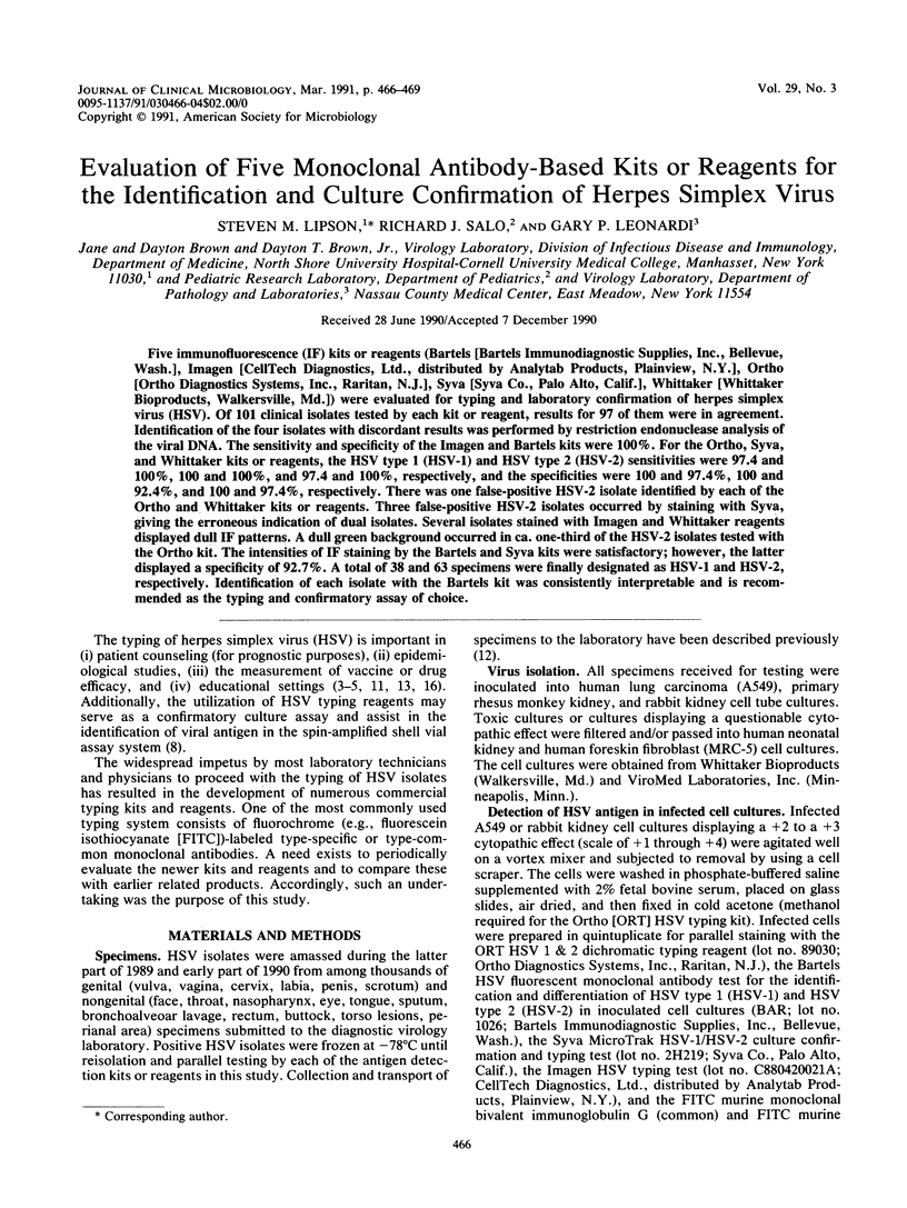
Five immunofluorescence (IF) kits or reagents (Bartels [Bartels Immunodiagnostic Supplies, Inc., Bellevue, Wash.], Imagen [CellTech Diagnostics, Ltd., distributed by Analytab Products, Plainview, N.Y.], Ortho [Ortho Diagnostics Systems, Inc., Raritan, N.J.], Syva [Syva Co., Palo Alto, Calif.], Whittaker [Whittaker Bioproducts, Walkersville, Md.]) were evaluated for typing and laboratory confirmation of herpes simplex virus (HSV). Of 101 clinical isolates tested by each kit or reagent, results for 97 of them were in agreement. Identification of the four isolates with discordant results was performed by restriction endonuclease analysis of the viral DNA. The sensitivity and specificity of the Imagen and Bartels kits were 100%. For the Ortho, Syva, and Whittaker kits or reagents, the HSV type 1 (HSV-1) and HSV type 2 (HSV-2) sensitivities were 97.4 and 100%, 100 and 100%, and 97.4 and 100%, respectively, and the specificities were 100 and 97.4%, 100 and 92.4%, and 100 and 97.4%, respectively. There was one false-positive HSV-2 isolate identified by each of the Ortho and Whittaker kits or reagents. Three false-positive HSV-2 isolates occurred by staining with Syva, giving the erroneous indication of dual isolates. Several isolates stained with Imagen and Whittaker reagents displayed dull IF patterns. A dull green background occurred in ca. one-third of the HSV-2 isolates tested with the Ortho kit. The intensities of IF staining by the Bartels and Syva kits were satisfactory; however, the latter displayed a specificity of 92.7%. A total of 38 and 63 specimens were finally designated as HSV-1 and HSV-2, respectively. Identification of each isolate with the Bartels kit was consistently interpretable and is recommended as the typing and confirmatory assay of choice.
Full text
PDF


Selected References
These references are in PubMed. This may not be the complete list of references from this article.
- Aarnaes S. L., Peterson E. M., de la Maza L. M. Evaluation of a new herpes simplex virus typing reagent for tissue culture confirmation. Diagn Microbiol Infect Dis. 1989 May-Jun;12(3):269–270. doi: 10.1016/0732-8893(89)90026-6. [DOI] [PubMed] [Google Scholar]
- Corey L. Laboratory diagnosis of herpes simplex virus infections. Principles guiding the development of rapid diagnostic tests. Diagn Microbiol Infect Dis. 1986 Mar;4(3 Suppl):111S–119S. doi: 10.1016/s0732-8893(86)80049-9. [DOI] [PubMed] [Google Scholar]
- Corey L., Whitley R. J., Stone E. F., Mohan K. Difference between herpes simplex virus type 1 and type 2 neonatal encephalitis in neurological outcome. Lancet. 1988 Jan 2;1(8575-6):1–4. doi: 10.1016/s0140-6736(88)90997-x. [DOI] [PubMed] [Google Scholar]
- De Clercq E., Desgranges C., Herdewijn P., Sim I. S., Jones A. S., McLean M. J., Walker R. T. Synthesis and antiviral activity of (E)-5-(2-bromovinyl)uracil and (E)-5-(2-bromovinyl)uridine. J Med Chem. 1986 Feb;29(2):213–217. doi: 10.1021/jm00152a008. [DOI] [PubMed] [Google Scholar]
- Dragavon J., Peterson E., Ashley R., Lafferty W., Corey L. Routine typing of herpes simplex virus isolates. Diagn Microbiol Infect Dis. 1985 May;3(3):269–270. doi: 10.1016/0732-8893(85)90040-9. [DOI] [PubMed] [Google Scholar]
- Gleaves C. A., Rice D. H., Bindra R., Hursh D. A., Curtis S. E., Lee C. F., Wendt S. F. Evaluation of a HSV specific monoclonal antibody reagent for laboratory diagnosis of herpes simplex virus infection. Diagn Microbiol Infect Dis. 1989 Jul-Aug;12(4):315–318. doi: 10.1016/0732-8893(89)90096-5. [DOI] [PubMed] [Google Scholar]
- Hursh D. A., Wendt S. F., Lee C. F., Gleaves C. A. Detection of herpes simplex virus by using A549 cells in centrifugation culture with a rapid membrane enzyme immunoassay. J Clin Microbiol. 1989 Jul;27(7):1695–1696. doi: 10.1128/jcm.27.7.1695-1696.1989. [DOI] [PMC free article] [PubMed] [Google Scholar]
- Lafferty W. E., Krofft S., Remington M., Giddings R., Winter C., Cent A., Corey L. Diagnosis of herpes simplex virus by direct immunofluorescence and viral isolation from samples of external genital lesions in a high-prevalence population. J Clin Microbiol. 1987 Feb;25(2):323–326. doi: 10.1128/jcm.25.2.323-326.1987. [DOI] [PMC free article] [PubMed] [Google Scholar]
- Lipson S. M., Schutzbank T. E., Szabo K. Evaluation of three immunofluorescence assays for culture confirmation and typing of herpes simplex virus. J Clin Microbiol. 1987 Feb;25(2):391–394. doi: 10.1128/jcm.25.2.391-394.1987. [DOI] [PMC free article] [PubMed] [Google Scholar]
- Lipson S. M., Szabo K., Lin J. H. Changing patterns of genital herpes simplex virus infections. Zentralbl Bakteriol Mikrobiol Hyg A. 1988 Mar;268(1):57–61. doi: 10.1016/s0176-6724(88)80115-9. [DOI] [PubMed] [Google Scholar]
- Lipson S. M., Zelinsky-Papez K. A. Comparison of four latex agglutination (LA) and three enzyme-linked immunosorbent assays (ELISA) for the detection of rotavirus in fecal specimens. Am J Clin Pathol. 1989 Nov;92(5):637–643. doi: 10.1093/ajcp/92.5.637. [DOI] [PubMed] [Google Scholar]
- Salo R. J., Ortega A. P. Effect of interferon on herpes simplex virus replication in murine macrophage-like cell lines. Antiviral Res. 1986 May;6(3):161–169. doi: 10.1016/0166-3542(86)90010-0. [DOI] [PubMed] [Google Scholar]


