Abstract
Objective
To determine the findings of various focal hepatic lesions at contrast-enhanced gray-scale ultrasound (US) using a coded harmonic angio (CHA) technique and emphasizing lesion characterization.
Materials and Methods
The study involved 95 patients with 105 focal hepatic lesions, namely 51 hepatocellular carcinomas (HCCs), 22 metastases, 22 hemangiomas, four cases of focal nodular hyperplasia (FNH), and six nontumorous nodules. After the injection of a microbubble contrast agent (SH U 508A), gray-scale harmonic US studies using a CHA technique were performed with a combination of continuous scanning to assess the intratumoral vasculature (vascular imaging) and interval-delay scanning to determine the sequential enhancement pattern (acoustic emission imaging). Each imaging pattern was categorized and analyzed.
Results
At vascular imaging, 69% of HCCs (35/51) showed irregular branching vessels, while in 91% of metastases (20/22) a peripherally stippled pattern was observed. Intratumoral vessels were absent in 95% of hemangiomas (21/22) and all nontumorous lesions (6/6), while in 75% of FNHs (3/4) a spoke-wheel pattern was evident. At acoustic emission imaging, 71% of HCCs (36/51) showed heterogeneous enhancement and 86% (19/22) of metastases showed rim- or flame-like peripheral enhancement during the early phase, with washout occurring in all HCCs and metastases (100%, 73/73) during the late phase. In hemangiomas, enhancement was either peripheral and nodular (19/22, 86%) or persistent and homogeneous (3/22, 14%), and 75% of FNHs (3/4) became isoechoic during the late phase.
Conclusion
At contrast-enhanced gray-scale US using a CHA technique, a period of continuous scanning depicted the intratumoral vasculature, and interval-delay scanning demonstrated the sequential enhancement pattern. The characteristic findings of various focal hepatic lesions were thus determined.
Keywords: Liver neoplasms, US; Ultrasound (US), contrast media; Ultrasound, harmonic study; Ultrasound, technology
The sensitive detection and characterization of tumor vascularity is crucial for the differential diagnosis of focal hepatic lesions, and there is increasing consensus that the addition of contrast agents has significantly expanded the current role of ultrasonography (US) into diagnostic territories where CT and MRI were previously dominant. Although the findings of recent reports are preliminary and further studies with a large patient population are needed to clarify the definite role of contrast-enhanced US, current results suggest that the modality has great potential for the detection and characterization of hepatic tumors. However, established techniques suffer certain limitations: Doppler studies in which microbubble agents are used are seriously affected by 'blooming' and motion artifacts, and are insensitive to very slow flow (1-4). More recently, various gray-scale harmonic US techniques have been reported to be useful in characterizing hepatic tumors (5-8). For visualization of sufficient stimulated acoustic emission (SAE) effect, these, however, usually require maximal mechanical index (MI), and strict intermittent scanning is thus necessary (9, 10). This causes technical difficulties, however, and generally provides less helpful information from vascular imaging, especially with a fragile air-based contrast agent such as SH U 508A.
Coded harmonic angio (CHA) is a new US technique that is specifically sensitive to signals from a contrast agent but does not give rise to contrast-related artifacts. Its extreme sensitivity to contrast agents provides sufficient SAE effect at medium MI and, at the same time, since there is less destruction of microbubbles, a period of continuous scanning is possible with excellent depiction of intratumoral microvasculature. We found that CHA in conjunction with a scan technique exploiting both vascular and acoustic emission imaging accurately represented the vascular characteristics of various focal hepatic lesions.
The purpose of this study was to describe the contrast-enhancement patterns of various focal hepatic lesions at CHA, emphasizing lesion characterization.
MATERIALS AND METHODS
Subjects
During a recent six-month period, 104 consecutive patients with 114 focal hepatic lesions and no history of treatment were examined using a CHA technique in conjunction with a microbubble contrast agent. Nine lesions in nine patients were excluded: five lesions were not properly scanned according to our protocol, and in four cases the diagnosis was not verified. Finally, 95 patients with 105 focal hepatic lesions were included: 51 patients with 51 hepatocellular carcinomas (HCCs), 16 with 22 hepatic metastases, 18 with 22 hemangiomas, four with focal nodular hyperplasia (FNH), three with focal eosinophilic necrosis (FEN), two with inflammatory nodules, and one with focal fatty lesion. Lesions ranged in size from 0.7 to 6.9 (mean, 2.6) cm, and at the time of patient recruitment the diagnosis was unknown. In four patients with metastases and four with hemangiomas, two or three lesions were included in one scanning plane. In each of the remaining patients, one lesion was scanned. Patients with HCCs were 30-74 (mean, 56.3) years old, those with metastases were 34-77 (mean, 60.5), those with hemangiomas were 28-75 (mean, 52.7), those with FNH were 33-62 (mean, 46.5), and the remaining patients with nontumorous lesions were 36-65 (mean, 51.2). Men accounted for 41 of the 51 patients with HCCs, 12 of the 16 with metastases, eight of the 18 with hemangiomas, all four with FNH, and three of the seven with nontumorous lesions. All patients gave their full informed consent to our study and the approval of our institutional review board was obtained.
Hemangiomas were proven by means of characteristic findings at triple-phase (hepatic arterial, portal venous, and equilibrium phase) contrast-enhanced helical CT (n=11) or dynamic contrast-enhanced MR imaging (n=10), with no growth at follow-up imaging for a minimum of six months, or by intraoperative biopsy (n=1). One focal fatty lesion was verified by MR imaging using the fat suppression technique, and all other lesions were proven by percutaneous needle biopsy (n=74) or resection (n=9). Pathologic proof was obtained for each nodule, except in four patients with metastases which were included in one scan plane. All metastatic lesions in these four patients showed the same enhancement pattern and were thus considered to be pathologically similar to the one which was proven. The histologic diagnoses of metastases were gastric carcinoma (n=7), colorectal carcinoma (n=6), pancreatic carcinoma (n=3), adenocarcinoma of unknown primary origin (n=3), gallbladder carcinoma (n=1), malignant stromal tumor of the duodenum (n=1), and non-small cell lung carcinoma (n=1).
CHA Imaging
The US contrast agent used in the present study, SH U 508 A (Levovist; Schering AG, Berlin, Germany), is a suspension of galactose microparticles in sterile water. In preparation for its injection, and prior to US, this agent was shaken for 5-10 seconds with 11 mL of sterile water. After standing for 2 minutes for equilibration, a 4-gm milky suspension of SH U 508 A at a concentration of 300 mg/mL was injected into the brachial vein as a bolus, using a 20-gauge peripheral intravenous cannula.
Prior to final diagnosis, CHA US was prospectively performed by either of two examiners (H.K.L. and H-J.J.). For all US examinations, a 2-4-MHz curved-array wide-band transducer (LOGIQ 700 MR EXPERT or LogiQ 9; General Electric Medical Systems, Milwaukee, Wis., U.S.A.) was used. The acoustic power of CHA US was set at the default setting - an MI of 0.6-0.8. Before injection of the contrast agent, the area including the tumor was focused by zooming, and with a single focal zone either at the bottom or at the center of the lesions, depending on the size of the lesions. After administration of the contrast agent, CHA US involving 'modified interval-delay scanning' was performed, with a combination of continuous scanning and interval- delay techniques, and the two different patterns of images observed were differentiated during the two scanning modes. The real-time assessment of intratumoral vascular flow during continuous scanning at each interval was designated 'vascular imaging' and assessment of the sequential enhancement pattern seen during the initial one or two frames of each interval scan was termed 'acoustic emission imaging'. During the first 60-second period after injection, before reaching the point of maximal parenchymal enhancement, lesions were scanned every 20 seconds for 5 to 10 seconds, and between each scan the display was frozen. Just after the end of each scanning period, a series of hard-copy images was obtained as images were reviewed in cine-mode. During this phase (vascular imaging), the intralesional vascular pattern was the main focus of evaluation, though the SAE effect was frequently apparent during the first few frames of each scanning period. Modified intermittent scanning was then performed, sweeping for 2-3 seconds with a 1- or 2-minute interval (at 1-, 2-, 3-, and 5- minute delay); between each scan the display was frozen, and for 2-3 seconds during each scan was unfrozen. During this phase, the chief concern was the sequential enhancement pattern, which relies mainly on the SAE effect (acoustic emission imaging phase). The time delay from injection and the time at which each image was obtained were automatically recorded by a timer function.
Analysis
US images were retrospectively reviewed on a 2K×2K picture archiving and communication system monitor (General Electric Medical Systems Integrated Imaging Solutions, Mt. Prospect, Ill., U.S.A.) by two abdominal radiologists (W.J.L., S.H.K.) experienced in contrast-enhanced US and blinded to the final diagnosis.
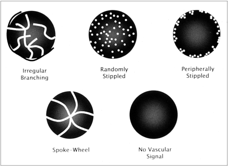
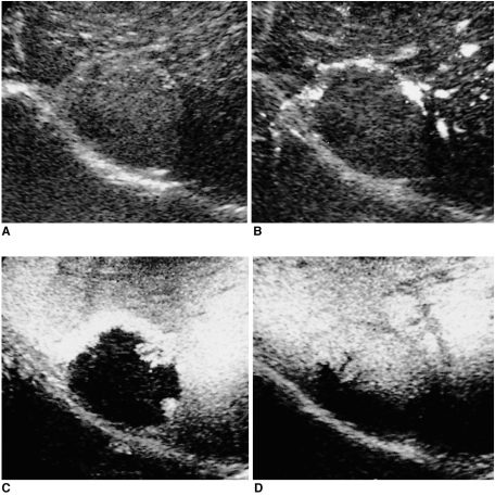
The study coordinator (H-J.J.) annotated vascular phase and acoustic emission images in advance on the sets of images. For vascular images, each reviewer was asked, in each case, to categorize the vascular pattern as one of the following five: irregular branching, randomly stippled, peripherally stippled, spoke-wheel, or no vascular signal (Fig. 1). As for acoustic emission images, the reviewers were asked to categorize the pattern seen during the early phase (start - 1 or 2 minute delay) as one of the following four: peripheral nodular, peripheral rim or flame, heterogeneous, or homogeneous (hyper-, iso-, or hypoechoic to surrounding parenchymal echogenicity). The evolving pattern was then classified as centripetal fill-in, washout (becoming hypo- or isoechoic to surrounding parenchymal echogenicity), or persistent. Discrepancies in the findings were resolved by consensus. The reviewers were not asked to diagnose the lesions studied. All observations of the enhancement patterns were totally subjective and not substantiated quantitatively.
Fig. 1.
The intratumoral vascular patterns seen during vascular phase imaging.
RESULTS
The intralesional vascular pattern seen at vascular imaging (Table 1) and the enhancement patterns at acoustic emission imaging (Table 2) are summarized in the tables.
Table 1.
Intratumoral Vascular Patterns Observed at Vascular Imaging
Note.-HCC= hepatocellular carcinoma, FNH= focal nodular hyperplasia
Table 2.
Serial Enhancement Patterns at Emission Imaging
Note.-HCC=hepatocellular carcinoma, Mets=metastasis, FNH=focal nodular hyperplasia
The patterns observed at acoustic emission imaging of nontumorous lesions were difficult to classify.
Hepatocellular Carcinomas
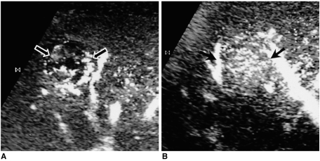
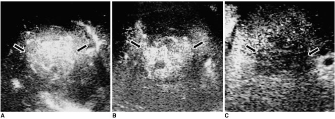
Tumor diameters as measured at US were 14-55 (mean, 25) mm. For HCCs, the most common intratumoral vascular pattern seen during the vascular imaging phase was irregular branching vessels (69%, 35/51), followed by randomly stippled vascularity (27%, 14/51) (Fig. 2). The intratumoral vessels of the remaining two HCCs (4%, 2/51) were judged to have a spoke-wheel appearance. During the acoustic emission imaging phase, 71% of HCCs (36/51) showed heterogeneous enhancement during the early phase, and the echogenicity of all HCCs diminished and became converse when compared with that of the parenchyma. This was, in other words, a washout pattern (Fig. 3).
Fig. 2.
Two characteristic vascular patterns of HCC (arrows).
A. Irregular branching pattern.
B. Randomly stippled pattern.
Fig. 3.
HCC (arrows). Serial acoustic emission images obtained at 20-sec (A), 2-min (B), and 5-min (C) delay show early enhancement (A) and heterogeneous washout (B, C).
Metastases
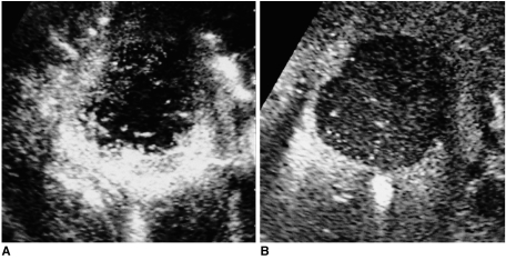
Tumor diameters as measured at US were 7-72 (mean, 23.5) mm. During the vascular imaging phase, 20 of 22 metastases (91%) showed a peripherally stippled pattern, while the remaining two demonstrated randomly stippled vascularity (2/22, 9%). During the early emission imaging phase (1-2 minute delay), 86% (19/22) showed rim- or flame-like peripheral enhancement (Fig. 4A) and the remaining three (3/22, 14%) showed homogeneous hypoechogenicity, with no intratumoral enhancement. A peripherally stippled vascular pattern was also observed for the few seconds remaining after emission imaging during this phase. The echo intensity of the enhancing areas of the tumor diminished (washout pattern), and the remaining areas with no early-phase enhancement were persistently hypoechoic (Fig. 4B). Accordingly, scans obtained after a delay of three or more minutes showed that all metastases became homogeneous cold lesions within the enhancing parenchyma.
Fig. 4.
Metastasis from duodenal carcinoma. Serial acoustic emission image depicts rim-or flame-like enhancement during the early phase (A, 40-sec delay) and a homogeneous perfusion defect during a later phase (B, 5-min delay).
Hemangiomas
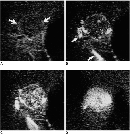
Tumor diameters as measured at US were 7-52 (mean, 23) mm. During the acoustic emission phase imaging, 19 of 22 hemangiomas (86%) showed typical peripheral nodular enhancement and progressive centripetal fill-in (Fig. 5), and the remaining three (3/22, 14%) showed persistent homogeneous enhancement, also one of the characteristic enhancement patterns of hemangiomas. Although the intensity of enhancing areas of the tumor diminished progressively after injection, areas of enhancement enlarged and persisted until the late phase of acoustic emission imaging. As for vascular phase imaging, no intratumoral flow signal was observed in 21 of 22 hemangiomas (95%) during the continuous vascular-phase scanning. Only one unusual hemangioma, with arterioportal shunt, showed diffuse intratumoral stippling during this phase scanning (Fig. 6).
Fig. 5.
Hemangioma. Serial CHA acoustic emission images obtained during the precontrast phase (A), and at 20-sec (B), 1-min (C), and 5-min (D) delay, clearly demonstrate typical peripheral nodular enhancement with progressive centripetal fill-in.
Fig. 6.
Unusual hemangioma with arterioportal shunt (A, precontrast; B, 20-sec delay; C, 42-sec delay; and D, 5-min delay). Only 20 sec after injection (B), CHA vascular phase imaging shows an early draining portal vein (arrows) and intratumoral vascular stippling. However, persistent homogeneous enhancement until 5-minute delay (D) led to the correct diagnosis.
Focal Nodular Hyperplasia
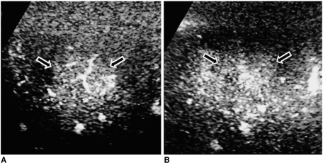
Tumor diameters as determined at US were 14-46 (mean, 28.5) mm. At vascular imaging, CHA clearly depicted the central arteries of the typical spoke-wheel pattern in three of four cases (3/4, 75%) (Fig. 7A). At acoustic emission imaging, the lesions showed a varying degree of homogeneous enhancement during the early phase and tended (3/4 cases, 75%) to be isoechoic during the late phase (Fig. 7B).
Fig. 7.
FNH (arrows). CHA images clearly depict the typical spoke-wheel pattern seen during the early vascular phase (A, 40-sec delay). The mass becomes isoechoic at late acoustic emission imaging (B, 3-min delay), a tendency which may help differentiate FNH from some HCCs showing a similar spoke-wheel vascular pattern but a washout pattern at late acoustic emission imaging.
Nontumorous Lesions
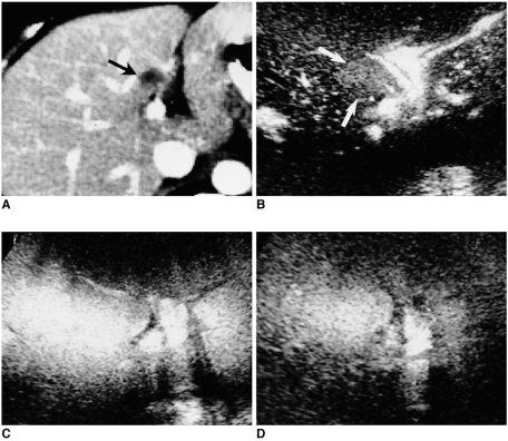
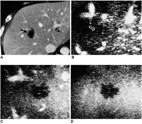
At US, lesion diameters were found to be 12-35 (mean, 23.3) mm. No lesions in this group (three FENs, two inflammatory nodules, and one focal fatty lesion) showed an intralesional vascular pattern at vascular phase imaging (Figs. 8, 9). At emission imaging, the focal fatty lesion was hyperechoic at precontrast scanning, but after contrast administration, its echo intensity became identical to that of surrounding parenchyma at any phase of acoustic emission imaging (Fig. 8). FENs showed a complete perfusion defect in their center and fuzzy enhancement at the periphery, but for the first one or two minutes, this was weaker than that of surrounding parenchyma. Inflammatory nodules showed a complete lack of enhancement at emission imaging (Fig. 9).
Fig. 8.
Focal fatty lesion in a breast cancer patient. Portal venous phase CT (A) shows a target-like hepatic nodule (arrow). At CHA vascular phase imaging (B, 40-sec delay), no vascular pattern is observed (arrows), and during both other phases of acoustic emission imaging (C, 3-min delay; D, 5-min delay), the lesion is completely obscured; its enhancement is the same as that of surrounding parenchyma. Subsequent MR imaging using the fat suppression technique (not shown) confirmed the diagnosis of focal fat deposition.
Fig. 9.
Inflammatory nodule in a rectal cancer patient. Portal venous phase CT (A) shows a hypoattenuating mass (arrow). CHA vascular phase imaging (B, 40-sec delay) reveals no vascular pattern within the mass (arrows), which appears as a perfusion defect at both other phases of acoustic emission imaging (C, 2-min delay; D, 5-min delay). Biopsy revealed chronic inflammation and necrosis.
DISCUSSION
CHA is a new contrast-specific gray-scale harmonic US technique based on digitally encoded US technology. To overcome the shortcomings of conventional harmonic imaging, various gray-scale harmonic imaging techniques have recently been developed (5, 7, 8, 11-13). Among these, digitally encoded harmonic US is the one that encodes the unwanted fundamental frequency component when transmitting pulses and then, through a decoding process when receiving signals, removes the pre-tagged fundamental echo without the loss of overlapping harmonic signals (14). The B-flow technique, optimized for direct visualization of blood cells at gray-scale imaging, uses codes to enhance weak blood echoes and equalize a non-moving tissue signal (15), and is thus free of Doppler artifact. Combining the advantages of the two techniques, CHA employs a technique that separates harmonic signals from tissue and those from contrast agents, and boosts harmonic return from the latter while suppressing not only fundamental but also harmonic signals from background tissue (14). Thus, CHA is specifically sensitive to signals from a contrast-agent, but without contrast-related artifacts. The resultant high sensitivity to a contrast agent provides sufficient SAE effect at medium MI, while other gray-scale harmonic US techniques usually require maximal MI to achieve this (5, 6, 8). Furthermore, since there is less destruction of microbubbles, a period of continuous scanning is possible. CHA's excellent sensitivity to contrast agents also permits direct visualization of mobile microbubbles per se, present in intratumoral microvasculature.
Previous studies in which contrast-enhanced harmonic US was used to depict hepatic lesions (5, 6, 8, 16) have mostly emphasized either the depiction of intratumoral vasculature by continuous scanning or the importance of a strict intermittent scanning technique to exploit the maximal SAE effect, according to the characteristics of the contrast agents and harmonic techniques used. Thus, in few studies have both vascular and acoustic emission imaging provided useful and detailed information. SH U 508A is currently the most widely available US contrast agent, and is known to be well tolerated in intravenous injection and to have a good safety profile (7, 16). It is, though, an air-based contrast agent which gives rise to weak harmonic signals at low MI and is therefore less suitable for continuous scanning (6). In a recent study (6) in which with SH U 508 A was used during vascular, interval-flash, and post-vascular imaging at different MIs, vascular imaging at low MI provided inconsistent data. On the other hand, contrast-agents containing higher density gas such as perfluorocarbon are more durable, providing acceptable harmonic imaging at low MI under continuous insonation (5). In our experience, CHA using SH U 508 A gave rise to harmonic signals which at low MI (0.2) were mostly inadequate for acceptable imaging, in accordance with previous results. In this study using SH U 508 A, we performed 'modified intermittent scanning', a combination of interval-delay and a short period of continuous scanning, both at medium MI. Before peak hepatic parenchymal enhancement was achieved, the 5-10-second period of continuous scanning was sufficient to observe the flow within intratumoral microvasculature, which was similar to that observed at conventional angiography, and we obtained an initial frame of acoustic emission images at the end of continuous vascular imaging. After the portal venous phase, our main concern was to observe the sequential dynamic enhancement patterns acquired at acoustic emission imaging, as well as those acquired at dynamic CT or MR imaging. The scans obtained during the late phase usually gave little additional information about vascular patterns.
The ability of both vascular and acoustic emission imaging to provide excellent depiction of vascular and enhancement patterns is noteworthy: combined information from both phases was very helpful for accurate characterization. In differentiating between HCC, metastasis and FNH, recognition of the characteristic vascular pattern of each tumor, as seen at vascular imaging, is very important; since most malignant tumors exhibit a washout pattern and cold defects at late acoustic emission imaging, the information obtained from such imaging alone may not be sufficient to differentiate HCC with central necrosis from metastasis. In addition, our results showed that the vascular patterns of HCC, metastasis and FNH could be potentially useful in differentiating these from inflammatory nodules; at late acoustic emission imaging, these latter sometimes appeared as a cold lesion, similar to a malignant tumor, but showed no vascular signal at vascular imaging. In many cases, the evolving pattern seen at acoustic emission imaging also provided helpful additional information. FNH tended to be isoechoic or hyperechoic at late acoustic emission imaging, and since all HCCs showed washout at acoustic emission imaging, this finding may help differentiate between unusual HCC with a spoke-wheel vascular pattern and FNH. The SAE effect frequently led to tumor enhancement even as early as 20 seconds after administration of a contrast agent, especially in hypervascular tumors. This early-phase acoustic emission imaging thereby provides images similar to those obtained at dynamic CT or MRI during the hepatic arterial phase.
All hemangiomas showed one of two characteristic patterns, which corresponded to the findings of dynamic CT or MRI: peripheral nodular enhancement with progressive centripetal fill-in (86%), or persistent homogeneous enhancement (14%). These findings were seen only during one or two initial frames of each interval scan, suggesting that these relied mostly on the SAE effect. In the next few minutes, hemangiomas - except for one unusual one with an arteriportal shunt - were completely unenhanced due to bubble destruction caused by extremely slow circulation. The detection of nodular or strong homogeneous enhancement during an initial frame of each interval delay is therefore very important. Once typical nodular enhancement was detected, we minimized the duration of continuous scanning in order to exploit efficient acoustic emission imaging during the late phase. Lengthening the interval delay rendered this enhancement toward the center of the lesion (8) and vascular imaging provided little additional information for the diagnosis of hemangioma. As suggested in our study and those of previous researchers (7, 8, 17), hemangiomas may be confidently diagnosed solely on the basis of contrast-enhanced gray-scale harmonic US findings, without further invasive or costly studies.
Another potential benefit of our CHA technique is that since there is time to adjust the scan field, it can be fairly easily applied to a small lesion. Kim et al. (8) previously reported the usefulness of pulse-inversion harmonic US in characterizing focal hepatic lesions, but they dealt with nodules at least 17 mm in diameter and their strictly intermittent scanning procedure is difficult to apply to smaller nodules. It is noteworthy that our cases included many nodules about 1 cm or even less in diameter, and our procedure was especially useful for characterizing nonspecific small hypoattenuating hepatic lesions detected at single-phase helical CT in oncologic patients. Most small hemangiomas or nontumorous lesions in our study were detected incidentally in patients with malignancy. Moreover, unlike Doppler techniques (1-4), even lesions located in the dome of the left hepatic lobe were clearly demonstrated without flash artifacts related to respiration, cardiac pulsation, or blooming, since CHA is free of Doppler or contrast-related artifacts. For the characterization of indeterminate focal hepatic lesions, CHA therefore appears to be a promising problem-solving tool, especially where CT or MR imaging has been inconclusive.
In spite of the many strong points described above, CHA has several drawbacks. Firstly, its inherent limitation is that the image quality of precontrast CHA is inferior to that of conventional gray-scale imaging: CHA suppresses stationary background tissue signals, both fundamental and harmonic, and is therefore unsuitable for routine hepatic examination and should be reserved for complementary study involving contrast enhancement. Secondly, it is highly dependent on the focal zone involved, and zooming in and accurate focusing on the region of interest of the lesion are thus important. Thirdly, harmonic signals from a deep-seated lesion are often insufficient for acceptable imaging, a general drawback of other harmonic techniques. Lastly, the additional time required for encoding and decoding results in a small drop in frame rates.
The usefulness of our findings is limited by the following problems. First, since baseline images may have included diagnostic clues such as underlying cirrhosis or the typical findings of a certain disease, the reviewers were not totally blinded to the diagnostic findings. Second, except for one malignant stromal tumor of the duodenum, no hypervascular metastasis was included in this study. In addition, in five cases, mostly involving deep-seated lesions, the proper scanning technique either was not or could not be employed, and this should be born in mind when interpreting our detection rate of almost 100% vis-á-vis vascularity in HCC, metastasis, and FNH.
In summary, owing to its excellent sensitivity to the contrast agent used, contrast-enhanced gray-scale US using a CHA technique permitted a period of continuous scanning for the depiction of intratumoral vasculature, as well as intermittent acoustic emission imaging, and thus provided characteristic findings of various focal hepatic lesions. To verify the potential clinical utility our study suggests, further investigation is warranted.
References
- 1.Goldberg BB, Merton DA, Forsberg F, Liu J-B, Rawool N. Color amplitude imaging: preliminary results using vascular sonographic contrast agents. J Ultrasound Med. 1996;15:127–134. doi: 10.7863/jum.1996.15.2.127. [DOI] [PubMed] [Google Scholar]
- 2.Kim TK, Han JK, Kim AY, Park SJ, Choi BI. Signal from hepatic hemangiomas on power Doppler US: real or artefactual? Ultrasound Med Biol. 1999;25:1055–1061. doi: 10.1016/s0301-5629(99)00058-7. [DOI] [PubMed] [Google Scholar]
- 3.Choi BI, Kim TK, Han JK, Kim AY, Seong CK, Park SJ. Vascularity of hepatocellular carcinoma: assessment with contrast-enhanced second-harmonic versus conventional power Doppler US. Radiology. 2000;214:381–386. doi: 10.1148/radiology.214.2.r00fe01381. [DOI] [PubMed] [Google Scholar]
- 4.Kim TK, Han JK, Kim AY, Choi BI. Limitations of characterization of hepatic hemangiomas using an ultrasound contrast agent (Levovist) and power Doppler ultrasound. J Ultrasound Med. 1999;18:737–743. doi: 10.7863/jum.1999.18.11.737. [DOI] [PubMed] [Google Scholar]
- 5.Wilson SR, Burns PN, Muradali D, Wilson JA, Lai X. Harmonic hepatic US with microbubble contrast agent: initial experience showing improved characterization of hemangioma, hepatocellular carcinoma, and metastasis. Radiology. 2000;215:153–161. doi: 10.1148/radiology.215.1.r00ap08153. [DOI] [PubMed] [Google Scholar]
- 6.Dill-Macky MJ, Burns PN, Khalili K, Wilson SR. Focal hepatic masses: enhancement patterns with SH U 508A and pulse-inversion US. Radiology. 2002;222:95–102. doi: 10.1148/radiol.2221010092. [DOI] [PubMed] [Google Scholar]
- 7.Tanaka S, Ioka T, Oshikawa O, Hamada Y, Yoshioka F. Dynamic sonography of hepatic tumors. AJR Am J Roentgenol. 2001;177:799–805. doi: 10.2214/ajr.177.4.1770799. [DOI] [PubMed] [Google Scholar]
- 8.Kim TK, Choi BI, Han JK, Hong HS, Park SH, Moo SG. Hepatic tumors: contrast agent-enhancement patterns with pulse-inversion harmonic US. Radiology. 2000;216:411–417. doi: 10.1148/radiology.216.2.r00jl21411. [DOI] [PubMed] [Google Scholar]
- 9.Burns PN, Wilson SR, Simpson DH, Chin CT, Lai X. Harmonic interval delay imaging: a new ultrasound contrast method for imaging the blood pool volume in the liver. Radiology. 1998;209(P):189. [Google Scholar]
- 10.Burns PN, Wilson SR, Muradali D, Powers JE, Fritzsch TT. Intermittent US harmonic contrast-enhanced imaging and Doppler improve sensitivity and longevity of vessel detection. Radiology. 1996;201(P):159. [Google Scholar]
- 11.Ding H, Kudo M, Onda H, et al. Evaluation of posttreatment response of hepatocellular carcinoma with contrast-enhanced coded phase-inversion harmonic US: comparison with dynamic CT. Radiology. 2001;221:721–730. doi: 10.1148/radiol.2213010358. [DOI] [PubMed] [Google Scholar]
- 12.Jang HJ, Lim HK, Lee WJ, Kim SH, Kim KA, Kim EY. Ultrasonographic evaluation of focal hepatic lesions: comparison of pulse inversion harmonic, tissue harmonic, and conventional imaging techniques. J Ultrasound Med. 2000;19:293–299. [PubMed] [Google Scholar]
- 13.Albrecht T, Hoffman CW, Schettler S, et al. B-mode enhancement at phase-inversion US with air-based microbubble contrast agent: initial experience in humans. Radiology. 2000;216:273–278. doi: 10.1148/radiology.216.1.r00jl27273. [DOI] [PubMed] [Google Scholar]
- 14.Kim JH, Kim TK, Kim BS, et al. Depiction of typical enhancement pattern of hepatic hemangiomas: coded harmonic angio US with microbubble contrast agent. J Ultrasound Med. 2002;21:141–148. doi: 10.7863/jum.2002.21.2.141. [DOI] [PubMed] [Google Scholar]
- 15.Furuse J, Maru Y, Mera K, et al. Visualization of blood flow in hepatic vessels and hepatocellular carcinoma using B-flow sonography. J Clin Ultrasound. 2001;29:1–6. doi: 10.1002/1097-0096(200101)29:1<1::aid-jcu1>3.0.co;2-f. [DOI] [PubMed] [Google Scholar]
- 16.Ding H, Kudo M, Onda H, Suetomi Y, Minami Y, Maekawa K. Hepatocellular carcinoma: depiction of tumor parenchymal flow with intermittent harmonic power Doppler US during the early arterial phase in dual-display mode. Radiology. 2001;220:349–356. doi: 10.1148/radiology.220.2.r01au07349. [DOI] [PubMed] [Google Scholar]
- 17.Lee JY, Choi BI, Han JK, Kim AY, Shin SH, Moon SG. Improved sonographic imaging of hepatic hemangioma with contrast-enhanced coded harmonic angiography: comparison with MR imaging. Ultrasound Med Biol. 2002;28:287–295. doi: 10.1016/s0301-5629(01)00511-7. [DOI] [PubMed] [Google Scholar]


