Abstract
Sequential proliferation, hypertrophy and maturation of chondrocytes are required for proper endochondral bone development and tightly regulated by cell signaling. The canonical Wnt signaling pathway acts through β-catenin to promote chondrocyte hypertrophy whereas PTHrP signaling inhibits it by holding chondrocytes in proliferating states. Here we show by genetic approaches that chondrocyte hypertrophy and final maturation are two distinct developmental processes that are differentially regulated by Wnt/β-catenin and PTHrP signaling. Wnt/β-catenin signaling regulates initiation of chondrocyte hypertrophy by inhibiting PTHrP signaling activity, but it does not regulate PTHrP expression. In addition, Wnt/β-catenin signaling regulates chondrocyte hypertrophy in a non-cell autonomous manner and Gdf5/Bmp signaling may be one of the downstream pathways. Furthermore, Wnt/β-catenin signaling also controls final maturation of hypertrophic chondrocytes, but such regulation is PTHrP signaling-independent.
Introduction
Mammalian skeletal development is controlled by two mechanisms, endochondral and intramembranous bone formation. Endochondral ossification occurs in most parts of the body and requires a cartilage model prior to bone formation. Cartilage is composed of chondrocytes and in the developing long bones, these chondrocytes are organized into zones with distinct cellular morphologies and proliferation properties. Such organization is essential for the proper directional growth and elongation and is subject to tight regulation by secreted signaling molecules and transcription factors [1]–[3]. Proliferating chondrocytes are located at both ends of the cartilage and express Sox9 and Col2a1. Closer to the middle of the cartilage, proliferating chondrocytes become elongated and line up in columns along the longitudinal axis. These columnar chondrocytes divide quickly and begin to express Indian hedgehog (Ihh) and Runx2 as they exit the cell cycle to become prehypertrophic chondrocytes. Prehypertrophic chondrocytes then differentiate into hypertrophic chondrocytes that no longer express Sox9 and Col2a1, but express Col10a1 and angiogenic factors such as vascular endothelial growth factor (Vegf). Hypertrophic chondrocyes can be further divided into two populations according to their differences in gene expression and mineralization levels. Ihh and parathyroid hormone receptor 1 (Pthr1) are expressed only in prehypertrophic and early hypertrophic chondrocytes. The mature population of hypertrophic chondrocytes express Osteopontin (Opn) [4] and matrix metalloproteinase 13 (Mmp13) that is required for invasion of blood vessels and osteoblasts to form the trabecular bone [5]. These most mature hypertrophic chondrocytes undergo apoptosis and are removed eventually. In addition, calcium deposition in the cartilage, which can be visualized by Von Kossa and Alizarin red staining, only occurs in the extracellular matrix of the final mature hypertrophic chondrocytes. Thus, both chondrocyte hypertrophy and final maturation are required for endochondral bone formation. However, it is not clear whether chondrocyte hypertrophy and final maturation are two consecutive developmental processes regulated by the same molecular pathways or they are actually developmentally separated and differentially regulated.
Ihh, parathyroid hormone related peptide (PTHrP), Wnts, Bone morphogenetic proteins (Bmps) and fibroblast growth factor (Fgfs) etc., regulate progressive chondrocyte proliferation and hypertrophy. PTHrP and Ihh are major regulators of chondrocyte hypertrophy by forming a negative feedback loop [6]. Ihh activates PTHrP expression and PTHrP then signals to proliferating chondrocytes to inhibit Ihh expression and chondrocyte hypertrophy by holding chondrocytes in a proliferating state. Wnt signaling pathways also regulate chondrocyte hypertrophy [7]–[10]. Among known Wnt signaling pathways, the canonical Wnt signaling pathway mediated by β-catenin [11] is the best understood and has been found to promote both chondrocyte hypertrophy and final maturation [9], [10]. Wnt/β-catenin signaling acts independently of Ihh signaling to promote chondrocyte hypertrophy [12], but it is unknown whether Wnt/β-catenin and PTHrP signaling pathways regulate each other to control chondrocyte hypertrophy and maturation.
Here we have investigated the regulation of chondrocyte hypertrophy and final maturation and the genetic relationship between the canonical Wnt and PTHrP signaling pathways in sequential chondrocyte differentiation. We uncovered that chondrocyte hypertrophy and final maturation are two distinct processes that are differentially regulated by Wnt/β-catenin and PTHrP signaling. Canonical Wnt signaling promotes chondrocyte hypertrophy by antagonizing PTHrP signaling activity. However, the final maturation of hypertrophy chondrocytes is controlled by Wnt/β-catenin signaling independently of PTHrP signaling.
Results
Wnt signaling controls chondrocyte hypertrophy by antagonizing PTHrP signaling
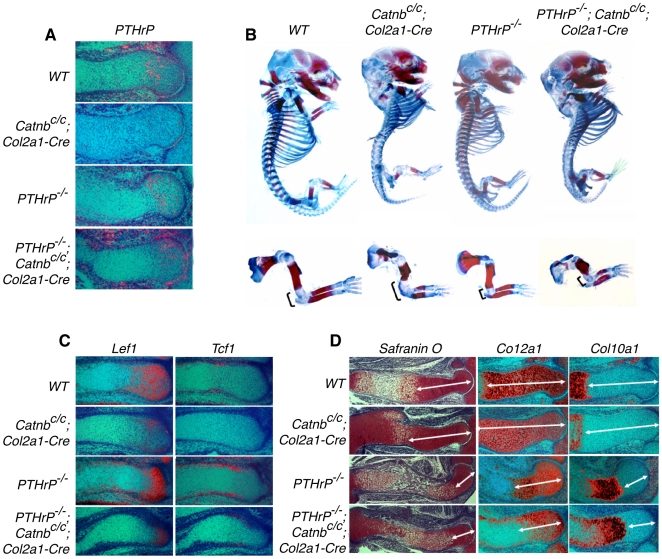
To test whether Wnt/β-catenin signaling acts through the PTHrP pathway in regulating chondrocyte hypertrophy, we first examined whether PTHrP expression was regulated by Wnt/β-catenin signaling in the developing long bone cartilage. β-catenin was removed in chondrocytes using the Col2a1-Cre mouse line [13]. The onset of chondrocyte hypertrophy at 12.5 days post coitum (dpc) were not altered [9]. However, continued initiation of chondrocyte hypertrophy by the remaining proliferating chondrocytes was slower in the Catnbc/c;Col2a1-Cre mouse embryos [9]. PTHrP expression in the β-catenin deficient cartilage was similar to that in the control, but weaker at 14.5dpc (Fig. 1A). This is likely secondary to weakened Ihh expression in the cartilage of the Catnbc/c; Col2a1-Cre mouse embryo [9], [14], since ligand-independent activation of Hedgehog (Hh) signaling in the Catnbc/c; Col2a1-Cre mice rescues PTHrP expression [12]. Furthermore, if Wnt/β-catenin signaling acts by regulating PTHrP expression to control chondrocyte hypertrophy, reduced PTHrP expression should lead to accelerated chondrocyte hypertrophy, the opposite of the observed phenotype for the Catnbc/c; Col2a1-Cre mouse embryo [9], [10]. These data suggest that Wnt/β-catenin signaling regulates chondrocyte hypertrophy independently of PTHrP expression.
Figure 1. Analysis of PTHrP and β-catenin mutant skeletons.
(A) Sections of developing humerus at 14.5 dpc were examined by in situ hybridization with PTHrP probes. Expression of PTHrP was reduced in periarticular chondrocytes in Catnbc/c; Col2a1-Cre single mutant embryos compared to that in wild type embryos. PTHrP expression was increased in the double mutant embryos compared to that in the Catnbc/c; Col2a1-Cre single mutant embryos. (B) Skeletal preparation of 16.5 dpc embryos. A forelimb of each embryo is shown in lower panel. Alizarin red stains the mineralized hypertrophic chondrocytes and bone. Alcian blue stains the unmineralized cartilage. Mineralization was accelerated in the PTHrP−/−; Catnbc/c; Col2a1-Cre double mutant and the PTHrP−/− single mutant. The bracket indicates the non-mineralized cartilage at the joint region. (C) Consecutive sections of the radius at 14.5 dpc were examined by in situ hybridization with probes of Lef1 and Tcf1. Lef1 and Tcf1 expression were downregulated in both Catnbc/c; Col2a1-Cre single mutant and PTHrP−/−; Catnbc/c; Col2a1-Cre double mutant embryos. (D) Consecutive sections of the developing humerus at 14.5 dpc were examined by Safranin O staining and in situ hybridization with the indicated riboprobes. Safranin O staining and expression of Col2a1 marked nonhypertrophic chondrocytes. The nonhypertrophic domain (double headed arrow) was expanded in Catnbc/c; Col2a1-Cre mutant embryos whereas it was shortened to the same degree in PTHrP−/− single mutant and PTHrP−/−; Catnbc/c; Col2a1-Cre double mutant embryos. Col10a1 expression marked hypertrophic region, which was accelerated to the same level in PTHrP−/− single mutant and PTHrP−/−; Catnbc/c; Col2a1-Cre double mutant embryos.
The possibility remains that β-catenin interacts with PTHrP signaling in PTHrP-responding chondrocytes. To test this, we generated the PTHrP−/−; Catnbc/c; Col2a1-Cre double mutant mouse embryos. If Wnt/β-catenin signaling acts downstream of PTHrP signaling, initiation of chondrocyte hypertrophy should be similarly delayed in both the Catnbc/c; Col2a1-Cre and PTHrP−/−; Catnbc/c; Col2a1-Cre embryos compared to that in the wild type embryo. Conversely, if Wnt/β-catenin signaling acts upstream of PTHrP signaling, initiation of chondrocyte hypertrophy should be similarly accelerated in the PTHrP−/− and PTHrP−/−; Catnbc/c; Col2a1-Cre embryos. Single and double mutant embryos were harvested together with their wild type control littermates and analyzed by skeletal preparations. The relative length of Alizarin red stained segment (shows mineralized cells) versus Alcian blue stained segment (shows unmineralized chondrocytes) was compared in the same cartilage elements (Fig. 1B). Long bone mineralization was increased in the PTHrP−/− single mutant, but reduced in the Catnbc/c; Col2a1-Cre and PTHrP−/−; Catnbc/c; Col2a1-Cre embryos (Fig. 1B). These results suggest that final maturation of hypertrophic chondrocytes was similarly delayed in the Catnbc/c; Col2a1-Cre and PTHrP−/−; Catnbc/c; Col2a1-Cre double mutant embryos.
Initiation and final maturation of chondrocyte hypertrophy were then examined more closely on histological sections of the limb at 14.5 dpc and 16.5 dpc. In both Catnbc/c; Col2a1-Cre and PTHrP−/−; Catnbc/c; Col2a1-Cre double mutant embryos, Wnt signaling activity was similarly reduced as shown by the reduction in the expression of two transcriptional targets of canonical Wnt signaling, Lef1 and Tcf1 (Fig. 1C) [15], [16]. Chondrocyte hypertrophy was then analyzed by Safranin O staining. Hypertrophic chondrocytes were those enlarged and lightly stained chondrocytes whereas proliferating chondrocytes were smaller and stained bright red. Surprisingly, although chondrocyte hypertrophy was delayed in the Catnbc/c; Col2a1-Cre embryo, loss of PTHrP signaling leads to an acceleration of chondrocyte hypertrophy in the PTHrP−/−; Catnbc/c; Col2a1-Cre embryo similar to that in the PTHrP−/− embryo (Fig. 1D). These morphological observations were further confirmed by molecular markers analysis. Col10a1 is expressed in hypertrophic chondrocytes whereas Col2a1 is expressed in proliferating chondrocytes. Col2a1 expression domain was reduced whereas Col10a1 expression domain was expanded to the same degree in the PTHrP−/− and PTHrP−/−; Catnbc/c; Col2a1-Cre embryos at 14.5 dpc (Fig. 1D). Similar acceleration of chondrocyte hypertrophy in PTHrP−/− and PTHrP−/−; Catnbc/c; Col2a1-Cre embryos was also observed in other skeletal elements such as the vertebral bodies (Fig. S1). These results indicate that Wnt/β-catenin signaling is dependent on PTHrP signaling in regulation of chondrocyte hypertrophy.
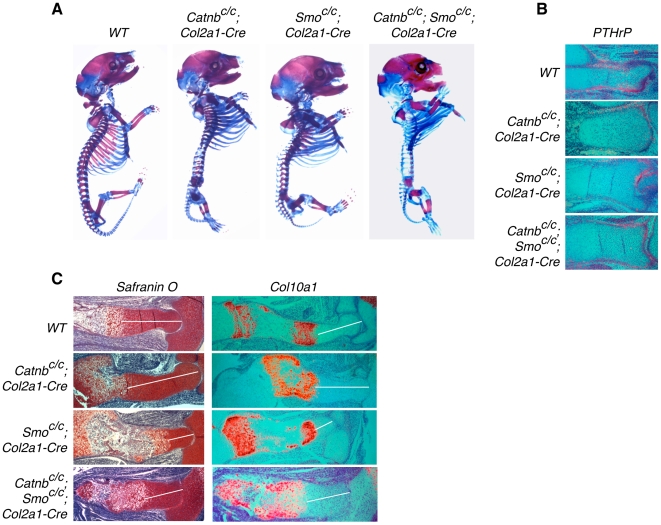
The above data suggest that active PTHrP signaling is required for Wnt/β-catenin signaling to regulate chondrocyte hypertrophy, but PTHrP signaling may not require β-catenin to regulate chondrocyte hypertrophy. This predicts that in the absence of β-catenin, PTHrP signaling levels can still determine the rate of chondrocyte hypertrophy. To test this, we need to partially reduce PTHrP signaling by reducing PTHrP expression. In this case, β-catenin removal should still enhance the residual PTHrP signaling, which will lead to a delay in chondrocyte hypertrophy. To achieve these genetic manipulations, we partially reduced PTHrP expression by partially reducing Ihh signaling as Ihh controls PTHrP expression. Ihh signaling was reduced by removing Smoothened (Smo), which encodes the signaling receptor for Ihh, in the cartilage in the Smoc/c; Col2a1-Cre embryo [17] and Fig. 2A). PTHrP expression was diminished in the cartilage, but still detectable in the joint region in the Smoc/c; Col2a1-Cre embryo (Fig. 2B). Chondrocyte hypertrophy was accelerated in the Smoc/c; Col2a1-Cre embryo, as indicated by both Safranin O staining and analysis of Col2a1 and Col10a1 expression (Fig. 2C). When β-catenin was also removed in the Smoc/c; Catnbc/c; Col2a1-Cre double mutant embryo, chondrocyte hypertrophy was delayed compared to that in the Smoc/c; Col2a1-Cre embryos (Fig. 2C), despite PTHrP expression levels being similarly reduced in both (Fig. 2B). In addition, we observed previously that when PTHrP expression was similarly upregulated in Ptch1c/−; Col2a1-Cre and Ptch1c/−; Catnbc/−; Col2a1-Cre embryos, there was a further delay of chondrocyte hypertrophy when β-catenin is removed [12]. Thus, in the absence of β-catenin, PTHrP signaling still inhibits chondrocyte hypertrophy in a dose-dependent manner, whereas in the absence of PTHrP signaling, β-catenin has no effect on chondrocyte hypertrophy.
Figure 2. Analysis of chondrocyte hypertrophy in β-catenin and Smo mutant embryos.
(A) Skeletal preparation of 18.5 dpc embryos. Mineralization (stained by Alizarin red) in the Catnbc/c; Smoc/c; Col2a1-Cre double mutant embryo was more advanced than that in the Catnbc/c; Col2a1-Cre mutant embryo, but less advanced than that in the Smoc/c; Col2a1-Cre mutant embryo. (B) Sections of the developing distal tibia at 15.5 dpc were examined by in situ hybridization with PTHrP probes. PTHrP expression was slightly reduced only in the periarticular chondrocytes in Catnbc/c; Col2a1-Cre mutant embryos compared to that in wild type embryos. Catnbc/c; Smoc/c; Col2a1-Cre and Smoc/c; Col2a1-Cre embryos have similar expression of PTHrP. (C) Safranin O staining and Col10a1 expression in the developing humerus at 15.5 dpc. Non-hypertrophic chondrocyte region (white line) was expanded in the Catnbc/c; Col2a1-Cre mutant, but reduced in the Smoc/c; Col2a1-Cre mutant embryo, compared that in the wild type control. Chondrocyte hypertrophy in the Catnbc/c; Smoc/c; Col2a1-Cre double mutant embryo was delayed than that in the Smoc/c; Col2a1-Cre mutant embryo, but accelerated than that in the Catnbc/c; Col2a1-Cre mutant embryo.
Delayed chondrocyte hypertrophy can be secondary to increased proliferation [18], [19]. Interestingly, chondrocyte proliferation was slightly increased in the PTHrP−/−; Catnbc/c; Col2a1-Cre embryos compared to that in the PTHrP−/− single mutants at 16.5 dpc (Fig. S2), although chondrocyte hypertrophy was similar in both mutants. These results indicate that Wnt/β-catenin signaling can affect the rate at which cells divide in the proliferating chondrocyte pool, but only PTHrP signaling controls the brake that determines when chondrocytes exit cell cycle to undergo hypertrophy.
Wnt/β-catenin signaling is required for maturation of hypertrophic chondrocytes independently of PTHrP signaling
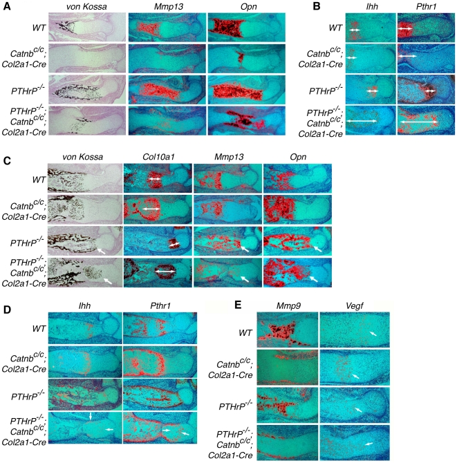
In the normal process of long bone development, chondrocyte hypertrophy is followed by hypertrophic chondrocyte maturation to allow the invasion of osteoblasts and blood vessels. Our observation that mineralization, but not hypertrophy, was similarly delayed in the PTHrP−/−; Catnbc/c; Col2a1-Cre and Catnbc/c; Col2a1-Cre embryos led us to think that formation of the early and late populations of hypertrophic chondrocytes are differentially regulated. To test this, we further examined chondrocyte maturation on histological sections. At 14.5 dpc, although Col10a1 expression domains were comparable between PTHrP−/− and PTHrP−/−; Catnbc/c; Col2a1-Cre embryos (Fig. 1D), in stark contrast to PTHrP−/− embryo, where maturation of hypertrophic chondrocytes was also accelerated, hypertrophic chondrocyte maturation detected by von Kossa staining was significantly delayed in the PTHrP−/−; Catnbc/c; Col2a1-Cre embryos (Fig. 3A). We then performed in situ hybridizations with probes specific to distinct stages of chondrocyte hypertrophy and maturation. At 14.5 dpc, expression domains of Mmp13 and Opn were much smaller in the PTHrP−/−; Catnbc/c; Col2a1-Cre embryos compared to those in the PTHrP−/− embryos (Fig. 3A). By contrast, expression domains of Ihh and Pthr1, which mark prehypertrophic chondrocytes and early hypertrophic chondocytes, were expanded in the PTHrP−/−; Catnbc/c; Col2a1-Cre embryos compared to that in the PTHrP−/− embryos (Fig. 3B). Thus, in the PTHrP−/−; Catnbc/c; Col2a1-Cre embryo, loss of PTHrP signaling accelerated chondrocyte hypertrophy, but these hypertrophic chondrocytes were not able to undergo further maturation due to the blockage of Wnt/β-catenin signaling. As a result, the Ihh and Pthr1 expressing early hypertrophic chondrocyte population was significantly expanded. This increased Ihh expression domain in the PTHrP−/−; Catnbc/c; Col2a1-Cre embryo likely caused increased osteoblast differentiation compared to that in the Catnbc/c; Col2a1-Cre embryo (Fig. 3A and S3).
Figure 3. Hypertrophic chondrocyte maturation was regulated by β-catenin independently of PTHrP.
Consecutive sections of the developing humerus at 14.5 dpc and 16.5 dpc were examined by Von Kossa staining and in situ hybridization with the indicated riboprobes. Mature hypertrophic chondrocytes were stained black by the Von Kossa method and expressed Mmp13 and Opn. (A) Von Kossa staining and Mmp13 and Opn expression were much reduced in both Catnbc/c; Col2a1-Cre and PTHrP−/−; Catnbc/c; Col2a1-Cre mutant embryos at 14.5 dpc. (B) Ihh and Pthr1 were expressed in the less mature hypertrophic chondrocytes (double-headed arrow). This domain was expanded significantly only in the PTHrP−/−; Catnbc/c; Col2a1-Cre mutant embryos at 14.5 dpc. (C) At 16.5 dpc, chondrocyte final maturation in the Catnbc/c; Col2a1-Cre and PTHrP−/−; Catnbc/c; Col2a1-Cre mutant was similar to that of the wild type control and PTHrP−/− mutant, respectively. (D) Expansion of Ihh and Pthr1 expression domains (white line) were still observed in the PTHrP−/−; Catnbc/c; Col2a1-Cre mutant at 16.5 dpc. (E) Expression of Mmp9 and Vegf in the developing humerus of the indicated genotypes at 14.5 dpc.
By 16.5 dpc, hypertrophic chondrocyte maturation, as indicated by von Kossa staining and expression of Mmp13 and Opn, caught up in the PTHrP−/−; Catnbc/c; Col2a1-Cre embryos with those in the PTHrP−/− embryos (Fig. 3C). However, at this stage more Col10a1-expressing hypertrophic chondrocytes were observed in the Catnbc/c; Col2a1-Cre and PTHrP−/−; Catnbc/c; Col2a1-Cre embryos compare to the wild type control and the PTHrP−/− embryos, respectively (Fig. 3C). This is likely due to reduced elimination of mature hypertrophic chondrocytes caused by delayed blood vessel invasion and bone formation when β-catenin was removed [9]. Consistent with this, mineralized hypertrophic chondrocytes domains were increased in both Catnbc/c; Col2a1-Cre and PTHrP−/−; Catnbc/c; Col2a1-Cre embryos compared to the wild type control and PTHrP−/− embryos, respectively (Fig. 3C). In addition, expression of Mmp9, which is a marker for osteoclasts and chondroclasts, was delayed in both Catnbc/c; Col2a1-Cre and PTHrP−/−; Catnbc/c; Col2a1-Cre embryos compared to the wild type control and PTHrP−/− embryos, respectively (Fig. 3E). However, Vegf expression, which was detected in hypertrophic chondrocytes was not decreased in Catnbc/c; Col2a1-Cre and PTHrP−/−; Catnbc/c; Col2a1-Cre embryos (Fig. 3E). There was also no significant change in RANKL expression (Fig. S4). These results suggest that delayed blood vessel invasion and trabecular bone formation in the PTHrP−/−; Catnbc/c; Col2a1-Cre embryo are at least in part a result of reduced expression of Mmps, not Vegf or RANKL. Interestingly, domains of Ihh/Pthr1 expressing- early hypertrophic chondrocytes were still expanded in the PTHrP−/−; Catnbc/c; Col2a1-Cre embryos at 16.5 dpc, particularly in the periphery of the cartilage compared to that in the PTHrP−/− embryos (Fig. 3D), which created a zone with a mixture of early and late mature hypertrophic chondrocytes. Taken together, these results indicate that the Wnt/β-catenin pathway regulates hypertrophic chondrocyte final maturation, at least partly and transiently, in a manner independent of PTHrP signaling.
Wnt signaling may regulate chondrocyte hypertrophy and maturation non-cell autonomously
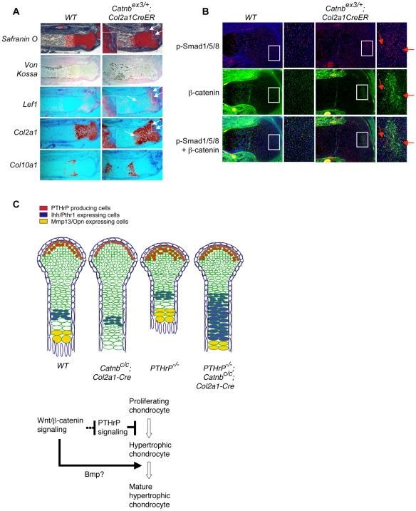
As PTHrP signaling controls chondrocyte hypertrophy cell autonomously [23], we tested whether the Wnt/β-catenin signaling pathway also acts cell autonomously. We expressed an activated form of β-catenin in subsets of chondrocytes using a mouse line with the exon 3 of β-catenin floxed (CatnbEx3) [24]. This mouse line was crossed with a tamoxifen (TM) inducible Cre line, Col2a1-CreER [25]. As the tamoxifen induced Cre activity is not robust in all cells, the activated form of β-catenin is only expressed in a subset of chondrocytes in the CatnbEx3/+; Col2a1-CreER embryos following TM injection. Since Lef1 is a transcription target of Wnt/β-catenin signaling [16], in these subset of chondrocytes, Lef1 expression was upregulated (Fig. 4A). In addition, bone formation was increased in the TM injected CatnbEx3/+; Col2a1-CreER embryos indicated by von Kossa staining (Fig. 4A), suggesting that Wnt/ β-catenin signaling in chondrocytes may also promote bone formation in the perichondrium non-cell autonomously. Interestingly, Safranin O staining and Col2a1 expression was significantly downregulated in the chondrocytes that expressed the activated form of β-catenin (Fig. 4A). However, these cells did not ectopically express Col10a1, a marker for hypertrophic chondrocytes (Fig. 4A). This is in sharp contrast to the normal process of chondrocyte hypertrophy in which Col2a1 expression is turned off, while Col10a1 expression is switched on. This is also drastically different from the Pthr1− /− clones in cartilage [23], in which loss of PTHrP signaling led to cell autonomous expression of Col10a1. In addition, while PTHrP signaling does not affect the determination of chondrocyte lineage [26], [27], activated Wnt signaling in the developing cartilage can change chondrocyte identity to the ones that resemble cells found in the joint interzone or joint [28]. These data suggest that activated β-catenin signaling acts cell autonomously to control chondrocyte identity, but non-cell autonomously in regulating chondrocyte hypertrophy. Among the secreted signaling molecules that can regulate chondrocyte hypertrophy, Growth differentiation factor 5 (Gdf5) and Bmp2 expression are upregulated by Wnt/β-catenin signaling [28]–[30]. To address the potential role of these genes, we examined Bmp signaling activities in our mutant mice (Fig. 4B). In and around chondrocytes that express activated β-catenin, we observed stronger phosphorylation of Smad1, 5 and 8, indicating increased Bmp signaling [31]. As Bmp/Gdf5 signaling has been shown to promote chondrocyte hypertrophy [32]–[34], Gdf5 and Bmp2 are likely to participate in mediating the non-cell autonomous function of Wnt/β-catenin signaling in chondrocyte hypertrophy.
Figure 4. β-catenin may control chondrocyte hypertrophy non-cell autonomously.
Tamoxifen was injected into pregnant females at 13.5 dpc. Embryos were harvested at 17.5 dpc and sections of the proximal tibia were analyzed by Safranin O staining, in situ hybridization with the indicated probes and immnohistochemistry. (A) Upregulated Wnt/β-catenin signaling was indicated by ectopic Lef1 expression and loss of Safranin O staining and Col2a1 expression (arrow). Pictures of higher magnification of the boxed area are shown as insets. Col10a1 expression was not ectopically detected in chondrocytes that had lost Col2a1 expression. (B) Increased p-Smad1/5/8 staining (red) was observed in chondrocyte patches of Catnbex3/+; Col2a1-CreER mouse embryos. Such increase in p-Smad1/5/8 staining was in the area with increased β-catenin staining (green). But p-Smad1/5/8 and β-catenin staining did not colocalize in many chondrocytes. Some cells with increased p-Smad1/5/8 staining (arrows) did not show increased β-catenin staining. (C) Distinct genetic interactions between Wnt/β-catenin and PTHrP signaling in regulating chondrocyte hypertrophy and maturation. The sequential process of chondrocyte hypertrophy and maturation in developing wild type and indicated mutant long bone cartilage were shown in the diagram in the upper panel. Hypertrophic chondrocytes are enlarged and undergo final maturation before they are replaced by the trabecular bone. Initiation of chondrocyte hypertrophy and final maturation are two separate processes that are differentially regulated by Wnt/β-catenin and PTHrP signaling. Wnt/β-catenin signaling controls chondrocyte hypertrophy by inhibiting PTHrP signaling activity. However, Wnt/β-catenin signaling promoted final maturation of hypertrophic chondrocyte independently of PTHrP signaling.
Discussion
We report here that during mouse embryonic cartilage development, Wnt/β-catenin signaling controls chondrocyte hypertrophy and final maturation by two distinct mechanisms. Wnt/β-catenin signaling regulates initiation of chondrocyte hypertrophy by antagonizing PTHrP signaling, whereas it acts independently of PTHrP signaling in controlling the final maturation of hypertrophic chondrocytes (Fig. 4C). Our results indicate that chondrocyte hypertrophy and final maturation are two separate developmental events that are regulated by distinct signaling interactions.
Our data suggests that delayed chondrocyte hypertrophy in the Catnbc/c; Col2a1-Cre mutant embryo is due to upregulated PTHrP signaling, not enhanced PTHrP expression. As β-catenin is required for activating the expression of downstream targets of the canonical Wnt signaling pathway, a possible scenario is that one of the transcription targets of the canonical Wnt signaling pathway in the chondrocytes suppresses the PTHrP signaling activity. Alternatively, the β-catenin protein may directly suppress PTHrP signaling activity by interacting with PTHrP signaling components. Interaction of β-catenin with components of other signaling pathways may be an important mechanism underlying Wnt signaling cross regulation with other pathways. For instance, a LEF/TCF independent regulatory role of β-catenin in cell lineage determination has been demonstrated during pituitary gland development, in which β-catenin binds a specific homeodomain factor, Prop1, to activate expression of the critical lineage-determining transcription factor, Pit1 [35]. However, if β-catenin inhibits PTHrP signaling by directly binding PTHrP signaling component(s), one may predict that PTHrP receptor and activated β-catenin both play a cell-autonomous role in chondrocyte hypertrophy. Although the PTHrP receptor acts cell autonomously in regulating chondrocyte hypertrophy [23], ectopic chondrocyte hypertrophy is not detected in chondrocytes expressing activated β-catenin as these cells have lost their chondrocyte identity because they express neither Col2a1 nor Col10a1. Interestingly, PTH activation of Pthr1 has been shown to activate the Wnt/β-catenin signaling directly by stabilizing β-catenin protein in osteoblasts [36]. However, in chondrocytes, it is unlikely that PTH/PTHrP signaling regulates chondrocyte hypertrophy through β-catenin as loss of PTH/PTHrP and Wnt/β-catenin signaling leads to opposite phenotypes.
Wnt/β-catenin signaling has two distinct roles in cartilage development. First, it inhibits chondrocyte cell lineage determination and maintenance [28]. Second, it promotes chondrocyte hypertrophy by inhibiting PTHrP signaling. However, such inhibition is likely to be incomplete. In addition, Wnt/β-catenin signaling may exert distinct functions in a dose dependent manner. It is conceivable that the two functions of Wnt/β-catenin signaling are also dose-dependent: Strong Wnt/β-catenin signaling (i.e., expression of activated β-catenin) inhibits chondrocyte cell fate determination and maintenance whereas weaker Wnt/β-catenin signaling promotes chondrocyte hypertrophy by reducing PTHrP signaling activities. Our results suggest that Wnt/β-catenin signaling may also interact with PTHrP signaling indirectly through a secondary signaling pathway. To this end, it will be interesting to further investigate genetically whether Gdf5/Bmp signaling control initiation chondrocyte hypertrophy by antagonizing PTHrP signaling and whether Gdf5/Bmp signaling also mediates the role of Wnt/β-catenin signaling in hypertrophic chondrocyte maturation.
Materials and Methods
Mice generation and Genotyping
Embryos were generated by crosses between mice carrying heterozygous PTHrP null allele PTHrP+/− [26], β-catenin conditional allele Catnbc/c or gain-of-function allele Catnbex3, and a transgene Cre which is under the regulation of Col2a1 promoter and enhancer. Embryos were collected at 14.5, 15.5 and 16.5 dpc and embryos were analyzed as described. Genotyping for the Col2a1-Cre transgene and β-catenin alleles were performed as previously described [9].
Skeletal Preparation
Alizarin red and Alcian blue staining of skeletons was conducted as previously described [12]. Embryos were eviscated, fixed for 48 hours in ethanol and then transferred to acetone for 24 hours. Embryos were stained overnight at 37°C followed by 2 additional days at room temperature. Then embryos were cleared in 1% KOH and stored in 80% glycerol.
Histology and in situ Hybridization
Limbs were fixed by 4% paraformaldehyde at 4°C overnight, embedded in paraffin and sectioned at 6 µm. Sections were stained by Safranin O and Von Kossa method. In situ hybridization was performed as previously described [8]. 35S-lablled riboprobes of Col 2a1, Col 10a1, Ihh, Pthr1, PTHrP, Opn, Mmp13, Mmp9 and Vegf were synthesized as previously [9], [12] and applied to limb sections. For immunnohistochemistry, primary antibodies used include anti β-catenin mouse monoclonal IgG (Transduction Lab) at 1∶50, anti-phospho-Smad1/5/8 rabbit polyclonal IgG (Cell signaling) at 1∶100 and anti-RANKL mouse monoclonal IgG (IMGENEX) at 1∶50. The signal was detected using FITC-conjugated secondary antibodies (Molecular Probes) or ABC kits (Vector Laboratories).
Tamoxifen preparation and injection
60 mg/ml Tamoxifen (Sigma) was dissolved in corn oil (Sigma) and sonicated until the solution became clear. The solution was filtered and 0.05 ml was injected intraperitoneally into the pregnant female mice at 13.5 dpc and the embryos were harvested at 17.5 dpc.
Supporting Information
Analysis of chondrocyte hypertrophy in vertebral skeletons. Vertebral skeletons were visualized by Alcian Blue / Alizarin Red staining at E14.5. Mineralization was greatly reduced in the ribs and vertebral bodies in both the Catnbc/c; Col2a1-Cre single mutant and the PTHrP−/−; Catnbc/c; Col2a1-Cre double mutant. Chondrocyte hypertrophy was revealed by Safranin O staining and the expression of Col2a1 and Col10a1 in sections of vertebral bodies at E14.5. Compared to the Catnbc/c; Col2a1-Cre single mutant, chondrocyte hypertrophy in the PTHrP−/−; Catnbc/c; Col2a1-Cre double mutant was much accelerated and similar to that in PTHrP−/− single mutant embryos indicated by the expression of Col10a1.
(3.47 MB TIF)
Analysis of chondrocyte proliferation in PTHrP and β-catenin mutant embryos. (A) BrdU-labeled chondrocytes were detected by immunohistochemistry on sections of distal tibia at 16.5dpc. Zone I cells are resting chondrocytes and Zone II are columnary proliferating chondrocytes. Zone I and II were greatly reduced and the difference between them was not clear in the PTHrP−/− single mutant and PTHrP−/−; Catnbc/c; Col2a1-Cre double mutant embryos. The entire proliferating region was marked as Zone I and II. (B) The percentage of BrdU labeled chondrocytes was counted from four different samples of each genotype and the average with standard deviations are shown. Significant difference with p<0.05 is shown.
(2.47 MB TIF)
Analysis of osteoblast differentiation in PTHrP and β-catenin mutant embryos. Consecutive sections of developing humerus at E14.5 and E16.5 were examined by in situ hybridization with indicated probes. (A) At E14.5, expression of early osteoblast marker Runx2 and Osx in PTHrP−/−; Catnbc/c; Col2a1-Cre double mutant embryos was accelerated to the same level as that in PTHrP−/− mutant embryos. (B) At E16.5, expression of Runx2 and Osx in double mutant embryos was accelerated whereas Osc expression was still delayed compared to that in PTHrP−/− single mutant embryos.
(4.01 MB TIF)
Analysis of RANKL expression in PTHrP and β-catenin mutant embryos. Proximal tibia sections at E16.5 were examined by immunohistochemistry with a RANKL monoclonal antibody. The expression of RANKL by osteoblasts is indicated by an arrow. RANKL expression in mature hypertrophic chondrocytes was very low and slightly increased in the Catnbc/c; Col2a1-Cre mutant but not in the PTHrP−/− and PTHrP−/−;Catnbc/c;Col2a1-Cre mutant embryos.
(3.75 MB TIF)
Acknowledgments
We thank members of the Yang lab for stimulating discussion during the work. We are grateful to Dr. Hank Kronenberg for sending us the PTHrP+/− mice and Dr. Susan Mackem for sharing with us the Col2a1-CreER transgenic mice. We also thank Ms. Julia Fekecs for help in preparing the figures.
Footnotes
Competing Interests: The authors have declared that no competing interests exist.
Funding: Work in the Yang lab is supported by the intramural research program of NIH, National Human Genome Research Institute. X. Guo was also partially supported by the National Grand Fundamental Research 973 Program of China under Grant 2007CB947301, the Chinese Ministry of Science and Technology. The funders had no role in study design, data collection and analysis, decision to publish, or preparation of the manuscript.
References
- 1.Kronenberg HM. Developmental regulation of the growth plate. Nature. 2003;423:332–336. doi: 10.1038/nature01657. [DOI] [PubMed] [Google Scholar]
- 2.Karsenty G. Transcriptional control of skeletogenesis. Annu Rev Genomics Hum Genet. 2008;9:183–196. doi: 10.1146/annurev.genom.9.081307.164437. [DOI] [PubMed] [Google Scholar]
- 3.Solomon LA, Berube NG, Beier F. Transcriptional regulators of chondrocyte hypertrophy. Birth Defects Res C Embryo Today. 2008;84:123–130. doi: 10.1002/bdrc.20124. [DOI] [PubMed] [Google Scholar]
- 4.Nakase T, Takaoka K, Hirakawa K, Hirota S, Takemura T, et al. Alterations in the expression of osteonectin, osteopontin and osteocalcin mRNAs during the development of skeletal tissues in vivo. Bone Miner. 1994;26:109–122. doi: 10.1016/s0169-6009(08)80056-6. [DOI] [PubMed] [Google Scholar]
- 5.Stickens D, Behonick DJ, Ortega N, Heyer B, Hartenstein B, et al. Altered endochondral bone development in matrix metalloproteinase 13-deficient mice. Development. 2004;131:5883–5895. doi: 10.1242/dev.01461. [DOI] [PMC free article] [PubMed] [Google Scholar]
- 6.Kronenberg HM. PTHrP and skeletal development. Ann N Y Acad Sci. 2006;1068:1–13. doi: 10.1196/annals.1346.002. [DOI] [PubMed] [Google Scholar]
- 7.Hartmann C, Tabin CJ. Dual roles of Wnt signaling during chondrogenesis in the chicken limb. Development. 2000;127:3141–3159. doi: 10.1242/dev.127.14.3141. [DOI] [PubMed] [Google Scholar]
- 8.Yang Y, Topol L, Lee H, Wu J. Wnt5a and Wnt5b exhibit distinct activities in coordinating chondrocyte proliferation and differentiation. Development. 2003;130:1003–1015. doi: 10.1242/dev.00324. [DOI] [PubMed] [Google Scholar]
- 9.Day TF, Guo X, Garrett-Beal L, Yang Y. Wnt/β-catenin signaling in mesenchymal progenitors controls osteoblast and chondrocyte differentiation during vertebrate skeletogenesis. Dev Cell. 2005;8:739–750. doi: 10.1016/j.devcel.2005.03.016. [DOI] [PubMed] [Google Scholar]
- 10.Akiyama H, Lyons JP, Mori-Akiyama Y, Yang X, Zhang R, et al. Interactions between Sox9 and β-catenin control chondrocyte differentiation. Genes Dev. 2004;18:1072–1087. doi: 10.1101/gad.1171104. [DOI] [PMC free article] [PubMed] [Google Scholar]
- 11.Huang H, He X. Wnt/β-catenin signaling: new (and old) players and new insights. Curr Opin Cell Biol. 2008;20:119–125. doi: 10.1016/j.ceb.2008.01.009. [DOI] [PMC free article] [PubMed] [Google Scholar]
- 12.Mak KK, Chen MH, Day TF, Chuang PT, Yang Y. Wnt/β-catenin signaling interacts differentially with Ihh signaling in controlling endochondral bone and synovial joint formation. Development. 2006;133:3695–3707. doi: 10.1242/dev.02546. [DOI] [PubMed] [Google Scholar]
- 13.Ovchinnikov DA, Deng JM, Ogunrinu G, Behringer RR. Col2a1-directed expression of Cre recombinase in differentiating chondrocytes in transgenic mice. Genesis. 2000;26:145–146. [PubMed] [Google Scholar]
- 14.Spater D, Hill TP, O'Sullivan RJ, Gruber M, Conner DA, et al. Wnt9a signaling is required for joint integrity and regulation of Ihh during chondrogenesis. Development. 2006;133:3039–3049. doi: 10.1242/dev.02471. [DOI] [PubMed] [Google Scholar]
- 15.Roose J, Huls G, van Beest M, Moerer P, van der Horn K, et al. Synergy between tumor suppressor APC and the β-catenin-Tcf4 target Tcf1. Science. 1999;285:1923–1926. doi: 10.1126/science.285.5435.1923. [DOI] [PubMed] [Google Scholar]
- 16.Hovanes K, Li TW, Munguia JE, Truong T, Milovanovic T, et al. β-catenin-sensitive isoforms of lymphoid enhancer factor-1 are selectively expressed in colon cancer. Nat Genet. 2001;28:53–57. doi: 10.1038/ng0501-53. [DOI] [PubMed] [Google Scholar]
- 17.Long F, Zhang XM, Karp S, Yang Y, McMahon AP. Genetic manipulation of hedgehog signaling in the endochondral skeleton reveals a direct role in the regulation of chondrocyte proliferation. Development. 2001;128:5099–5108. doi: 10.1242/dev.128.24.5099. [DOI] [PubMed] [Google Scholar]
- 18.Cobrinik D, Lee MH, Hannon G, Mulligan G, Bronson RT, et al. Shared role of the pRB-related p130 and p107 proteins in limb development. Genes Dev. 1996;10:1633–1644. doi: 10.1101/gad.10.13.1633. [DOI] [PubMed] [Google Scholar]
- 19.Yan Y, Frisen J, Lee MH, Massague J, Barbacid M. Ablation of the CDK inhibitor p57Kip2 results in increased apoptosis and delayed differentiation during mouse development. Genes Dev. 1997;11:973–983. doi: 10.1101/gad.11.8.973. [DOI] [PubMed] [Google Scholar]
- 20.Ohbayashi N, Shibayama M, Kurotaki Y, Imanishi M, Fujimori T, et al. FGF18 is required for normal cell proliferation and differentiation during osteogenesis and chondrogenesis. Genes Dev. 2002;16:870–879. doi: 10.1101/gad.965702. [DOI] [PMC free article] [PubMed] [Google Scholar]
- 21.Liu Z, Xu J, Colvin JS, Ornitz DM. Coordination of chondrogenesis and osteogenesis by fibroblast growth factor 18. Genes Dev. 2002;16:859–869. doi: 10.1101/gad.965602. [DOI] [PMC free article] [PubMed] [Google Scholar]
- 22.Minina E, Kreschel C, Naski MC, Ornitz DM, Vortkamp A. Interaction of FGF, Ihh/Pthlh, and BMP signaling integrates chondrocyte proliferation and hypertrophic differentiation. Dev Cell. 2002;3:439–449. doi: 10.1016/s1534-5807(02)00261-7. [DOI] [PubMed] [Google Scholar]
- 23.Chung UI, Lanske B, Lee K, Li E, Kronenberg H. The parathyroid hormone/parathyroid hormone-related peptide receptor coordinates endochondral bone development by directly controlling chondrocyte differentiation. Proc Natl Acad Sci U S A. 1998;95:13030–13035. doi: 10.1073/pnas.95.22.13030. [DOI] [PMC free article] [PubMed] [Google Scholar]
- 24.Harada N, Tamai Y, Ishikawa T, Sauer B, Takaku K, et al. Intestinal polyposis in mice with a dominant stable mutation of the β-catenin gene. EMBO J. 1999;18:5931–5942. doi: 10.1093/emboj/18.21.5931. [DOI] [PMC free article] [PubMed] [Google Scholar]
- 25.Nakamura E, Nguyen MT, Mackem S. Kinetics of tamoxifen-regulated Cre activity in mice using a cartilage-specific CreER(T) to assay temporal activity windows along the proximodistal limb skeleton. Dev Dyn. 2006;235:2603–2612. doi: 10.1002/dvdy.20892. [DOI] [PubMed] [Google Scholar]
- 26.Karaplis AC, Luz A, Glowacki J, Bronson RT, Tybulewicz VL, et al. Lethal skeletal dysplasia from targeted disruption of the parathyroid hormone-related peptide gene. Genes Dev. 1994;8:277–289. doi: 10.1101/gad.8.3.277. [DOI] [PubMed] [Google Scholar]
- 27.Lanske B, Karaplis AC, Lee K, Luz A, Vortkamp A, et al. PTH/PTHrP receptor in early development and Indian hedgehog-regulated bone growth. Science. 1996;273:663–666. doi: 10.1126/science.273.5275.663. [DOI] [PubMed] [Google Scholar]
- 28.Guo X, Day TF, Jiang X, Garrett-Beal L, Topol L, et al. Wnt/β-catenin signaling is sufficient and necessary for synovial joint formation. Genes Dev. 2004;18:2404–2417. doi: 10.1101/gad.1230704. [DOI] [PMC free article] [PubMed] [Google Scholar]
- 29.Hartmann C, Tabin CJ. Wnt-14 plays a pivotal role in inducing synovial joint formation in the developing appendicular skeleton. Cell. 2001;104:341–351. doi: 10.1016/s0092-8674(01)00222-7. [DOI] [PubMed] [Google Scholar]
- 30.Zhu M, Tang D, Wu Q, Hao S, Chen M, et al. Activation of β-Catenin Signaling in Articular Chondrocytes Leads to Osteoarthritis-Like Phenotype in Adult β-Catenin Conditional Activation Mice. J Bone Miner Res. 2008 doi: 10.1359/JBMR.080901. [DOI] [PMC free article] [PubMed] [Google Scholar]
- 31.Attisano L, Wrana JL. Signal transduction by the TGF-β superfamily. Science. 2002;296:1646–1647. doi: 10.1126/science.1071809. [DOI] [PubMed] [Google Scholar]
- 32.Tsumaki N, Tanaka K, Arikawa-Hirasawa E, Nakase T, Kimura T, et al. Role of CDMP-1 in skeletal morphogenesis: promotion of mesenchymal cell recruitment and chondrocyte differentiation. J Cell Biol. 1999;144:161–173. doi: 10.1083/jcb.144.1.161. [DOI] [PMC free article] [PubMed] [Google Scholar]
- 33.Tsumaki N, Nakase T, Miyaji T, Kakiuchi M, Kimura T, et al. Bone morphogenetic protein signals are required for cartilage formation and differently regulate joint development during skeletogenesis. J Bone Miner Res. 2002;17:898–906. doi: 10.1359/jbmr.2002.17.5.898. [DOI] [PubMed] [Google Scholar]
- 34.Kobayashi T, Lyons KM, McMahon AP, Kronenberg HM. BMP signaling stimulates cellular differentiation at multiple steps during cartilage development. Proc Natl Acad Sci U S A. 2005;102:18023–18027. doi: 10.1073/pnas.0503617102. [DOI] [PMC free article] [PubMed] [Google Scholar]
- 35.Olson LE, Tollkuhn J, Scafoglio C, Krones A, Zhang J, et al. Homeodomain-mediated β-catenin-dependent switching events dictate cell-lineage determination. Cell. 2006;125:593–605. doi: 10.1016/j.cell.2006.02.046. [DOI] [PubMed] [Google Scholar]
- 36.Wan M, Yang C, Li J, Wu X, Yuan H, et al. Parathyroid hormone signaling through low-density lipoprotein-related protein 6. Genes Dev. 2008;22:2968–2979. doi: 10.1101/gad.1702708. [DOI] [PMC free article] [PubMed] [Google Scholar]
Associated Data
This section collects any data citations, data availability statements, or supplementary materials included in this article.
Supplementary Materials
Analysis of chondrocyte hypertrophy in vertebral skeletons. Vertebral skeletons were visualized by Alcian Blue / Alizarin Red staining at E14.5. Mineralization was greatly reduced in the ribs and vertebral bodies in both the Catnbc/c; Col2a1-Cre single mutant and the PTHrP−/−; Catnbc/c; Col2a1-Cre double mutant. Chondrocyte hypertrophy was revealed by Safranin O staining and the expression of Col2a1 and Col10a1 in sections of vertebral bodies at E14.5. Compared to the Catnbc/c; Col2a1-Cre single mutant, chondrocyte hypertrophy in the PTHrP−/−; Catnbc/c; Col2a1-Cre double mutant was much accelerated and similar to that in PTHrP−/− single mutant embryos indicated by the expression of Col10a1.
(3.47 MB TIF)
Analysis of chondrocyte proliferation in PTHrP and β-catenin mutant embryos. (A) BrdU-labeled chondrocytes were detected by immunohistochemistry on sections of distal tibia at 16.5dpc. Zone I cells are resting chondrocytes and Zone II are columnary proliferating chondrocytes. Zone I and II were greatly reduced and the difference between them was not clear in the PTHrP−/− single mutant and PTHrP−/−; Catnbc/c; Col2a1-Cre double mutant embryos. The entire proliferating region was marked as Zone I and II. (B) The percentage of BrdU labeled chondrocytes was counted from four different samples of each genotype and the average with standard deviations are shown. Significant difference with p<0.05 is shown.
(2.47 MB TIF)
Analysis of osteoblast differentiation in PTHrP and β-catenin mutant embryos. Consecutive sections of developing humerus at E14.5 and E16.5 were examined by in situ hybridization with indicated probes. (A) At E14.5, expression of early osteoblast marker Runx2 and Osx in PTHrP−/−; Catnbc/c; Col2a1-Cre double mutant embryos was accelerated to the same level as that in PTHrP−/− mutant embryos. (B) At E16.5, expression of Runx2 and Osx in double mutant embryos was accelerated whereas Osc expression was still delayed compared to that in PTHrP−/− single mutant embryos.
(4.01 MB TIF)
Analysis of RANKL expression in PTHrP and β-catenin mutant embryos. Proximal tibia sections at E16.5 were examined by immunohistochemistry with a RANKL monoclonal antibody. The expression of RANKL by osteoblasts is indicated by an arrow. RANKL expression in mature hypertrophic chondrocytes was very low and slightly increased in the Catnbc/c; Col2a1-Cre mutant but not in the PTHrP−/− and PTHrP−/−;Catnbc/c;Col2a1-Cre mutant embryos.
(3.75 MB TIF)


