Abstract
When two different strains of swarming Proteus mirabilis encounter one another on an agar plate, swarming ceases and a visible line of demarcation forms. This boundary region is known as the Dienes line and is associated with the formation of rounded cells. While the Dienes line appears to be the product of distinction between self and nonself, many aspects of its formation and function are unclear. In this work, we studied Dienes line formation using clinical isolates labeled with fluorescent proteins. We show that round cells in the Dienes line originate exclusively from one of the swarms involved and that these round cells have decreased viability. In this sense one of the swarms involved is dominant over the other. Close cell proximity is required for Dienes line formation, and when strains initiate swarming in close proximity, the dominant Dienes type has a significant competitive advantage. When one strain is killed by UV irradiation, a Dienes line does not form. Killing of the dominant strain limits the induction of round cells. We suggest that both strains are actively involved in boundary formation and that round cell formation is the result of a short-range killing mechanism that mediates a competitive advantage, an advantage highly specific to the swarming state. Dienes line formation has implications for the physiology of swarming and social recognition in bacteria.
The gram-negative bacterium Proteus mirabilis is well known for its ability to differentiate into hyperflagellated, motile, and elongated swarmer cells that rapidly spread over a surface. When cultured on a nutrient agar plate, a strain of P. mirabilis typically is able to colonize the whole plate within 24 h. This phenomenon is both of interest in terms of the differentiation and survival strategy of the organism and of practical importance, as contamination of agar plates by swarming P. mirabilis is a common problem in diagnostic microbiology. When two different strains of P. mirabilis swarm on the same agar plate, a visible demarcation line with lower cell density forms at the intersection, and this line is known as a Dienes line (5, 6). A Dienes line is seen when both strains are swarming; it is not a property of the smaller vegetative cells (5). When two identical isolates meet, the swarming edges merge without formation of a Dienes line. This phenomenon has been used in epidemiological typing of clinical isolates (4, 20, 23, 28) and raises interesting questions concerning its mechanism and biological importance. Dienes typing in the clinical environment suggests that the number of Dienes types is large; 81 types were found in one study alone (25). Incompatibility between swarming strains may not be unique to P. mirabilis; a comparable process also appears to occur in Pseudomonas aeruginosa (19). Like many other bacteria, some P. mirabilis strains produce bacteriocins, termed proticines (3). It has been shown that Dienes line formation is not directly caused by a proticine, nor has any other secreted substance or cellular lysate been found to contribute to this phenomenon (27). There does, however, seem to be a circumstantial link between proticine production and Dienes line formation. In the 1970s Senior typed strains of P. mirabilis based on their proticine production and sensitivity (23, 24). He found that proticine production is not related to proticine sensitivity (apart from strains being resistant to their own proticine) but that there is a good correlation between a combined proticine production-sensitivity (P/S) type and Dienes line formation. Strains with the same P/S type do not form a Dienes line, whereas strains with different P/S types do. The more closely related the P/S types of two strains, the less clearly defined the Dienes line is, suggesting that relatedness of strains plays a central role. In contrast, a strain of Proteus vulgaris has been shown to be Dienes compatible with a strain of P. mirabilis with the same P/S type (24). Furthermore, Dienes incompatibility between otherwise identical strains can be triggered by phage lysogeny (2). In contrast to P/S type, the polysaccharide (O) or flagellar (H) serotype has been shown to have no relationship to Dienes line formation (25, 27).
Research into the mechanism governing Dienes line formation in Proteus has revealed some important features. The Dienes line region contains many large and often rounded cells (5). The nature of these cells remains controversial. Dienes suggested that round cells originated from both strains but that viable round cells always originated from one of the two intersecting strains. He concluded from this that nonviable round cells should therefore be cells of the other strain (6). In contrast, Wolstenhome suggested that the round cells originated mostly from one of the two swarms involved and that only 50% of these cells were viable (32). It has also been noted that round cells occasionally seem to develop into stable L forms lacking a full cell wall and that they can grow to form tiny L-type colonies (5). Additionally, extracellular DNase has been found at the site of the Dienes line (1). The presence of this enzyme has been interpreted to be a result of cell lysis, as P. mirabilis is known to contain large amounts of DNase (22, 26). Recently, a cluster of six genes, termed ids (identification of self), has been linked to the incompatibility in interpenetration between two strains (and therefore formation of the visible demarcation line), although the function and expression of these genes are not understood yet (8).
Despite the fact that Dienes line formation has been known for over 50 years, many questions concerning this phenomenon remain unanswered. Recent advances in imaging, molecular biology, and genomics offer new ways of investigating it. In this work, clinical isolates expressing fluorescent proteins were used to observe the cells involved in Dienes line formation in real time, to evaluate the fate of the round cells, and to test the role of extracellular material and direct cell-cell contact in this phenomenon. Furthermore, the biological role of the Dienes phenomenon in the competition between strains in different situations was investigated.
MATERIALS AND METHODS
Strains and media.
Four clinical isolates of P. mirabilis (strains 1 to 4), identified by using VITEK2 (bioMérieux, Marcy l'Etoile, France), were used throughout this study. These strains were all ampicillin sensitive.
Luria broth (10 g/liter peptone, 5 g/liter yeast extract, 10 g/liter NaCl) and Luria agar (Luria broth with 1.5% [wt/vol] agar) were used as culture media for all experiments involving transformation. Tryptic soy agar (TSA) (40 g/liter; Difco, United Kingdom), Mueller-Hinton agar (MH agar) (38 g/liter; Difco, United Kingdom), and sheep blood agar (Oxoid, Cambridge, United Kingdom) were used for swarming experiments. When necessary for plasmid maintenance, plates and media were supplemented with ampicillin (100 mg/liter).
Construction of strains expressing fluorescent proteins.
Strains 1 to 4 were transformed to express the green fluorescent protein (GFP) or a red fluorescent protein (DsRed). The plasmids used were pBAC001 (12) and pGT3dsRed (30). Plasmid pBAC001 contains a ColE1 origin and lac and flaA (flagellin) promoters, both driving expression of gfpmut2 (GFP) and an ampicillin resistance gene. Plasmid pGT3dsRed contains a plac promoter driving expression of DsRed and an ampicillin resistance gene. The P. mirabilis strains were transformed with these plasmids using standard electroporation protocols, resulting in strains 1G to 3G for the GFP-expressing variants and strains 1R to 4R for the DsRed-expressing variants. Strain 4G was excluded as it repeatedly lost the ability to swarm after transformation. GFP and DsRed were constitutively produced in strains 1G to 3G and strains 1R to 4R, respectively. In these strains, the production of fluorescent proteins had no major effects on the growth rate, swarming, or the ability to form Dienes lines.
Direct observation of Dienes line formation.
Using the fluorescently tagged strains, pairs of strains were inoculated onto TSA plates containing ampicillin in all combinations to examine Dienes line formation. The plates were incubated at 37°C until the swarm edges intersected and were subsequently examined by fluorescence microscopy. TSA was used because it is nearly transparent and contains no autofluorescent components.
Quantification of round cells.
To determine the ratio of the round cells of the two strains in a Dienes line, strains were inoculated in order to form Dienes lines on TSA plates containing ampicillin. A sterile toothpick was used to recover cells from the Dienes line region, which were mounted on microscope slides for observation.
SEM visualization of Dienes line formation.
Formation of a Dienes line between strains 2R and 3G was observed by in situ scanning electron microscopy (SEM). Strains were cultured in shake flasks containing MH broth with 100 mg/liter ampicillin. Spots of the cultures (1 μl) were placed on Anopore strips (obtained from PamGene International, The Netherlands, and manufactured by Whatman UK) resting on MH plates without ampicillin and incubated (10). After 3 to 4 h, if swarming was observed, the Anopore strips were transferred (bacterial inoculum up) to MH agar plates containing 3% (wt/vol) glutaraldehyde (Sigma, The Netherlands) and fixed for 3 h. Anopore is an inert ceramic, an aluminum oxide that is formed in thin sheets by a high-pressure and etching technique that creates a porous planar material; up to 50% of the volume is pores. The Anopore strips used were 8 by 36 mm and 60 μm thick and had 3 × 10⁹ 0.2-μm-diameter pores/cm2. Strips were transferred before the two strains met (control), 1 or 2 h after they met (Dienes lines forming), and 6 h after they met (very clear, stable lines). The area of Dienes line formation was then imaged by SEM using osmium tetroxide treatment to preserve extracellular polymers during critical point drying (11).
L-form induction using tap water.
Unstable L forms were generated from strains containing fluorescent proteins by osmotic shock, as described by Dienes (5). Swarmer cells from actively swarming edges on TSA containing 100 mg/liter ampicillin were transferred into 1 ml of tap water using a sterile toothpick. The preparation was incubated for 10 to 60 min at 37°C, after which the medium was changed to 50% (vol/vol) tryptic soy broth (TSB). The preparations were observed by fluorescence microscopy using wet mounts on poly-l-lysine-coated microscope slides.
Live-dead staining.
Dienes lines were formed on MH agar using a combination of strains 2 and 3, both lacking fluorescent proteins. The regions in which rounded cells formed were targeted and recovered with a sterile toothpick guided by bright-field light microscopy. The recovered cells were resuspended in MH broth and double stained with Syto 9 and propidium iodide using dye concentrations described previously (10), with incubation for 20 min at room temperature (Live/Dead staining kit; Invitrogen, Carlsbad, CA). Stained cells were visualized by fluorescence microscopy of wet mounts and by phase-contrast light microscopy to detect any poorly stained cells. Large, rounded cells were defined as cells that were clearly separated from other cells, were 1.1 to 5.1 μm wide, and had an aspect ratio (length/width ratio) of <2.2. Swarmer cells were <0.8 μm wide and had an aspect ratio of >5. Vegetative cells were <0.8 μm wide and had an aspect ratio <5. At least 200 cells were analyzed for each data point.
Time-lapse series using fluorescence microscopy.
Cells from Dienes lines formed by strains 2G and 3R were collected using a sterile toothpick and inoculated onto Luria agar plates. Rounded cells and vegetative cells of strain 3R, without any 2G cells in the vicinity, were examined using a time-lapse series over 8 h with a Zeiss Axiovert 200 Marianas inverted microscope equipped with a motorized stage (stepper motor z-axis increments, 0.1 μm) and with Cy-3 and differential interference contrast cubes. Three independent experiments were performed with at least eight cells of each type in the field of view. A cooled color charge-coupled device camera (1280 by 1024 pixels; Cooke Sensicam; Cooke, Tonawanda, NY) was used to record images. Exposures, objective settings, montage, and pixel binning were automatically recorded, and each image was stored in memory. The microscope, camera, and all other aspects of data acquisition, as well as data processing, were controlled by SlideBook software (SlideBook, version 3.1; Intelligent Imaging Innovations, Denver, CO). All live microscopy was performed with a custom ×40 air objective lens (Zeiss, The Netherlands).
Inhibition of cell-cell contact.
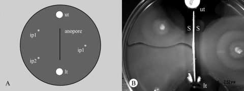
Two strains of P. mirabilis were allowed to swarm on different sides of a sheep blood agar plate separated by a sterile barrier embedded in the agar, a highly porous Anopore membrane that was permeable to most proteins and macromolecules but not to bacteria. Both untreated and poly-l-lysine-treated Anopore strips were tested (33). To prevent bacteria from bypassing the membrane, amoxicillin-clavulanate tablets (Rosco, Taastrup, Denmark) were placed at each end of the 36-mm-long barrier. Swarming bacteria were unable to pass this barrier. Strains were incubated for 24 h at 37°C in pairwise combinations. Strains 1 to 4, not expressing fluorescent proteins, were tested with each other in this setup in every combination. A second setup was used with the same model. Instead of inoculating one strain on each side of the membrane, on one side of the membrane two strains were inoculated so that a Dienes line would form perpendicular to the membrane. On the other side of the membrane, one of these strains was inoculated (Fig. 1A and B). Again, using strains 1 to 4, all possible permutations of pairs were tested. With both setups observations were made by eye and by using low-power microscopy to identify an inhibition zone, and microscopic observations of a Gram-stained smear of the growth closest to the membrane were made to look for round cells.
FIG. 1.
(A) Experimental design: sheep blood agar plate with two halves separated by an Anopore membrane embedded in the agar to create a barrier. To prevent swarming bacteria from bypassing the membrane, amoxicillin-clavulanate tablets (ut, upper tablet; lt, lower tablet) were placed at the ends of the Anopore membrane (vertical line). Two inoculation points for the first strain (ip1) and one inoculation point for the second strain (ip2) are indicated. (B) Two nonidentical strains were inoculated on one side of the Anopore membrane, and after swarming a Dienes line formed perpendicular to the membrane on the left side of the membrane. On the other side of the membrane one of the two strains was inoculated and was able to swarm right up to the membrane. Due to the illumination conditions the vertical Anopore membrane cast a shadow on both sides (S); this does not indicate reduced growth near the barrier. The upper antibiotic tablet is at position ut, and the lower antibiotic tablet is just below position lt.
Dead swarm experiment.
For the dead swarm experiment, all strains were allowed to swarm on individual TSA plates. When swarms covered approximately one-third of a plate, cells were killed in situ by inverting the plate over a UV transilluminator for 5, 20, and 30 min (eight 16-W bulbs, 312 nm). These experiments were performed in triplicate; three plates were used for further experiments, and three plates were used for testing the viability of UV-treated swarms. The latter plates were evaluated to determine viability in three ways. First, approximately 109 treated cells were inoculated into 30 ml Luria broth and incubated for 8 h with shaking at 37°C. Second, UV-treated swarms were incubated at 37°C for 12 h to determine whether swarming restarted. Third, viable cell counting on TSA was performed. On the plates not used to check for viability, a different strain was inoculated and allowed to swarm into the dead swarm. This experiment was performed for all combinations of transformed strains. Controls were included to show that UV irradiation of uninoculated TSA plates did not inhibit subsequent swarming or Dienes line formation. The resulting interactions in all experiments were evaluated by eye and by fluorescence microscopy.
Growth competition.
A number of different situations were used to evaluate competition between fluorescently tagged strains, including growth on agar without swarming, growth on agar with swarming, growth in liquid culture, and a biofilm model. For competition on semisolid media, TSA plates with and without p-nitrophenylglycerol (PNPG) (100 μg/ml) to inhibit swarming were used (16). Cells from liquid cultures (TSB) were mixed at different ratios (from 1:10,000 to 10,000:1), and 10-μl portions of the mixtures (106 cells) were then spotted in the center of the TSA plates, which were incubated for 24 h at 37°C. An experiment to examine competition in liquid culture was performed by incubating the same amount of mixtures in 20 ml of TSB shaken for 24 h at 37°C. The biofilm model used cultured proteus biofilms on glass coverslips in both Luria broth and TSB (in both cases with and without 100 μg/ml PNPG) and imaging after 24 and 48 h. Broth was recirculated over the coverslips by shaking individual coverslips in petri dishes to obtain flow rates equivalent to that described previously (14). For all experiments, cells were collected by harvesting the contents of entire plates or by sampling selected regions. The ratio of red to green was determined by scanning monolayers of cells on microscope slides and quantifying the cells using the ImageJ software package. A Leica dissecting microscope was used to visualize the segregation of the two strains on agar plates, taking advantage of the fact that the GFP and DsRed were sufficiently intense in the expressing strains that the two strains could be distinguished using reflected white light when they were viewed at a low magnification (×8). For these images, saturation values were enhanced by 60% (across all images equally). Finally, additional competition experiments were performed by transferring swarming cells of one strain labeled with one color to an actively swarming mass of a second, incompatible strain labeled with the other color and then incubating the culture and using fluorescence microscopy to determine the outcome after 2 and 18 h of incubation at 37°C.
RESULTS
Real-time visualization of Dienes lines and the formation of round cells. (i) Direct observation of Dienes line formation.
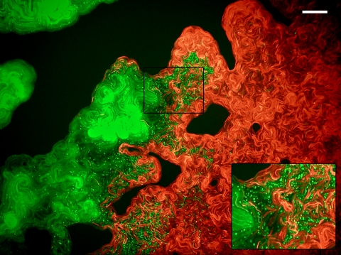
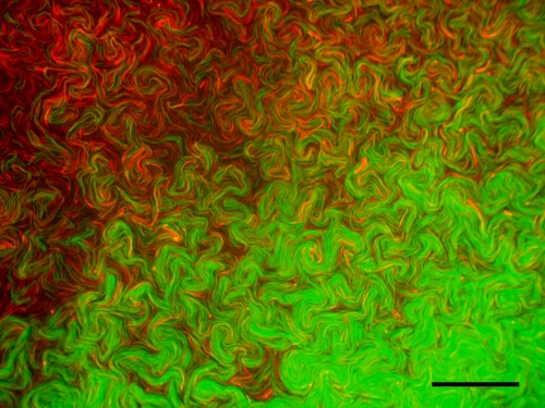
To study the sequence of events leading to Dienes line formation and the formation of round cells, four strains of P. mirabilis were transformed with plasmids allowing expression of either GFP or DsRed. These strains were fluorescent at all stages of development (swarmer and vegetative cells), and the expression of these fluorescent proteins in the strains shown in Table 1 had no effect on Dienes line formation. Additionally, these brightly colored proteins allowed masses of red and green strains to be distinguished by eye. As observed for their nonfluorescent counterparts, the frequency of spontaneous formation of rounded cells was negligible (<0.01%). Fluorescence microscopy of Dienes line formation showed a zone containing large round cells at the site where two nonidentical swarms intersected. These round cells started to appear 1 to 2 h after the initial contact between the opposing swarms. The abundance of the round cells that formed differed for different combinations of swarms, whereas the time of appearance was consistent. The appearance, timing, and abundance of these cells were similar whether the Dienes line was formed on TSA, MH agar, or sheep blood agar. For any combination of nonidentical strains, rounded cells originated from only one of the two strains (Fig. 2). Formation of round cells is thus an asymmetric process that occurs in only one strain in any given combination of strains. When the same strain was inoculated twice and allowed to swarm, the swarms merged without formation of any round cells (Fig. 3). Strains 3 and 4 formed only very indistinct Dienes lines, and in this case round cells were rarely observed (<1%). Table 1 shows the results for inoculation of strains in all combinations of pairs. We also confirmed previous observations that no round cells formed when swarmer cells reached nonswarming cells. Furthermore, after initial formation of the Dienes line, differentiation into swarmer cells ceased in the regions bordering the Dienes line. There was no mixing of strains across the Dienes line, and strains remained separated from each other.
TABLE 1.
Results of microscopic observation of intersecting swarms of fluorescent P. mirabilis strains tested in pairs (GFP-expressing strain A interacting with DsRed-expressing strain B)
| Strain B | Strain A
|
||
|---|---|---|---|
| 1G | 2G | 3G | |
| 1R | Xa | 1R | 3G |
| 2R | 1G | X | 3G |
| 3R | 3R | 3R | X |
| 4R | 1G | 4R | W |
A strain designation indicates the strain that formed large round cells when a Dienes line was formed. X indicates that there was no Dienes line (including no rounded cells), and W indicates that the Dienes line was weak and indistinct with few rounded cells (<0.5%).
FIG. 2.
Strain 2R intersecting with strain 3G. Strain 3G produces round cells, whereas strain 2R produces no round cells. The dark areas are agar with no growth. Magnification, ×400. Scale bar = 50 μm. (Inset) Intersection and rounded cells in more detail (magnification, ×800).
FIG. 3.
Intersection zone for strains 2G and 2R without boundary formation or rounded cells (magnification, ×1,000). Scale bar = 50 μm.
(ii) Quantification of round cells.
Because quantification of round cells in situ within the Dienes line was difficult, cells were harvested with a toothpick (targeted by viewing the plate by fluorescence microscopy), mounted on slides, and then observed by microscopy. This analysis confirmed the in situ microscopic observations described above. For strains 2G and 3R, which formed very clear Dienes lines, the proportion of round cells that were green was 99.6% (n = 471) 2 h after the initial contact of strains and 98.7% (n = 307) after 8 h. The same phenomenon was observed for all other interactions shown in Table 1: >98% of the rounded cells came from one population.
(iii) SEM visualization of Dienes line formation.
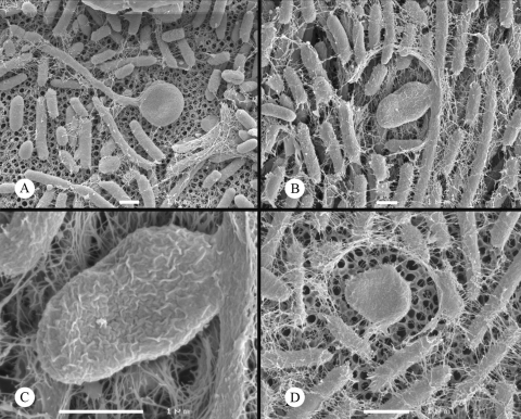
To gain better general insight into the morphology of the round cells, their formation, and their fate, SEM was performed for Dienes lines formed between strains 2R and 3G. This was done in situ; the Dienes lines were formed by swarming on a porous ceramic, which permitted fixation from below with minimal disruption of the cells. When strains were swarming but had not yet intersected, no round cells were found in 10 fields of view. From 1 to 2 h after the strains met, formation of a Dienes line could be seen macroscopically. At this stage round cells were most common and had an invaginated surface. Extracellular polymers and flagella were also observed. Often elongated swarmer cells in apparent transition to round cells were found (Fig. 4A to D). After 6 h round cells were rare, and almost all of them had a rough, invaginated morphology (Fig. 5A and B). For 14 to 21% of the round cells found after 8 h, lysis had clearly occurred (Fig. 5C); no lysed round cells were found after 2 h. These data suggested that there was progressive damage to the rounded cells over time.
FIG. 4.
SEM images of swarming P. mirabilis 1 to 2 h after intersection of swarms. (A and B) Round cells forming from swarmer cells. (C) Close-up of round cell from panel B. (D) Fully formed round cell. Scale bars = 1 μm.
FIG. 5.
SEM images of swarming P. mirabilis 6 h after intersection of swarms. (A and B) Highly invaginated rounded cells. (C) Flattened and lysed round cell. (A) Scale bar = 100 nm. (B and C) Scale bars =1 μm.
Fate of round cells in Dienes lines. (i) Live-dead staining.
In order to assess the fate of the round cells, live-dead staining was performed with cells recovered from the Dienes lines after 2, 4, and 8 h (Table 2). Cells stained with propidium iodide are considered to be dead, and cells staining with Syto 9 are considered to be alive. Within 8 h after contact between two Dienes line-forming strains, there was a progressive reduction in the proportion of living rounded cells from 81% to 52%. At all time points, slightly over 10% of round cells stained with both Syto 9 and propidium iodide (Table 2). For these cells, there was asymmetry in the regions of the cells stained preferentially with each dye. After 8 h almost one-third of round cells did not stain with either dye but could be visualized by phase-contrast microscopy. Nonstaining cells were not found up to 4 h after contact between the swarming strains. The appearance and abundance of these cells after 4 h suggest that they were lysed cells, as observed by SEM (Fig. 5C). We suggest that the high-nuclease environment of the Dienes line (1) resulted in rapid destruction of DNA so that many dead cells were not stained with propidium iodide. Where cells were caught in transition to rounded cells at 2 h, >90% of the cells stained as live cells, both in the elongated cell body and in the nascent rounded cell. During the experiment, swarmer cells progressively transformed into vegetative cells, and these cells were not found after 8 h.
TABLE 2.
Staining pattern for cell types in Dienes lines formed between strains 2 and 3a
| Time (h)b | Cell typec | % of cells
|
|||
|---|---|---|---|---|---|
| Lived | Deadd | Dual staininge | No stainf | ||
| 2 | Rounded | 81.25 | 6.25 | 12.5 | 0 |
| 2 | Swarmer | 94.25 | 5.75 | 0 | 0 |
| 2 | Vegetative | 97.5 | 2.5 | 0 | 0 |
| 4 | Rounded | 74.75 | 9.25 | 14.5 | 1.5 |
| 4 | Swarmer | 86.25 | 13.75 | 0 | 0 |
| 4 | Vegetative | 92 | 8 | 0 | 0 |
| 8 | Rounded | 52 | 4.5 | 11.5 | 32 |
| 8 | Swarmer | NDg | |||
| 8 | Vegetative | 96.25 | 3.75 | 0 | 0 |
Cells of each type at a time point were categorized by live-dead staining, and the results were expressed as percentages. The data are averages of two independent experiments (n = 200 for each experiment).
Time after contact. After 8 h there were not enough swarmers to score.
The three cell types were not necessarily equally abundant.
Live cells were stained only with Syto 9, and dead cells were stained only with propidium iodide.
Cells were strongly stained with both Syto 9 and propidium iodide.
Cells were visible by phase-contrast microscopy but not fluorescently stained.
ND, not determined.
(ii) Time-lapse series using fluorescence microscopy.
The live-dead staining experiment can provide information only on the state of round cells present at different time points. It does not provide information about the appearance and disappearance of round cells throughout the time period. Therefore, we investigated the fate of individual round cells. Twenty-seven strain 3G rounded cells (in three independent experiments) were collected from a Dienes line, put on a fresh TSA plate, and observed for 8 h. During this period none of the round cells divided, whereas all vegetative cells subjected to the same treatment divided repeatedly. Thirteen of the rounded cells lysed during the time-lapse series. These results suggest that round cells are in general not immediately viable and that some of them are damaged cells that are in the process of lysis.
Cell-cell contact versus extracellular substances. (i) Inhibition of cell-cell contact.
As no extracellular diffusible factor has yet been found to be responsible for Dienes line formation (26), it was important to establish whether cell-cell contact was required. When swarms of P. mirabilis cells were inoculated on either side of an Anopore membrane permeable to most proteins and macromolecules but not to bacteria (30), Dienes line formation was not observed for any combination of strains. Furthermore, the large round cells typical of Dienes line formation were not found close to the membrane for any strain in any combination. When both strains were inoculated on the same side of the Anopore membrane along with one of the two strains on the other side (Fig. 1), Dienes lines perpendicular to the Anopore membrane were observed for all strain combinations. However, the Dienes line had no effect on the strain swarming on the other side of the membrane, as observed by eye, nor were round cells detected by microscopy. This was true for both untreated Anopore and poly-l-lysine-treated Anopore membranes. These observations indicate that Dienes lines and rounded cells are not induced or propagated by a low-molecular-weight secreted substance. If secreted compounds are involved, they operate only at extremely short range (the thickness of an Anopore membrane is only 60 μm). An alternative is a complex that cannot pass through an Anopore membrane, although experiments with unfiltered extracts from Dienes lines by us and other workers have not identified such a factor.
(ii) Dead swarm experiment.
To determine whether cells have to be alive to induce or undergo round cell formation, another setup was used, employing UV light to kill one of the strains in a Dienes interaction. Exposure to UV light for 5 min prevented reswarming and reduced viability by >5 log10 units (as determined by viable counting). Longer irradiation (20 to 30 min) reduced viability >7 log10 units, and inoculation of 109 cells into 30 ml of Luria broth resulted in no increase in turbidity after shake cultures were incubated overnight. Furthermore, additional incubation of dead swarms (for up to 12 h) did not result in reinitiation of swarming. The UV treatment did not affect the medium in one respect: uninoculated TSA plates that were UV treated for up to 30 min could still support rapid swarming and Dienes line formation when they were subsequently inoculated with two incompatible strains.
In the main series of experiments, swarming colonies of strains 2R and 3G were killed in situ using UV light. Antagonistic strains (3G antagonistic for 2R and vice versa) were then allowed to swarm into the dead cell mass. Rather than forming a Dienes line, the live strains swarmed over the dead strains (and the position where a Dienes line would normally form) by at least 1 cm. Live strains then halted, and swarming did not restart; the results were similar for 5 to 30 min of exposure to UV light. Fluorescence microscopy of the interpenetrating region showed that live 3G swarms formed round cells when they were confronted with dead 2R swarmer cells, but they did so much less frequently than after exposure to live 2R swarmer cells (>6-fold reduction with 5 min of UV treatment). The effect was similar when strain 2R was exposed to UV light for 30 min, making it very unlikely that surviving 2R cells were triggering the response. In the complementary experiment, 2R swarmed over dead 3G cells to a similar extent, but no rounded cells that were either color were found. Formation of a visible Dienes line, in which swarming of both strains ceases and strains do not mix, therefore requires both strains to be alive. Triggering of round cell formation does not strictly require the majority of the triggering cells to be alive (only the strain forming the rounded cells has to be alive), but the effect is stronger when they are.
Effect of Dienes incompatibility on competition between strains.
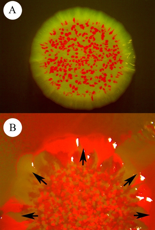
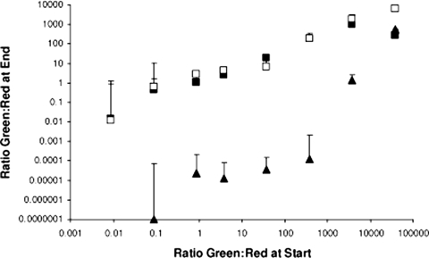
The Dienes phenomenon has usually been studied in a situation where two strains have colonized a significant fraction of an agar plate and a Dienes line forms at the limited interface. To evaluate whether Dienes incompatibility confers a competitive benefit in other settings, red and green strains were mixed at different ratios and in different combinations and spotted as mixtures (containing a total of 106 cells) in the center of agar plates. In this situation, multiple foci of swarming cells developed in close proximity, as observed by fluorescence microscopy. After incubation overnight the ratio of the two strains colonizing each plate was evaluated (Fig. 6 and 7). On TSA containing PNPG, no swarmer cells or round cells developed, and the ratios of red and green bacteria remained largely unchanged after incubation. There was some asymmetry in the positions of the two strains in the inoculated area (Fig. 6A), but this did not correlate with the Dienes type. For mixtures of strains 2R and 3G spotted on TSA without PNPG and so actively swarming, 2R dominated the plates almost completely when it was inoculated with 3G at a ratio of 1:1. Only at very high ratios was 3G able to colonize any significant area (Fig. 6B and 7) when it was competing with 2R. An initial 3G/2R ratio of >1,000:1 was required for strain 3G to colonize at least one-half of the plate area after swarming. Where cells of strain 3G were detectable, many of them were rounded. In “dye swap” experiments (using strains 2G and 3R) it was clear that the expression of GFP and DsRed was not a major factor in this interaction.
FIG. 6.
Low-power light microscopy images (magnification, ×10) showing competition between strains 2R and 3G on agar plates. (A) Coculture of the two strains at a 1:1 ratio on TSA containing PNPG to prevent swarming. The region of growth is 9 mm across, and there is equal growth but asymmetric distribution. (B) Inoculation of the two strains at a 1:1,000 ratio (ratio of 2R to 3G) in the central region (8 mm across), followed by swarming outward. Despite the asymmetry, both strains colonize areas of the plate. Outside the field of view only strain 2R reached the edge of the plate. When 2R and 3G are plated at a 1:1 ratio under these conditions, only 2R is detectable.
FIG. 7.
Quantification for competition experiments with strains 2R and 3G. The ratio of green cells to red cells at the start of each experiment is plotted against the ratio at the end of the experiment. Open squares, liquid culture; filled squares, TSA with 100 μg/ml PNPG; filled triangles, TSA without PNPG. For TSA without PNPG, no green cells were detectable at the end of the experiment when they were initially present at a ratio of <0.1:1. The error bars indicate the upper limit of the standard deviation of the mean for triplicate assays.
The competitive nature of the Dienes phenomenon was further analyzed by removing 2R swarmers with toothpicks (hundreds of cells) and depositing them into an actively swarming front of strain 3G cells. This resulted in cell growth and pinpoint red colonies after 8 h and in the cessation of swarming. The converse experiment, depositing 3G swarmers at the edge of 2R swarms, did not result in growth of 3G. The same trends were observed for other combinations of strains; the cells forming the rounded cells had a competitive disadvantage when they were mixed with inducing strains when they were swarming. Generally, a 100- to 10,000-fold excess of the strain forming the rounded cells was required for colonization of a plate when it was in competition with an inducing (dominant) strain. When strains were cocultured in liquid media, the ratios of red and green bacteria remained largely unchanged after incubation. It has been suggested by Jones and coworkers (14) that swarmers are found in Proteus biofilms. If this is true, Dienes incompatibility may be relevant to survival when two different strains form a mixed biofilm (all experiments, three replicates). The competition between the two strains was tested using a biofilm model on glass. The biofilms were similar to those reported previously and contained predominantly vegetative cells and <1% elongated cells, which one group has suggested are swarmers (14). In this series of experiments, asymmetry was observed in the distribution and abundance of the two strains within biofilms (formed in both Luria broth and TSB and observed after 24 and 48 h). However, there was no significant survival advantage that correlated with the dominant Dienes type, and any biases noted were not affected by the addition of PNPG. For example, strain 2G outcompeted 1R by more than 500-fold in a Luria broth biofilm after 48 h. This result was similar whether PNPG was present or not present. In both cases no rounded cells resembling those from Dienes lines were seen by fluorescence microscopy. Strain 2G induces rounded cells in 1R, so it was dominant both in a biofilm model and in a Dienes line. Strain 3G outcompeted 1R by more than 1,000-fold in a Luria broth biofilm, and again no rounded cells were seen. However, it was 3G that formed the rounded cells when the two strains formed a Dienes line, so the strain that was nondominant based on Dienes type had a competitive advantage in a biofilm. This suggests that the Dienes phenomenon does not provide a decisive competitive advantage in a two-strain biofilm.
Taken together, this set of experiments suggests that in a complex situation (where different strains can initiate swarming in close proximity with a large interface) the Dienes type is a decisive competitive factor. In other situations, including nonswarming growth on agar, in liquid culture, and in biofilm models, the Dienes type appears to be relatively unimportant.
Induction of unstable L forms using tap water.
Dienes suggested that the rounded bodies formed were L forms (6). Given that only one strain formed rounded cells, it was important to check if one or both strains involved in Dienes line formation were capable of producing unstable L forms. Swarmer cells of all strains (not expressing fluorescent proteins) were suspended in tap water, a method of producing L forms described by Dienes (6). After this osmotic shock, all strains produced large amounts of round cells (13 to 41% of the population within 10 to 30 min). These rounded cells were scored as living as determined by live-dead staining (>81% in all cases). All strains were thus apparently equally capable of producing rounded L forms from swarmers.
DISCUSSION
We have studied the nature and timing of Dienes line formation and strain recognition by P. mirabilis. This study concentrated on imaging the events that occur when two strains meet, including both the prevention of interpenetration and the appearance of rounded cells. We used recent clinical isolates because laboratory-adapted bacteria grown in nonnatural monocultures for many generations have been shown to lose important aspects of social behavior (31). We show that Dienes incompatibility is the result of a dominant strain inducing round cells with reduced viability in a nondominant strain. This strain dominance is very important in competitive colonization of a territory. Dienes line formation also provides a way of establishing boundaries between strains, effectively preventing mixing. This phenomenon is highly specific to swarmers; vegetative cells (from liquid or solid culture) do not show competition by Dienes type. We were also unable to find evidence for this competition in biofilms composed of two strains with incompatible Dienes types. In traditional Dienes assays, where two strains are inoculated at different starting positions (Fig. 1), colonization of the plate is largely determined by the rate and initiation of swarming. Any competition or exclusion due to the Dienes effect is confined to a narrow border region between the two strains. Spotting mixtures in the center of a plate and allowing them to swarm, as we did, creates a more complex interface region that may resemble competition between multiple strains in microniches (e.g., the soil). This assay highlights the competitive advantage possessed by the dominant strain more effectively; despite being outnumbered by 500:1 or more, the dominant strain can colonize most of the plate, and the strain forming the rounded cells must be present in excess to have any success (Fig. 7). We suggest that boundary formation is not sufficient to explain this competition but rather that a killing effect related to rounded cell formation is involved.
Dienes line formation between unrelated strains is associated with formation of rounded cells. However, it is clear that the inability of incompatible strains to interpenetrate can be at least partially uncoupled from rounded cell formation. In this work, UV irradiation of one strain was shown to allow overswarming of one strain by another strain. This was the case regardless of whether the irradiated strain was otherwise capable of triggering formation of rounded cells or forming them. Induction of rounded cells was less sensitive to UV irradiation of the triggering strain. The fact that boundary formation can be distinct from rounded cell formation is also indicated by the identification of ids mutations by Gibbs and coworkers (8). Mutations in the ids cluster prevent interpenetration of otherwise isogenic strains but do not induce rounded cells. The ids cluster does not obviously encode a toxin-antitoxin or other killing system, but similarities to antigenic variation systems and hypervariable regions may provide clues as to how a wide variety of Dienes types can be generated (8). There are differences in strain interactions within the Dienes line that are not explained by the ids system, particularly the apparent killing system discussed below.
That the rounded cells are formed almost exclusively by only one of the two swarms has now been unambiguously shown in this work by direct visualization and quantification experiments. Dienes suggested that viable rounded cells came from one strain and nonviable rounded cells came from the other (6), Wolstenholme suggested that the process was unidirectional, and this suggestion appears to be correct (32). Whether round cells in Dienes lines are related to L forms as Dienes suggested (5) is hard to test, especially as there is still not a clear definition of L forms. The osmotic shock experiments in this work showed that swarmers of all strains were able to form unstable L forms, so if the rounded cells are L forms, then the trigger is unidirectional.
The numbers of round cells in a Dienes line declined progressively with time. This can be at least partially attributed to cell lysis, as shown by SEM and supported by live-dead staining (Table 2) and as also suggested by time-lapse imaging. This is in line with previous reports indicating that round cell forms of Helicobacter pylori and Campylobacter jejuni are in the process of dying (9, 15, 17, 18). Rounded cells were also seen when mixtures of incompatible P. mirabilis strains were spotted in the center of a petri dish and allowed to swarm outward, and it is likely that whatever mechanism underlies rounded cell formation contributes to the competitive advantage seen in this situation. Competition is extremely tightly regulated; no competition that was consistent with the hierarchy of dominance of Dienes strains operated in vegetative cells (in liquid culture or when PNPG blocked swarmers on agar) or in a biofilm. This may suggest that when a new niche opens up, the competition to exclude other P. mirabilis strains is most intense. It will be interesting to look for microenvironments dominated by specific strains that may have succeeded because of the Dienes effect.
The absence of Dienes line formation and rounded cells when direct cell-cell contact was inhibited implied that a simple diffusible substance is not involved. This notion is further substantiated by the fact that no supernatant of a swarm or extract of a Dienes region has been shown to have any effect on swarming cells (24), a finding which we have been able to confirm and extend. The UV swarm experiment showed that intact dead cells of a strain with a dominant Dienes type induce rounded cells, albeit significantly less strongly than living cells. However, a (weak) response to dead cells occurs only when the strain that forms round cells is alive. Taken together, the cell-cell inhibition and dead swarm experiments are consistent with a requirement for close cell proximity. If a particle or complex is involved in rounded cell formation, then it does not pass through an Anopore membrane and is not detectable by other assays using cell extracts (24). For boundary formation, both strains appeared to be equal parties in the sense that both strains ceased swarming in the Dienes interaction in response to the other. The Anopore membrane also inhibited this boundary formation, and again no extracellular extract was found to be effective.
There is growing interest in microorganisms as social organisms and in the evolution of competitive and cooperative behavior; for example, “green beard” genes that facilitate altruistic behavior between individuals that both carry these genes and recognize each other as carriers have been described (13, 29). In the microbial world, examples of green beard effects include bacterial toxin-antitoxin systems and the cdsA adhesion gene in Dictyostelium (21). It is not clear if the Dienes system completely fits such a model, although the ids genes do mediate self-recognition. Mathematical modeling suggests that in mobile populations with a high level of kinship, cooperative or altruistic behavior is selected for (7). In Proteus, swarming is indeed a cooperative activity of a single strain. Competition between different Dienes compatibility groups occurs during swarming, but not in other modes of existence. The Dienes interaction appears to be a fascinating system for social microbiology and should be investigated further to understand the physiology and genetics of swarming more fully.
Footnotes
Published ahead of print on 27 February 2009.
REFERENCES
- 1.Chambers, T. J. 1975. Deoxyribonuclease release by incompatible strains of Proteus. Zentralbl. Bakteriol. Orig. A 233228-231. [PubMed] [Google Scholar]
- 2.Coetzee, J. N. 1961. Lysogenic conversion in the genus Proteus. Nature 189946-947. [DOI] [PubMed] [Google Scholar]
- 3.Cradock-Watson, J. E. 1961. The production of bacteriocines by Proteus species. Zentralbl. Bakteriol. Orig. 196385-388. [Google Scholar]
- 4.de Louvois, J. 1969. Serotyping and the Dienes reaction on Proteus mirabilis from hospital infections. J. Clin. Pathol. 22263-268. [DOI] [PMC free article] [PubMed] [Google Scholar]
- 5.Dienes, L. 1946. Reproductive processes in Proteus cultures. Proc. Soc. Exp. Biol. Med. 63265-270. [DOI] [PubMed] [Google Scholar]
- 6.Dienes, L. 1947. Further observations on the reproduction of bacilli from large bodies in Proteus cultures. Proc. Soc. Exp. Biol. Med. 6697-98. [DOI] [PubMed] [Google Scholar]
- 7.Gardner, A., and S. A. West. 2006. Demography, altruism, and the benefits of budding. J. Evol. Biol. 191707-1716. [DOI] [PubMed] [Google Scholar]
- 8.Gibbs, K. A., M. L. Urbanowski, and E. P. Greenberg. 2008. Genetic determinants of self identity and social recognition in bacteria. Science 321256-259. [DOI] [PMC free article] [PubMed] [Google Scholar]
- 9.Hazeleger, W. C., J. D. Janse, P. M. Koenraad, R. R. Beumer, F. M. Rombouts, and T. Abee. 1995. Temperature-dependent membrane fatty acid and cell physiology changes in coccoid forms of Campylobacter jejuni. Appl. Environ. Microbiol. 612713-2719. [DOI] [PMC free article] [PubMed] [Google Scholar]
- 10.Ingham, C. J., M. van den Ende, D. Pijnenburg, P. C. Wever, and P. M. Schneeberger. 2005. Growth and multiplexed analysis of microorganisms on a subdivided, highly porous, inorganic chip manufactured from Anopore. Appl. Environ. Microbiol. 718978-8981. [DOI] [PMC free article] [PubMed] [Google Scholar]
- 11.Ingham, C. J., M. van den Ende, P. C. Wever, and P. M. Schneeberger. 2006. Rapid antibiotic sensitivity testing and trimethoprim-mediated filamentation of clinical isolates of the Enterobacteriaceae assayed on a novel porous culture support. J. Med. Microbiol. 551511-1519. [DOI] [PubMed] [Google Scholar]
- 12.Jansen, A. M., C. V. Lockatell, D. E. Johnson, and H. L. Mobley. 2003. Visualization of Proteus mirabilis morphotypes in the urinary tract: the elongated swarmer cell is rarely observed in ascending urinary tract infection. Infect. Immun. 713607-3613. [DOI] [PMC free article] [PubMed] [Google Scholar]
- 13.Jansen, V. A., and M. van Baalen. 2006. Altruism through beard chromodynamics. Nature 440663-666. [DOI] [PubMed] [Google Scholar]
- 14.Jones, S. M., J. Yerly, Y. Hu, H. Ceri, and R. Martinuzzi. 2007. Structure of Proteus mirabilis biofilms grown in artificial urine and standard laboratory media. FEMS Microbiol. Lett. 26816-21. [DOI] [PubMed] [Google Scholar]
- 15.Kusters, J. G., M. M. Gerrits, J. A. Van Strijp, and C. M. Vandenbroucke-Grauls. 1997. Coccoid forms of Helicobacter pylori are the morphologic manifestation of cell death. Infect. Immun. 653672-3679. [DOI] [PMC free article] [PubMed] [Google Scholar]
- 16.Liaw, S. J., H. C. Lai, S. W. Ho, K. T. Luh, and W. B. Wang. 2000. Inhibition of virulence factor expression and swarming differentiation in Proteus mirabilis by p-nitrophenylglycerol. J. Med. Microbiol. 49725-731. [DOI] [PubMed] [Google Scholar]
- 17.Moran, A. P., and M. E. Upton. 1986. A comparative study of the rod and coccoid forms of Campylobacter jejuni ATCC 29428. J. Appl. Bacteriol. 60103-110. [DOI] [PubMed] [Google Scholar]
- 18.Moran, A. P., and M. E. Upton. 1987. Factors affecting production of coccoid forms by Campylobacter jejuni on solid media during incubation. J. Appl. Bacteriol. 62527-537. [DOI] [PubMed] [Google Scholar]
- 19.Munson, E. L., M. A. Pfaller, and G. V. Doern. 2002. Modification of Dienes mutual inhibition test for epidemiological characterization of Pseudomonas aeruginosa isolates. J. Clin. Microbiol. 404285-4288. [DOI] [PMC free article] [PubMed] [Google Scholar]
- 20.Pfaller, M. A., I. Mujeeb, R. J. Hollis, R. N. Jones, and G. V. Doern. 2000. Evaluation of the discriminatory powers of the Dienes test and ribotyping as typing methods for Proteus mirabilis. J. Clin. Microbiol. 381077-1080. [DOI] [PMC free article] [PubMed] [Google Scholar]
- 21.Queller, D. C., E. Ponte, S. Bozzaro, and J. E. Strassman. 2003. Single gene greenbeard effects in the social amoeba Dictyostelium discoideum. Science 299105-106. [DOI] [PubMed] [Google Scholar]
- 22.Salikhova, Z. Z., R. B. Sokolova, A. Z. Ponomareva, and D. V. Iusupova. 2001. Endonuclease from Proteus mirabilis. Prikl. Biokhim. Mikrobiol. 3743-47. (In Russian.) [PubMed] [Google Scholar]
- 23.Senior, B. W. 1977. Typing of Proteus strains by proticine production and sensitivity. J. Med. Microbiol. 107-17. [DOI] [PubMed] [Google Scholar]
- 24.Senior, B. W. 1977. The Dienes phenomenon: identification of the determinants of compatibility. J. Gen. Microbiol. 102235-244. [DOI] [PubMed] [Google Scholar]
- 25.Senior, B. W., and P. Larsson. 1983. A highly discriminatory multi-typing scheme for Proteus mirabilis and Proteus vulgaris. J. Med. Microbiol. 16193-202. [DOI] [PubMed] [Google Scholar]
- 26.Sokolova, R. B., M. I. Beliaeva, and D. V. Iusupova. 1976. Intracellular nucleodepolymerase of bacteria, representatives of the Proteus and Providencia groups. Its isolation and properties. Vopr. Med. Khim. 22488-493. (In Russian.) [PubMed] [Google Scholar]
- 27.Sourek, J. 1968. On some findings concerning Dienes's phenomenon in swarming Proteus strains. Zentralbl. Bakteriol. Orig. 208419-427. [PubMed] [Google Scholar]
- 28.Tracy, O., and E. J. Thomson. 1972. An evaluation of three methods of typing organisms of the genus Proteus. J. Clin. Pathol. 2569-72. [DOI] [PMC free article] [PubMed] [Google Scholar]
- 29.van Baalen, M., and V. A. Jansen. 2003. Common language or Tower of Babel? On the evolutionary dynamics of signals and their meanings. Proc. Biol. Sci. 27069-76. [DOI] [PMC free article] [PubMed] [Google Scholar]
- 30.van der Sar, A. M., R. J. Musters, F. J. van Eeden, B. J. Appelmelk, C. M. Vandenbroucke-Grauls, and W. Bitter. 2003. Zebrafish embryos as a model host for the real time analysis of Salmonella typhimurium infections. Cell. Microbiol. 5601-611. [DOI] [PubMed] [Google Scholar]
- 31.Velicer, G. J., R. E. Lenski, and L. Kroos. 2002. Rescue of social motility lost during evolution of Myxococcus xanthus in an asocial environment. J. Bacteriol. 1842719-2727. [DOI] [PMC free article] [PubMed] [Google Scholar]
- 32.Wolstenholme, J. 1973. Evidence that the production of swollen bodies in the Dienes phenomenon may be confined to one of two interacting strains of Proteus mirabilis. J. Gen. Microbiol. 74343-344. [DOI] [PubMed] [Google Scholar]
- 33.Wu, Y., P. de Kievit, L. Vahlkamp, D. Pijnenburg, M. Smit, M. Dankers, D. Melchers, M. Stax, P. J. Boender, C. Ingham, N. Bastiaensen, R. de Wijn, Alewijk, D., H. van Damme, A. K. Raap, A. B. Chan, and R. van Beuningen. 2004. Quantitative assessment of a novel flowthrough porous microarray for the rapid analysis of gene expression profiles. Nucleic Acids Res. 32e123. [DOI] [PMC free article] [PubMed] [Google Scholar]


