Abstract
Repeated bottleneck passages of RNA viruses result in accumulation of mutations and fitness decrease. Here, we show that clones of foot-and-mouth disease virus (FMDV) subjected to bottleneck passages, in the form of plaque-to-plaque transfers in BHK-21 cells, increased the thermosensitivity of the viral clones. By constructing infectious FMDV clones, we have identified the amino acid substitution M54I in capsid protein VP1 as one of the lesions associated with thermosensitivity. M54I affects processing of precursor P1, as evidenced by decreased production of VP1 and accumulation of VP1 precursor proteins. The defect is enhanced at high temperatures. Residue M54 of VP1 is exposed on the virion surface, and it is close to the B-C loop where an antigenic site of FMDV is located. M54 is not in direct contact with the VP1-VP3 cleavage site, according to the three-dimensional structure of FMDV particles. Models to account for the effect of M54 in processing of the FMDV polyprotein are proposed. In addition to revealing a distance effect in polyprotein processing, these results underline the importance of pursuing at the biochemical level the biological defects that arise when viruses are subjected to multiple bottleneck events.
As a consequence of the quasispecies population structure, when a virus is subjected to an extreme bottleneck regime, such as successive plaque-to-plaque transfers, it accumulates deleterious mutations that result in fitness loss (reviewed in references 15, 21, and 33). These observations constitute experimental support for the Muller's ratchet hypothesis, which states that asexual populations of organisms tend to acquire deleterious mutations unless compensatory mechanisms (such as sex or recombination) intervene (39, 41). Several lines of evidence indicate that population bottlenecks are abundant in the life cycle of viruses, both during host-to-host transmission and during intrahost replication (2, 8, 10, 22, 26, 32, 42, 45-48, 52, 53). Most studies have addressed the effects of bottlenecks on the reduction of intramutant spectrum diversity in relation to virus survival and persistence, effects on fitness, or as promoters of stochastic processes and drift in viral evolution. Yet the possible biological effects of specific mutations fixed as a result of bottleneck events remain largely unexplored.
Experimental designs consisting of many successive plaque-to-plaque transfers, without intervening large-population passages, are ideal for obtaining viral clones that are debilitated by the occurrence of mutations because negative selection is highly attenuated (15, 21, 33). The deleterious nature of some mutations that become fixed in viral genomes subjected to repeated bottlenecks can be inferred from their position in the viral genome and then confirmed experimentally. For example, an internal tract of four oligoadenylate residues that precede the second functional AUG initiation codon of foot-and-mouth disease virus (FMDV) was invariant among natural isolates of the virus or among populations subjected to large-population passages. Yet this oligoadenylate tract was extended in several clones subjected to plaque-to-plaque transfers (17). This lesion, unique to clones that had undergone multiple bottleneck transfers, was associated with a decrease in replicative fitness (4, 17), and some of the clones displayed reduced levels of Lb, the form of the leader proteinase L synthesized from the second functional AUG initiation codon (17). However, the effect of other mutations that accumulate as a result of bottleneck transfers cannot be easily anticipated. Some mutations will likely be neutral while others are deleterious, and there is experimental and in silico evidence that a few mutations are advantageous or compensatory, thereby allowing the virus to survive despite continuous accumulation of mutations (21, 28).
Nonsynonymous mutations in coding regions may perturb the structure and function of viral proteins. Despite evidence that such mutations can affect viral fitness, in very few cases the biochemical effect of a lesion associated with the operation of Muller's ratchet has been identified. Here, we report that the accumulation of mutations in FMDV subjected to plaque-to-plaque passages results in a gradual increase in the thermosensitivity of infectious progeny production, with a several-logarithm decrease in progeny production at 42°C relative to 37°C at plaque transfer 230. Part of the thermosensitivity at early transfers could be traced to a single amino acid substitution, M54I, located at the B-C loop of capsid protein VP1. This loop corresponds to antigenic site 3 of FMDV (30, 36). We show that the M54I mutation decreases the proteolytic cleavage between capsid proteins VP3 and VP1 and that the impairment is manifested more severely at high temperatures. This cleavage is catalyzed by proteinase 3Cpro (31, 50, 51, 56), which does not show any substitution in the mutant FMDV clone that harbored the M54I mutation in VP1. Thus, a distant amino acid located at an antigenic site of a virus can affect a protein processing step catalyzed by wild-type 3Cpro. We discuss possible models to explain the link between two disparate phenotypic traits, antigenicity and protein processing, in the life cycle of a virus.
MATERIALS AND METHODS
Cells, viruses, and infections.
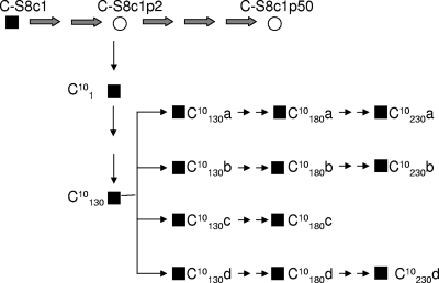
Procedures to grow BHK-21 cells, to infect them with FMDV, to carry out large-population passages and plaque-to-plaque transfers on BHK-21 cell monolayers, and to isolate virus from individual plaques have been previously described (14, 17, 54). FMDV clone C-S8c1 was derived from natural isolate C-Santa Pau-Spain/70 and cloned in BHK-21 cells (54). Clone C101 was obtained from a single plaque derived from population C-S8c1 subjected to two large-population passages in BHK-21 cells (17). The characterization of FMDV clones derived from C101 has been previously described (20) (Fig. 1). Virus from clone C10130 (where the subscript indicates the number of plaque-to-plaque transfers) was diluted and plated, and four subclones, termed C10130 a, C10130 b, C10130 c, and C10130 d (abbreviated as sublineages a, b, c, and d), were subjected to additional plaque-to-plaque transfers (Fig. 1).
FIG. 1.
Scheme of the origin of FMDV clones C101, C10130, and sublineages a, b, c, and d subjected to 180 or 230 plaque-to-plaque transfers, and of population C-S8c1p50. Squares, biological clones; circles, uncloned populations; thick arrows, large-population passages (2 × 10⁶ cells infected with 2 × 10⁶ to 5 × 10⁶ PFU of C-S8c1); thin arrows, isolation of virus from a single plaque. For sublineage c, clones became noncytopathic after transfer 180 (20). The origin of FMDV C-S8c1 and procedures for infections and plaque-to-plaque transfers are detailed in Materials and Methods.
The thermosensitivity of FMDV is defined as the ratio of infectious progeny production at 37°C and 43°C in a standard infectivity assay carried out at a multiplicity of infection (MOI) of 0.5 to 1 PFU/cell. In some assays the restrictive temperature used was 42°C instead of 43°C because when clone C101 was subjected to 230 plaque-to-plaque transfers, it yielded detectable infectious progeny at 42°C but not at 43°C. The procedure is identical to that used to quantify the increase in thermosensitivity of FMDV in the course of persistent infections in BHK-21 cells (12). Calibrated thermometers were used to ensure that the restrictive temperature was maintained at 42°C ± 0.1°C or 43°C ± 0.1°C throughout the experiments. The thermosensitivities of clones C10180 (sublineages a, b, c, and d) and C10230 (sublineages a, b, and d) were determined on different days, and clone C10130 (the parental clone from which sublineages a, b, c, and d were derived) was included as a control. Infectivity at 37°C and at 42°C was determined by triplicate platings of each biological clone.
To determine the thermosensitivity of viral particles, aliquots of FMDV populations were incubated at 43°C in cell culture medium (Dulbecco's modified Eagle's medium [DMEM]), chilled at 0°C at defined time intervals, and titrated on BHK-21 cell monolayers, as previously described (14).
FMDV genomic nucleotide sequences.
Nucleotide sequencing of genomic RNA of the FMDV clones and populations described in the present study were determined using the ABI 373 sequencer, as previously described (18); all sequences were determined at least in duplicate from independent sequencing reaction mixtures. The sequences of FMDV C-S8c1 and derived clones and populations have been previously reported (17, 19-21, 23, 55).
Plasmids, infectious transcripts, and electroporation of cells.
Plasmid pMT28, which encodes an infectious transcript that produces C-S8c1, has been previously described (23, 55). Specific mutations were introduced into plasmid pMT28 using oligonucleotides containing the desired changes (3). To generate C-S8c1 or mutant C-S8c1 from the corresponding plasmids, pMT28 or its mutant versions were linearized with NdeI. The linearized plasmid DNA was purified by Wizard PCR Preps DNA purification resin (Promega) and dissolved in RNase-free water. Infectious FMDV RNA was transcribed from the linearized plasmids by using a Riboprobe in vitro transcription system (Promega). The mixture contained transcription buffer (Promega), 10 mM dithiothreitol, 1 mg/ml bovine serum albumin, 0.48 units/μl RNasin, 1 mM of each ribonucleoside triphosphate, 4 to 15 ng/μl linearized plasmid DNA, and 0.3 or 0.4 units/μl SP6 RNA polymerase; incubation was for 2 h at 37°C. The RNA concentration was estimated by agarose gel electrophoresis, with known amounts of rRNA as markers.
To electroporate BHK-21 cells with RNA transcribed in vitro, subconfluent cells were harvested, washed with ice-cold phosphate-buffered saline (PBS), and resuspended in PBS at a density of about 2.5 × 10⁶ cells/ml. Aliquots (50 to 80 μl) of transcription mixture with the appropriate amount of RNA were added to 0.4 ml of cell suspension, and the mixtures were transferred to 2-mm electroporation cuvettes (Bio-Rad). Electroporation was performed at room temperature by two consecutive pulses of 1.5 kV and 25 μF using a Gene Pulser apparatus (Bio-Rad), as described previously (44). As a control, BHK cells were electroporated with 50 to 80 μl of transcription mixture (without template DNA) in PBS to monitor the absence of contamination. The cells were then resuspended in DMEM with 10% fetal calf serum and transferred to culture plates. At 2, 3, and 4 h postelectroporation, cells were metabolically labeled with [35S]Met-Cys, as described in the next section. Mock-electroporated cultures were treated in parallel and served as controls for the experiments of determination of viral infectivity. No evidence of viral contamination was obtained in any of the experiments. In some experiments, BHK-21 cells were transfected with FMDV RNA transcripts using Lipofectin, as previously described (3).
Transcripts from plasmid pMT28 and from plasmid pMT28 encoding the amino acid substitution M54I in VP1 give rise to viruses that have been termed pMT28-WT (where WT is wild type) and pMT28-M1054I (where 1 indicates VP1 and 054 is the amino acid position in VP1), respectively, throughout this report.
Metabolic labeling of proteins.
Viral protein synthesis was analyzed by metabolic labeling with [35S]Met-Cys, followed by sodium dodecyl sulfate-polyacrylamide gel electrophoresis (SDS-PAGE) and fluorography, as previously described (44). Proteins were labeled by the addition to BHK-21 cells (grown in M-24 wells) of 60 μCi of [35S]Met-Cys (Amersham) per ml in methionine-free DMEM. After 1 h of incubation of the cell monolayers with the radioactive medium, the medium was removed, and the cells were harvested in 0.1 ml of sample buffer (160 mM Tris-HCl, pH 6.8, 2% SDS, 11% glycerol, 0.1 M dithiothreitol, 0.033% bromophenol blue). The amount of cell extract used for electrophoretic analysis was normalized to a constant amount of cellular actin, measured by reactivity with monoclonal antibody (anti-β-actin clone AC-15; Sigma) (44). In pulse-chase experiments, the metabolic labeling was terminated by washing the cell monolayers with fresh DMEM and maintaining the cells in DMEM with unlabeled methionine during the chosen chase period. The samples were boiled for 5 min, and aliquots were analyzed by SDS-PAGE with 8 M urea, run at 200 V. Fluorography and autoradiography of the gels were carried out as described previously (44).
Antibodies and Western blot analysis.
To identify virus-specific proteins by Western blotting using monoclonal antibodies (37), proteins were separated electrophoretically by SDS-PAGE and transferred to a nitrocellulose membrane (Bio-Rad) in transfer buffer (25 mM Tris-HCl, pH 8.3, 190 mM glycine, 20% methanol, 0.1% SDS) at 200 mA for 15 h. The membrane was saturated with a solution of nonfat powdered milk (5%, wt/vol) in PBS for 1 h with gentle stirring. Then an adequate dilution in PBS (with 0.1% powdered milk) of the primary antibody was added, and the membrane was incubated for 2 h. Then the membrane was washed three times for 15 min in TPBS buffer (Tween 20 at 0.05% [vol/vol] in PBS) and incubated with the corresponding secondary antibody conjugated to peroxidase (1:10,000 dilution in TPBS). After 1 h of incubation, three washes with PBS were carried out, and the membrane was developed by chemiluminescence (ECL kit; Amersham) (27, 44). FMDV proteins were detected using mouse monoclonal antibodies that recognize capsid proteins VP1, VP3, 2C (kindly provided by E. Brocchi), monoclonal antibody anti-β-actin clone AC-15 (Sigma), and a polyclonal antibody against the polymerase 3D (38, 44). The amount of cell extract used for Western blot analysis was normalized to a constant amount of cellular actin and corresponded to a concentration of protein in the linear region of the relationship between the Western blot signal and the protein concentration (44).
RESULTS
Thermosensitivity of FMDV clones subjected to plaque-to-plaque transfers.
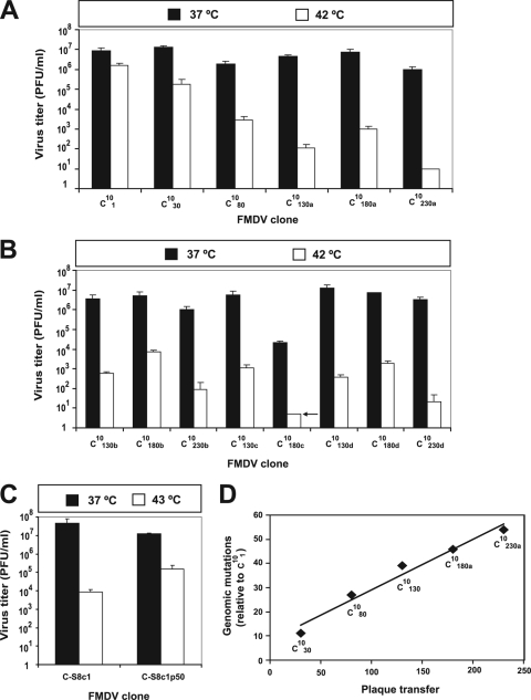
Clone C101, a clone of FMDV C-S8c1 (17, 54), was subjected to 130 plaque-to-plaque transfers in BHK-21 cells. The resulting clone (C10130) was then plated to obtain subclones C10130 a, C10130 b, C10130 c, and C10130 d, which were subjected to additional plaque-to-plaque transfers (Fig. 1). The clones studied in the present report were generated and described previously (20). To test whether repeated plaque-to-plaque transfers resulted in a temperature-sensitive (ts) phenotype, infectious progeny production of clonal populations C101, C1030, C1080, C10130 (sublineages a, b, c, and d), C10180 (a, b, c, and d), and C10230 (a, b, and d) was measured at 37°C and 42°C. The results show an increase of up to 10,000-fold in thermosensitivity as a result of the extensive plaque-to-plaque transfers (Fig. 2A and B). Similar results were observed in the parallel sublineages, including the decrease in thermosensitivity at plaque transfer 180 relative to transfers 130 and 230 for lineages a, b, and d. (Fig. 2B). Sublineage c was extinguished at transfer 190 (20), and, therefore, its thermosensitivity could be measured only at transfer 130, in which infectious progeny production at 42°C was below the limit of detection (Fig. 2B). At plaque transfers higher than 230, no infectious progeny could be detected in infections carried out at a temperature higher than 40°C (<2 × 10⁻⁵-fold the infectivity produced at 37°C). No increase in thermosensitivity was observed in populations of C-S8c1 subjected to large-population passages, with thermosensitivity measured at 43°C (Fig. 2C). Thus, plaque-to-plaque transfers of FMDV lead to a pronounced ts phenotype.
FIG. 2.
Thermosensitivity and accumulation of mutations upon subjecting FMDV clone C101 to plaque-to-plaque transfers. The origin of C101 and the experimental design are described in Fig. 1. (A) Progeny production at 37°C and 42°C in parallel infections of 2 × 10⁶ BHK-21 cells, with the clones indicated in the abscissa, at MOIs of 0.1 to 0.01 PFU/cell. After 1 h of adsorption at 37°C, the monolayers were incubated at 37°C or at 42°C for 22 h to 44 h until cytopathology at 37°C was complete. Titers in the cell culture supernatants were determined at 37°C in triplicate; standard deviations are given. (B) Progeny production at 37°C and 42°C of virus from C10180 and C10230 for sublineages b, c, and d (compare Fig. 1). Virus from sublineage c at transfer 180 yielded an undetectable number of plaques at 42°C (below the limit of detection, as indicated by the arrow); virus from this sublineage became noncytophatic at plaque transfer 190 (20), and therefore thermosensitivity at transfer 230 could not be measured. Titrations for the three lineages were carried out in separate experiments, but for each lineage the titrations of plaques in C10180 and C10230 were carried out in parallel, and C10130 (the parental clone of sublineages a, b, c, and d) (Fig. 1) was included as a control. The procedure is as described for panel A. (C) Progeny production at 37°C and 43°C of C-S8c1 and C-S8c1 subjected to 50 serial passages in BHK-21 cells (MOI of 1 to 2 PFU/cell) at 37°C. Note that in this control the restrictive temperature was 43°C and that the thermosensitivity of C-S8c1 need not be identical to that of C101 due to passage history (Fig. 1) and to FMDV population heterogeneity. (D) Accumulation of mutations, relative to the genomic sequence of C101, as a function of the plaque transfer number. Values are based on the nucleotide sequence of the entire FMDV genome, as previously described (23). Procedures are described in Materials and Methods.
Mapping of mutations associated with thermosensitivity of FMDV clones.
In the course of the serial plaque transfers, the consensus genomic sequence of successive clones accumulated point mutations at an average rate of 0.21 mutations per transfer (Fig. 2D). The genome of C1030 acquired one mutation in the 5′ untranslated region (U54C), five synonymous mutations, and four nonsynonymous mutations relative to C101; the mutations in the last group were C1283U and U1538A, which lead to amino acid mutations I83V and F167Y, respectively, in the L protein; G3369C, which leads to M54I in VP1; and C5876U, which leads to A17V in VPg2 (genomic residues are numbered according to reference 20). To evaluate the contribution of the mutation at the 5′ untranslated region and the nonsynonymous mutations to the ts phenotype of C1030, each mutation was introduced separately (except the two mutations in the L-coding region, which were introduced together) into plasmid pMT28 to produce infectious transcripts. FMDV progeny production in BHK-21 cells by each of the mutant transcripts was measured at 37°C and 43°C. The results (Fig. 3A) show that M54I is the main mutation responsible for the increased thermosensitivity of C1030 relative to that of C101. The decrease in progeny production of pMT28-M1054I at 43°C relative to that at 37°C was observed at all times after infection (Fig. 3B).
FIG. 3.
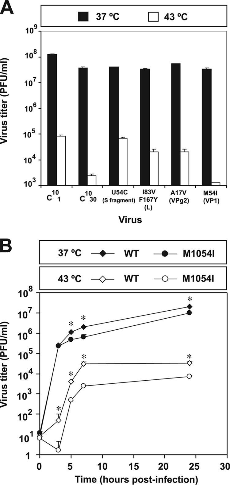
Viral yield of FMDV mutants at 37°C and 43°C. (A) The mutations that distinguish C1030 from C101 were introduced separately into pMT28, the plasmids were linearized and transcribed, and the infectious transcripts were transfected into cells to produce virus progeny. The mutations or amino acid substitutions present in each mutant are indicated in the abscissa. C101, C1030, and the pMT28 mutant derivatives were used to infect BHK-21 cells at an MOI of 0.1 PFU/cell. The 1-h adsorption period was at 37°C. Then, the infection was continued at either 37°C or 43°C until cytopathology was complete in the infections carried out at 37°C (21 h postinfection). Titrations were carried out in triplicate, and standard deviations are indicated. (B) Growth curve of viral progeny of pMT28-WT (WT) and pMT28-M1054I (M1054I) at 37°C and 43°C. BHK-21 cell monolayers were infected at an MOI of about 0.1 PFU/cell under the conditions indicated for panel A. At the indicated times postinfection, aliquots of the cell culture supernatants were withdrawn, and viral titers were determined at 37°C. Titrations were carried out in triplicate, and standard deviations are given. Asterisks indicate statistically significant differences between the titers produced by pMT28-WT and pMT28-M1054I either at 37°C or 43°C at each time point (P < 0.025; analysis of variance test). Procedures are detailed in Materials and Methods.
The effect of temperature on virus production was also studied in the absence of intracellular viral competition. To this aim, BHK-21 cells were infected with either pMT28-WT or pMT28-M1054I at an MOI of 0.1 PFU/cell, and the infection was allowed to proceed for only 3 h to prevent subsequent rounds of infection in which cells would be infected by multiple infectious particles. FMDV C-S8c1 entry into BHK-21 cells was completed in 40 min at 37°C, and the initial burst of FMDV infectious progeny production occurred after 2 h postinfection (13). After the 1-h adsorption period at 37°C, parallel cultures were incubated under four different conditions, as follows: either 3 h a 37°C or 1 h at 37°C and then 2 h at 43°C or 2 h at 43°C and then 1 h at 37°C or 3 h at 43°C. The results (Fig. 4A) indicate that elevated temperature at late times of infection had a more severe effect on progeny production by the FMDV M54I mutant than elevated temperature at early times postinfection. In a single round of infection, the ratio of wild-type to mutant progeny production was 9.3 when the second and third hours of infection took place at 43°C; the corresponding ratio was 1.5 when the first and second hours of infection took place at 43°C. The results were compatible with two explanations: either a late function of the mutant virus is sensitive to high temperature or the mutant viral particles are less stable at high temperature. This second possibility was ruled out because both pMT28-WT and pMT28-M1054I followed very similar kinetics of inactivation at 43°C (half-life of infectivity of 18.2 and 17.5 min, respectively) (Fig. 4B). The two slopes are not statistically different (P = 0.7898; Student's t test; 6 degrees of freedom). Thus, a late function appeared to be involved in the increased thermosensitivity of FMDV harboring the amino acid substitution M54I in capsid protein VP1 relative to the wild-type virus.
FIG. 4.
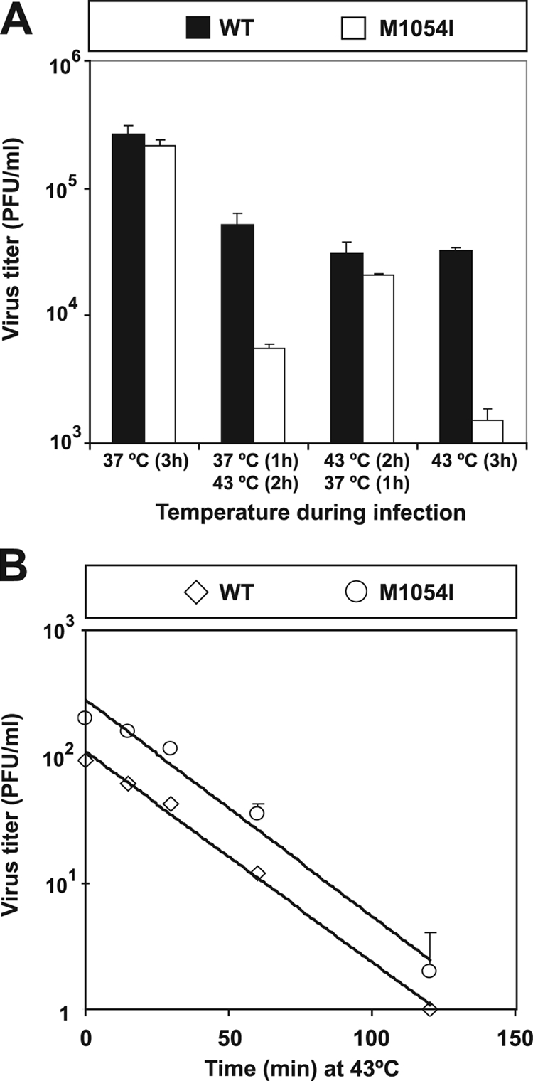
Infectious progeny production and stability of pMT28-WT (WT) and pMT28-M1054I (M1054I) at 37°C and 43°C. (A) pMT28-WT or pMT28-M1054I was adsorbed onto BHK-21 cells for 1 h at 37°C at an MOI of 0.1 PFU/cell. Infections were continued for either 3 h at 37°C or 1 h at 37°C, followed by 2 h at 43°C or 2 h at 43°C and then by 1 h at 37°C or 3 h at 43°C, as indicated in the abscissa. After the 3-h period the virus was titrated at 37°C. Titrations were carried out in triplicate; standard deviations are given. (B) Aliquots containing 100 to 200 PFU of virus pMT28-WT or pMT28-M1054I, respectively, were incubated at 43°C for the times indicated in the abscissa and titrated at 37°C. Titrations were carried out in triplicate, and standard deviations are given. The standard deviations for times 0, 15, 30, 60, and 120 min were 5.3, 1.2, 6.5, 1.5, and 0, respectively, for pMT28-WT and 39.8, 20.8, 10.4, 7, and 2, respectively, for pMT28-M1054I. The half-life of infectivity was calculated based on the regression lines for pMT28-WT (y = 109.84 e−0.0384x; r2 = 0.9937) and pMT28-M1054I (y = 282.24 e−0.0395x; r2 = 0.9772). Procedures are described in Materials and Methods.
Protein processing is affected in FMDV with the amino acid substitution M54I.
Since the ts phenotype affected a late viral function (Fig. 4), it was not expected that viral RNA synthesis was impaired at high temperatures. This was confirmed by comparing the amount of virus-specific RNA produced by FMDV pMT28-WT and pMT28-M1054I at 0, 1, 1.5, 1.75, and 2 h postinfection by real-time reverse transcription-PCR. No difference was observed in the amounts of RNA produced by the two viruses at 37°C and 43°C (results not shown).
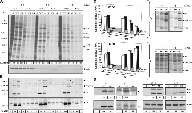
To address whether the defect of pMT28-M1054I at 43°C could lie in viral protein synthesis, the expression of viral and cellular proteins was analyzed in BHK-21 cells electroporated with infectious transcripts of either pMT28-WT or pMT28-M1054I at 37°C or 43°C. The intracellular proteins were metabolically labeled with [35S]Cys-Met and analyzed electrophoretically, using actin as a control (Fig. 5). Quantification of cellular proteins relative to actin (measured by Western blotting) indicated a four- to sixfold decrease in the level of cellular proteins expressed at 43°C relative to 37°C (Fig. 5A, compare lanes C at 37°C and 43°C) but a comparable level of shutoff of host cell protein synthesis by viral infection at both temperatures relative to the label in the actin band (determined by pulse-chase with [35S]Met-Cys, as detailed in Materials and Methods) (Fig. 5A).
FIG. 5.
Protein expression by wild type RNA and mutant M54I RNA. (A) Pattern of protein expression in BHK-21 cells electroporated with transcripts encoding pMT28-WT (WT) and pMT28-M1054I (MI) at 37°C and 43°C. BHK-21 cells were either mock electroporated (C lanes) or electroporated with about 20 μg of RNA transcripts from the indicated plasmids. Transfected cells were labeled with [35S]Met-Cys at 2 to 3, 3 to 4, or 4 to 5 h postelectroporation (HPE). Cell extracts were analyzed by SDS-PAGE, followed by fluorography and autoradiography. The extent of shutoff of host cell protein synthesis was determined by the amount of 35S label in the actin band (as a percentage of the C lane, taken as 100%; values are given in the boxes below each lane). The total amount of protein was determined by Western blotting using a monoclonal antibody specific for β-actin (bottom panel). The positions of viral proteins 3CD, 3D, VP3, and VP1 are indicated on the right. Mr values are in thousands. (B) Western blot analysis of the gel shown in panel A using monoclonal antibody SD6, specific for VP1 of FMDV. The positions of P1, VP3-VP1, and VP1 are indicated. P1 and VP3-VP1 were also positive with monoclonal antibody 6C2, which is specific for VP3 (38). The second blot is a longer exposure of the gel region around VP1; the percentage of pMT28-M1054I VP1 relative to pMT28-WT VP1, given at the bottom, was determined by densitometry of the VP1 bands using the appropriate exposure for each temperature (−, no density above the background was detected). Indication of FMDV proteins and molecular mass markers are as in panel A. (Our analysis did not determine whether the bands assigned to P1 and VP3-VP1 included 2A [50, 51, 56]). (C) A three-dimensional representation of the data shown in panel B at 3 to 4 (4 HPE) and 4 to 5 (5 HPE) h postelectroporation. The percentages of precursor proteins for the wild type (WT) and mutant M54I (MI) viruses at 37°C and 43°C were determined by densitometry of a Western blot with the appropriate exposure (shown on the right); the results are relative to the VP1 level, taken as 100%. (D) Western blot analysis using antibodies specific for VP3, 2C, and 3D of FMDV and β-actin at 4 h postelectroporation. The percentages at the bottom of the blots were determined by densitometry of the specific bands with the appropriate exposure for each temperature (−, no density above the background was detected). Procedures are described in Materials and Methods.
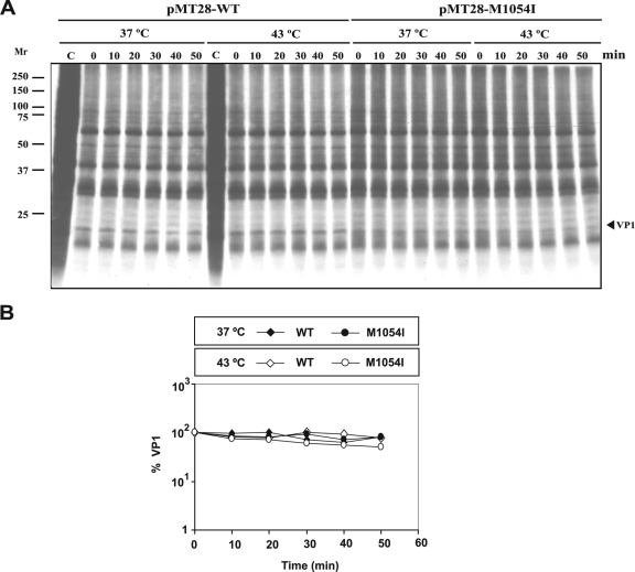
Capsid VP1 levels for mutant M54I relative to the wild type at 37°C and 43°C were quantitated by densitometry of the protein band obtained after Western blot analysis using a specific monoclonal antibody against VP1 protein (Fig. 5B). Expressed relative to the amount of VP1 for wild-type virus (which is taken as 100%), the decrease in VP1 expression in pMT28-M1054I was 10% at 37°C and about 90% at 43°C. Restriction of VP1 expression correlated with an increase at 43°C of the precursor proteins VP3-VP1 and P1 (Fig. 5C). The levels of 2C and 3D were less affected than the levels of VP1 and VP3 in pMT28-M1054I relative to pMT28-WT at 43°C (Fig. 5B and D). Once VP1 was produced, its amount remained constant at 37°C and 43°C both in pMT28-WT and pMT28-M1054I, as revealed by pulse-chase experiments (Fig. 6). Thus, an FMDV mutant with the substitution M54I in VP1 as the only mutation has a defect in the cleavage of the P1 precursor at the VP1-VP3 cleavage site, and the defect is enhanced at high temperatures.
FIG. 6.
Stability in BHK-21 cells of VP1 expressed from pMT28-WT or pMT28-M1054I. (A) BHK-21 cells were either mock electroporated (C lanes) or electroporated with about 20 μg of RNA transcripts from pMT28-WT or pMT28-M1054I at 37°C or 43°C. Transfected cells were labeled with [35S]Met-Cys at 3 h postelectroporation for 60 min, and then they were chased with unlabeled Met-Cys at the indicated times (min). Cell extracts were analyzed by SDS-PAGE, followed by fluorography and autoradiography, as detailed in the legend for Fig. 5 and in Materials and Methods. Mr values are in thousands. The position of VP1 is indicated. (B) The percentage of label in the band corresponding to capsid VP1, relative to the label at time zero taken as 100%, is indicated. VP1 was identified by its reactivity with monoclonal antibody SD6 (37). Procedures are described in Materials and Methods.
DISCUSSION
High mutation rates and rapid generation of complex mutant spectra during RNA virus replication underlie the profound biological consequences that population bottlenecks can have on virus fitness, adaptability, and pathogenesis (2, 8, 15, 21, 22, 26, 28, 32, 42, 44, 46-48, 52, 53). Clinical consequences of bottleneck events have been manifested in hepatitis C virus infections in which high inoculation doses predict chronicity of the infection while small-dose infections are often associated with virus clearance (reviewed in reference 46).
The numbers and types of mutations that are associated with a fitness decrease as a result of repeated plaque-to-plaque transfers (operation of Muller's ratchet) have been studied in detail with FMDV in our laboratory. Studies have included measurement of the rate of accumulation of mutations (0.26 to 0.34 mutations per transfer) (20, 21); the identification of unusual genetic lesions associated with repeated bottlenecks (17); the demonstration of a biphasic and fluctuating fitness decrease in the course of bottleneck transfers, with resistance of the virus to extinction prompted by compensatory mutations (19, 28, 29); the adherence of the fluctuating fitness values to a statistical Weibull function (29); and the striking evolution toward noncytopathic forms of FMDV (20). In the present study we have shown that an FMDV clone subjected to plaque-to-plaque transfers undergoes a gradual increase in thermosensitivity. While the ratio of infectious progeny production at 37°C versus 42°C was about 10 for the parental clone C101, it reached 4 × 10⁶ at the end of 230 plaque-to-plaque transfers (Fig. 2). Interestingly, four parallel sublineages that were initiated at passage 130 (Fig. 1) maintained their ts character, including a transient decrease in thermosensitivity at transfer 180 in sublineages a, b, and d, for which thermosensitivity could be measured (Fig. 2B). Such striking phenotypic similarity in three evolutionary lineages suggests a deterministic behavior (16), as previously documented in some processes with FMDV and other RNA viruses (26, 27, 46, 49). It is difficult to venture a molecular interpretation of the transient decrease in thermosensitivity at plaque transfer 180 that was observed in the three parallel lineages for two reasons: (i) the ts character is cumulative as a result of the addition of mutations at a constant rate in the viral genome (Fig. 2), and (ii) the mutations introduced from plaque transfer 130 until plaque transfer 230 are different in the four sublineages. Thus, some constraints might guide the virus toward a number of alternative mutational changes compatible with survival that by chance reduce thermosensitivity in a transient way; the molecular basis of such constraints is unknown. One possibility is that the observed increased thermosensitivity could be related to decreased robustness, as suggested by theoretical considerations (9, 40, 57).
One of the mutations that contributed to the ts character of FMDV clone C1030 is transversion G3369C, which leads to the amino acid substitution M54I in VP1 (Fig. 3). This mutation was acquired during the first 30 transfers of clone C101 (17) and was maintained at least up to transfer 230 (20). Therefore, M54I was present in the four sublineages that increased their thermosensitivity in parallel. The remarkable increase in thermosensitivity with additional plaque transfers of C1030 indicates that other viral functions must be affected as a result of the accumulation of mutations (Fig. 2).
Comparison of the pattern of FMDV protein expression using infectious transcripts that differed exclusively in transversion G3369C indicated that the mutant expressing VP1 with M54I yielded lower levels of mature VP1 than wild-type FMDV, and the difference was much more pronounced at 43°C than at 37°C (Fig. 5). In FMDV, as well as in other picornaviruses, capsid proteins are produced by the processing of a precursor P1 protein by 3Cpro (7, 25, 51, 56). Concomitantly with a decreased amount of VP1, the mutant virus, but not the wild-type, showed accumulation of the uncleaved VP3-VP1 and P1 precursors at 43°C. Therefore, FMDV with the mutation M54I in capsid protein VP1 as the only mutation in the FMDV genome shows a defect in processing of capsid protein precursor P1, even though the substitution is 54 amino acids away from the VP3-VP1 processing site in the primary structure. Our analysis did not allow us to distinguish whether the capsid precursor also included 2A (50, 51, 56).
Residue M1054 is located 6 residues downstream of the B-C loop of capsid protein VP1 and belongs to a stretch of exposed amino acids (residues 38 to 61) on the capsid surface (accessible to a surface probe of 0.2-nm radius) (35, 36). The B-C loop of VP1 is close to the fivefold axis and constitutes antigenic site 3 of FMDV (1, 30, 35, 36). M1054 is conserved in FMDV of serotype C even though it is surrounded by amino acids that are replaced in monoclonal antibody escape mutants (11, 34, 36). Conservation of M1054 may relate to its involvement in P1 processing. The α-carbons of M1054 and Q3219 (the C-terminal residue of VP3 at the cleavage site) are 12 Å apart, and direct interactions between these two amino acids are unlikely. However, Q3219 is in direct contact with the B-C loop of VP1, in particular, with H1057. Both M1054 and H1057 establish contacts with Q1055 and A1056 (1, 30). Therefore, an indirect influence of M1054 on the processing site cannot be excluded. However, the structure of VP3-VP1-containing precursors need not include VP1 and VP3 with the same conformation as in viral particles. Furthermore, in an analysis of the processing of truncated forms of the FMDV polyprotein by FMDV-infected cell extracts in a rabbit reticulocyte lysate system, removal of the C-terminal regions of the P1-2A and 2BC precursors impaired their cleavage by 3Cpro (50). Since the three-dimensional structure of the capsid precursors of FMDV, as they act as 3Cpro substrates, is not known, it cannot be excluded that M1054 can exert a short-distance effect on the cleavage site.
The VP3-VP1 cleavage site in pMT28 has the amino acid sequence ARQQ/T (where the slash represents the cleavage site), which is one of several alternative sequences found at this VP3-VP1 cleavage site among different FMDV isolates (51). The sequence is in agreement with the proposed picornavirus consensus sequence for 3Cpro cleavage (P4-X-X-P1/P1′, in which P4 is a hydrophobic amino acid, X is any amino acid, P1 is Q, and P1′ is a small amino acid) (31). Therefore, there is no unusual feature in the amino acid sequence of FMDV C-S8c1 at the VP3-VP1 junction that could render this cleavage more sensitive to perturbation by neighboring or distant sites. Effects at a distance within a viral particle or a viral protein are not uncommon. They have been observed among distant antigenic sites of FMDV (6, 35, 43) and in the poliovirus polymerase in connection with sites that can influence catalysis and template-copying fidelity (5). It should also be considered that the effect of M1054 on P1 processing might be exerted indirectly through an altered interaction with a host factor involved in positioning or folding of the P1 substrate, as previously shown for other picornaviruses (24, 31).
Independently of the molecular mechanism involved, the results reported here link two seemingly unrelated phenotypic traits: a surface domain of a capsid involved in antigenicity and a protein processing event essential to complete the virus life cycle. The defect associated with M54I affects secondary processing cleavages in trans, which are slower than the primary polyprotein processing reactions. Consistent with this, the ts phenotype associated with mutation M54I is manifested at late times during FMDV infection (Fig. 4A). Thus, the mutational instruction prompted by the operation of Muller's ratchet under limited negative selection has unveiled an unexpected molecular interaction that critically affects the picornavirus life cycle. More work is needed to investigate whether other residues around antigenic site 3 or residues at other antigenic sites may also influence polyprotein processing and to identify functions responsible for the increased thermosensitivity associated with additional plaque transfers. The results illustrate the benefits of pursuing at the biochemical level the basis of decreases in virus fitness that are associated with repeated bottleneck events.
Acknowledgments
We are indebted to M. G. Mateu for valuable comments and to M. Dávila and A. I. de Ávila for expert technical assistance. We thank A. Arias, V. Rincón, and N. Verdaguer for help with structural analysis and H. Tejero for help with the statistics.
This work was supported by grant BFU2008-02816/BMC and Fundación Ramón Areces. The Centro de Investigación Biomédica en Red de Enfermedades Hepáticas y Digestivas (CIBERehd) is funded by the Instituto de Salud Carlos III. C.P. was supported by a contract from CIBERehd.
Footnotes
Published ahead of print on 29 April 2009.
REFERENCES
- 1.Acharya, R., E. Fry, D. Stuart, G. Fox, D. Rowlands, and F. Brown. 1989. The three-dimensional structure of foot-and-mouth disease virus at 2.9 Å resolution. Nature 337709-716. [DOI] [PubMed] [Google Scholar]
- 2.Ali, A., H. Li, W. L. Schneider, D. J. Sherman, S. Gray, D. Smith, and M. J. Roossinck. 2006. Analysis of genetic bottlenecks during horizontal transmission of Cucumber mosaic virus. J. Virol. 808345-8350. [DOI] [PMC free article] [PubMed] [Google Scholar]
- 3.Arias, A., R. Agudo, C. Ferrer-Orta, R. Perez-Luque, A. Airaksinen, E. Brocchi, E. Domingo, N. Verdaguer, and C. Escarmis. 2005. Mutant viral polymerase in the transition of virus to error catastrophe identifies a critical site for RNA binding. J. Mol. Biol. 3531021-1032. [DOI] [PubMed] [Google Scholar]
- 4.Arias, A., C. M. Ruiz-Jarabo, C. Escarmis, and E. Domingo. 2004. Fitness increase of memory genomes in a viral quasispecies. J. Mol. Biol. 339405-412. [DOI] [PubMed] [Google Scholar]
- 5.Arnold, J. J., M. Vignuzzi, J. K. Stone, R. Andino, and C. E. Cameron. 2005. Remote site control of an active site fidelity checkpoint in a viral RNA-dependent RNA polymerase. J. Biol. Chem. 28025706-25716. [DOI] [PMC free article] [PubMed] [Google Scholar]
- 6.Baranowski, E., C. M. Ruiz-Jarabo, F. Lim, and E. Domingo. 2001. Foot-and-mouth disease virus lacking the VP1 G-H loop: the mutant spectrum uncovers interactions among antigenic sites for fitness gain. Virology 288192-202. [DOI] [PubMed] [Google Scholar]
- 7.Bedard, K. M., and B. L. Semler. 2004. Picornaviridae: the viruses and their replication, p. 795-838. In D. M. Knipe, P. M. Howley, D. E. Griffin, R. A. Lamb, M. A. Martin, B. Roizman, and S. E. Straus (ed.), Fields virology, 5th ed. Lippincott Williams & Wilkins, Philadelphia, PA.
- 8.Betancourt, M., A. Fereres, A. Fraile, and F. Garcia-Arenal. 2008. Estimation of the effective number of founders that initiate an infection after aphid transmission of a multipartite plant virus. J. Virol. 8212416-12421. [DOI] [PMC free article] [PubMed] [Google Scholar]
- 9.Bloom, J. D., J. J. Silberg, C. O. Wilke, D. A. Drummond, C. Adami, and F. H. Arnold. 2005. Thermodynamic prediction of protein neutrality. Proc. Natl. Acad. Sci. USA 102606-611. [DOI] [PMC free article] [PubMed] [Google Scholar]
- 10.Carrillo, C., Z. Lu, M. V. Borca, A. Vagnozzi, G. F. Kutish, and D. L. Rock. 2007. Genetic and phenotypic variation of foot-and-mouth disease virus during serial passages in a natural host. J. Virol. 8111341-11351. [DOI] [PMC free article] [PubMed] [Google Scholar]
- 11.Carrillo, C., E. R. Tulman, G. Delhon, Z. Lu, A. Carreno, A. Vagnozzi, G. F. Kutish, and D. L. Rock. 2005. Comparative genomics of foot-and-mouth disease virus. J. Virol. 796487-6504. [DOI] [PMC free article] [PubMed] [Google Scholar]
- 12.de la Torre, J. C., M. Davila, F. Sobrino, J. Ortin, and E. Domingo. 1985. Establishment of cell lines persistently infected with foot-and-mouth disease virus. Virology 14524-35. [DOI] [PubMed] [Google Scholar]
- 13.de la Torre, J. C., E. Martínez-Salas, J. Diez, A. Villaverde, F. Gebauer, E. Rocha, M. Dávila, and E. Domingo. 1988. Coevolution of cells and viruses in a persistent infection of foot-and-mouth disease virus in cell culture. J. Virol. 622050-2058. [DOI] [PMC free article] [PubMed] [Google Scholar]
- 14.Domingo, E., M. Davila, and J. Ortin. 1980. Nucleotide sequence heterogeneity of the RNA from a natural population of foot-and-mouth-disease virus. Gene 11333-346. [DOI] [PubMed] [Google Scholar]
- 15.Domingo, E., C. Escarmís, L. Menéndez-Arias, C. Perales, M. Herrera, I. S. Novella, and J. J. Holland. 2008. Viral quasispecies: dynamics, interactions and pathogenesis, p. 87-118. In E. Domingo, C. Parrish, and J. J. Holland (ed.), Origin and evolution of viruses, 2nd ed. Elsevier, Oxford, United Kingdom.
- 16.Eigen, M., and P. Schuster. 1979. The hypercycle: a principle of natural self-organization. Springer, Berlin, Germany. [DOI] [PubMed]
- 17.Escarmís, C., M. Dávila, N. Charpentier, A. Bracho, A. Moya, and E. Domingo. 1996. Genetic lesions associated with Muller's ratchet in an RNA virus. J. Mol. Biol. 264255-267. [DOI] [PubMed] [Google Scholar]
- 18.Escarmís, C., M. Dávila, and E. Domingo. 1999. Multiple molecular pathways for fitness recovery of an RNA virus debilitated by operation of Muller's ratchet. J. Mol. Biol. 285495-505. [DOI] [PubMed] [Google Scholar]
- 19.Escarmís, C., G. Gómez-Mariano, M. Dávila, E. Lázaro, and E. Domingo. 2002. Resistance to extinction of low fitness virus subjected to plaque-to-plaque transfers: diversification by mutation clustering. J. Mol. Biol. 315647-661. [DOI] [PubMed] [Google Scholar]
- 20.Escarmís, C., E. Lazaro, A. Arias, and E. Domingo. 2008. Repeated bottleneck transfers can lead to non-cytocidal forms of a cytopathic virus: implications for viral extinction. J. Mol. Biol. 376367-379. [DOI] [PubMed] [Google Scholar]
- 21.Escarmís, C., E. Lázaro, and S. C. Manrubia. 2006. Population bottlenecks in quasispecies dynamics. Curr. Top. Microbiol. Immunol. 299141-170. [DOI] [PubMed] [Google Scholar]
- 22.Foy, B. D., K. M. Myles, D. J. Pierro, I. Sanchez-Vargas, M. Uhlirova, M. Jindra, B. J. Beaty, and K. E. Olson. 2004. Development of a new Sindbis virus transducing system and its characterization in three Culicine mosquitoes and two Lepidopteran species. Insect Mol. Biol. 1389-100. [DOI] [PubMed] [Google Scholar]
- 23.García-Arriaza, J., S. C. Manrubia, M. Toja, E. Domingo, and C. Escarmís. 2004. Evolutionary transition toward defective RNAs that are infectious by complementation. J. Virol. 7811678-11685. [DOI] [PMC free article] [PubMed] [Google Scholar]
- 24.Geller, R., M. Vignuzzi, R. Andino, and J. Frydman. 2007. Evolutionary constraints on chaperone-mediated folding provide an antiviral approach refractory to development of drug resistance. Genes Dev. 21195-205. [DOI] [PMC free article] [PubMed] [Google Scholar]
- 25.Grubman, M. J., M. Zellner, G. Bablanian, P. W. Mason, and M. E. Piccone. 1995. Identification of the active-site residues of the 3C proteinase of foot-and-mouth disease virus. Virology 213581-589. [DOI] [PubMed] [Google Scholar]
- 26.Haaland, R. E., P. A. Hawkins, J. Salazar-Gonzalez, A. Johnson, A. Tichacek, E. Karita, O. Manigart, J. Mulenga, B. F. Keele, G. M. Shaw, B. H. Hahn, S. A. Allen, C. A. Derdeyn, and E. Hunter. 2009. Inflammatory genital infections mitigate a severe genetic bottleneck in heterosexual transmission of subtype A and C HIV-1. PLoS Pathog. 5e1000274. [DOI] [PMC free article] [PubMed] [Google Scholar]
- 27.Herrera, M., A. Grande-Perez, C. Perales, and E. Domingo. 2008. Persistence of foot-and-mouth disease virus in cell culture revisited: implications for contingency in evolution. J. Gen. Virol. 89232-244. [DOI] [PubMed] [Google Scholar]
- 28.Lázaro, E., C. Escarmís, E. Domingo, and S. C. Manrubia. 2002. Modeling viral genome fitness evolution associated with serial bottleneck events: evidence of stationary states of fitness. J. Virol. 768675-8681. [DOI] [PMC free article] [PubMed] [Google Scholar]
- 29.Lazaro, E., C. Escarmis, J. Perez-Mercader, S. C. Manrubia, and E. Domingo. 2003. Resistance of virus to extinction on bottleneck passages: Study of a decaying and fluctuating pattern of fitness loss. Proc. Natl. Acad. Sci. USA 10010830-10835. [DOI] [PMC free article] [PubMed] [Google Scholar]
- 30.Lea, S., J. Hernández, W. Blakemore, E. Brocchi, S. Curry, E. Domingo, E. Fry, R. Abu-Ghazaleh, A. King, J. Newman, D. Stuart, and M. G. Mateu. 1994. The structure and antigenicity of a type C foot-and-mouth disease virus. Structure 2123-139. [DOI] [PubMed] [Google Scholar]
- 31.Leong, L. E.-C., C. T. Cornell, and B. L. Semler. 2002. Processing determinants and functions of cleavage products of picornaviruses poly-proteins, p. 187-197. In B. L. Semler and E. Wimmer (ed.), Molecular biology of picornaviruses. ASM Press, Washington, DC.
- 32.Li, H., and M. J. Roossinck. 2004. Genetic bottlenecks reduce population variation in an experimental RNA virus population. J. Virol. 7810582-10587. [DOI] [PMC free article] [PubMed] [Google Scholar]
- 33.Manrubia, S. C., C. Escarmis, E. Domingo, and E. Lazaro. 2005. High mutation rates, bottlenecks, and robustness of RNA viral quasispecies. Gene 347273-282. [DOI] [PubMed] [Google Scholar]
- 34.Martinez, M. A., J. Hernandez, M. E. Piccone, E. L. Palma, E. Domingo, N. Knowles, and M. G. Mateu. 1991. Two mechanisms of antigenic diversification of foot-and-mouth disease virus. Virology 184695-706. [DOI] [PubMed] [Google Scholar]
- 35.Mateu, M. G. 1995. Antibody recognition of picornaviruses and escape from neutralization: a structural view. Virus Res. 381-24. [DOI] [PubMed] [Google Scholar]
- 36.Mateu, M. G., J. Hernández, M. A. Martínez, D. Feigelstock, S. Lea, J. J. Pérez, E. Giralt, D. Stuart, E. L. Palma, and E. Domingo. 1994. Antigenic heterogeneity of a foot-and-mouth disease virus serotype in the field is mediated by very limited sequence variation at several antigenic sites. J. Virol. 681407-1417. [DOI] [PMC free article] [PubMed] [Google Scholar]
- 37.Mateu, M. G., M. A. Martínez, E. Rocha, D. Andreu, J. Parejo, E. Giralt, F. Sobrino, and E. Domingo. 1989. Implications of a quasispecies genome structure: effect of frequent, naturally occurring amino acid substitutions on the antigenicity of foot-and-mouth disease virus. Proc. Natl. Acad. Sci. USA 865883-5887. [DOI] [PMC free article] [PubMed] [Google Scholar]
- 38.Mateu, M. G., E. Rocha, O. Vicente, F. Vayreda, C. Navalpotro, D. Andreu, E. Pedroso, E. Giralt, L. Enjuanes, and E. Domingo. 1987. Reactivity with monoclonal antibodies of viruses from an episode of foot-and-mouth disease. Virus Res. 8261-274. [DOI] [PubMed] [Google Scholar]
- 39.Maynard-Smith, J. 1976. The evolution of sex. Cambridge University Press, Cambridge, United Kingdom.
- 40.McBride, R. C., C. B. Ogbunugafor, and P. E. Turner. 2008. Robustness promotes evolvability of thermotolerance in an RNA virus. BMC Evol. Biol. 8231. [DOI] [PMC free article] [PubMed] [Google Scholar]
- 41.Muller, H. J. 1964. The relation of recombination to mutational advance. Mut. Res. 12-9. [DOI] [PubMed] [Google Scholar]
- 42.Novella, I. S. 2004. Negative effect of genetic bottlenecks on the adaptability of vesicular stomatitis virus. J. Mol. Biol. 33661-67. [DOI] [PubMed] [Google Scholar]
- 43.Parry, N., G. Fox, D. Rowlands, F. Brown, E. Fry, R. Acharya, D. Logan, and D. Stuart. 1990. Structural and serological evidence for a novel mechanism of antigenic variation in foot-and-mouth disease virus. Nature 347569-572. [DOI] [PubMed] [Google Scholar]
- 44.Perales, C., R. Mateo, M. G. Mateu, and E. Domingo. 2007. Insights into RNA virus mutant spectrum and lethal mutagenesis events: replicative interference and complementation by multiple point mutants. J. Mol. Biol. 369985-1000. [DOI] [PubMed] [Google Scholar]
- 45.Pfeiffer, J. K., and K. Kirkegaard. 2006. Bottleneck-mediated quasispecies restriction during spread of an RNA virus from inoculation site to brain. Proc. Natl. Acad. Sci. USA 1035520-5525. [DOI] [PMC free article] [PubMed] [Google Scholar]
- 46.Quer, J., A. Martell, R. Rodriguez, A. Bosch, R. Jardi, M. Buti, and J. I. Esteban. 2008. The impact of rapid evolution of hepatitis viruses, p. 303-350. In E. Domingo, C. Parrish, and J. J. Holland (ed.), Origin and evolution of viruses, 2nd ed. Elsevier, Oxford, United Kingdom.
- 47.Quer, J., J. I. Esteban, J. Cos, S. Sauleda, L. Ocana, M. Martell, T. Otero, M. Cubero, E. Palou, P. Murillo, R. Esteban, and J. Guardia. 2005. Effect of bottlenecking on evolution of the nonstructural protein 3 gene of hepatitis C virus during sexually transmitted acute resolving infection. J. Virol. 7915131-15141. [DOI] [PMC free article] [PubMed] [Google Scholar]
- 48.Roossinck, M. J., and W. L. Schneider. 2006. Mutant clouds and occupation of sequence space in plant RNA viruses. Curr. Top. Microbiol. Immunol. 299337-348. [DOI] [PubMed] [Google Scholar]
- 49.Ruiz-Jarabo, C. M., E. Miller, G. Gómez-Mariano, and E. Domingo. 2003. Synchronous loss of quasispecies memory in parallel viral lineages: a deterministic feature of viral quasispecies. J. Mol. Biol. 333553-563. [DOI] [PubMed] [Google Scholar]
- 50.Ryan, M. D., G. J. Belsham, and A. M. King. 1989. Specificity of enzyme-substrate interactions in foot-and-mouth disease virus polyprotein processing. Virology 17335-45. [DOI] [PubMed] [Google Scholar]
- 51.Ryan, M. D., M. L. L. Donnelly, M. Flint, V. M. Cowton, G. Luke, L. E. Hughes, C. Knock, and P. de Felipe. 2004. Foot-and-mouth disease virus proteinases, p. 53-76. In F. Sobrino and E. Domingo (ed.), Foot-and-mouth disease: current perspectives. Horizon Bioscience, Wymondham, United Kingdom.
- 52.Scholle, F., Y. A. Girard, Q. Zhao, S. Higgs, and P. W. Mason. 2004. trans-Packaged West Nile virus-like particles: infectious properties in vitro and in infected mosquito vectors. J. Virol. 7811605-11614. [DOI] [PMC free article] [PubMed] [Google Scholar]
- 53.Smith, D. R., A. P. Adams, J. L. Kenney, E. Wang, and S. C. Weaver. 2008. Venezuelan equine encephalitis virus in the mosquito vector Aedes taeniorhynchus: infection initiated by a small number of susceptible epithelial cells and a population bottleneck. Virology 372176-186. [DOI] [PMC free article] [PubMed] [Google Scholar]
- 54.Sobrino, F., M. Dávila, J. Ortín, and E. Domingo. 1983. Multiple genetic variants arise in the course of replication of foot-and-mouth disease virus in cell culture. Virology 128310-318. [DOI] [PubMed] [Google Scholar]
- 55.Toja, M., C. Escarmis, and E. Domingo. 1999. Genomic nucleotide sequence of a foot-and-mouth disease virus clone and its persistent derivatives. Implications for the evolution of viral quasispecies during a persistent infection. Virus Res. 64161-171. [DOI] [PubMed] [Google Scholar]
- 56.Vakharia, V. N., M. A. Devaney, D. M. Moore, J. J. Dunn, and M. J. Grubman. 1987. Proteolytic processing of foot-and-mouth disease virus polyproteins expressed in a cell-free system from clone-derived transcripts. J. Virol. 613199-3207. [DOI] [PMC free article] [PubMed] [Google Scholar]
- 57.Wilke, C. O., J. D. Bloom, D. A. Drummond, and A. Raval. 2005. Predicting the tolerance of proteins to random amino acid substitution. Biophys. J. 893714-3720. [DOI] [PMC free article] [PubMed] [Google Scholar]


