Abstract
Phylogenetic analysis of West Nile virus in North America has identified replacement of the originally introduced clade, Eastern United States (NY99), by the North American clade. In addition, the transient emergence of other clades and genetic variants has also been observed. In this study, we investigated the potential role of the mosquito in the selection of these clades and genetic variants. We determined the relative susceptibility of Culex quinquefasciatus to infection with isolates from the Eastern U.S. clade, the North American clade, and the Southeast coastal Texas clade. Although significant differences were observed in 50% oral infectious dose values between the Eastern U.S. and two attenuated North American genetic variants compared with the North American and Southeast coastal Texas clade viruses, these differences did not correlate with persistence of the genotype in nature. These results indicate that selection of these viral genotypes was independent of viral oral infectivity in the mosquito.
The spread of West Nile virus (WNV) throughout North America has allowed detailed studies of how the virus has evolved after its introduction in 1999 and how it becomes established in new areas. This virus is typically transmitted in an avian and ornithophilic mosquito cycle, with the principle vectors being mosquitoes of the genus Culex.1 Three Culex species have been identified as the primary vectors of WNV in the United States: Cx. pipiens pipiens, Cx. tarsalis, and Cx. quinquefasciatus.2,3
Using phylogenetic tools, Davis and others demonstrated that three distinct clades have evolved in the United States since 1999.4 The New York 1999 (NY99), or the Eastern U.S. clade was originally introduced into New York. This virus was determined to be most closely related to a WNV isolate obtained from a dead goose in Israel in 1998.5 As the virus moved westward, this clade was replaced by the North American clade in 2001–2002.4 By 2004, the NY99 genotype was no longer observed in nature.6 A third clade, Southeast coastal Texas, was identified in 2002. However, this clade has not been detected in subsequent years, which suggested that the clade has been displaced or has become extinct.4 To test the hypothesis that the selection of viral genotypes is determined by infection of mosquito vectors, we determined the oral 50% infectious dose (ID50) required to infect Cx. quinquefasciatus with five different isolates of WNV representing the three clades and two isolates that are naturally attenuated in their mouse neuroinvasive phenotype.7
The American prototype strain NY 382-99 belongs to the Eastern U.S. clade and was isolated from a flamingo in 1999 (GenBank accession no. AF196835).5 The WNV 114 isolate belongs to the North American clade and was isolated from a bluejay in June 2002 from Harris County, Texas (GenBank accession no. AY185907).8 The WNV 476 isolate, from the Bolivar peninsula located north of Galveston, Texas, was isolated in August 2002 from a bluejay and is representative of the Southeast coastal Texas clade (GenBank accession no. AY185914).8 These three isolates have a large plaque in vitro phenotype and are non-attenuated for mouse neuroinvasiveness.7 Two additional viruses, WNV v4369 and WNV 1153, which are genetic variants within the North American clade, were also examined. These isolates have a predominantly small plaque phenotype in vitro and are attenuated for mouse neuroinvasiveness. The WNV v4369 isolate was obtained from a Cx. quinquefasciatus pool (GenBank accession no. AY712948),7 and the WNV 1153 isolate was obtained from a mourning dove (GenBank accession no. AY712945) in Harris County, Texas in 2003.7,9
The well-characterized colony of Cx. quinquefasciatus (Sebring strain) mosquitoes were used for this study. These mosquitoes were collected in Sebring County Florida in 1988 and have oral infectivity to WNV similar to that of field-collected Cx. quinquefasciatus.10 This mosquito colony was maintained as previously described.11 Groups of 125 four-day-old adult female Cx. quinquefasciatus mosquitoes were fed a blood meal containing dilutions of one of the five isolates of WNV to be analyzed. Vero cell culture was used to grow fresh virus from stock and the virus was harvested when 75% of the cells showed a cytopathic effect.
Serial 10-fold dilutions of viral supernatant and an equal volume of defibrinated sheep blood were mixed, heated to 37°C in a Hemotek feeding apparatus (Discovery Workshops, Accrington, United Kingdom) and presented to mosquitoes in an isolation glove box in an Arthropod Containment Level 3 insectary.12 Fully engorged females were placed into new cartons and maintained until 14 days post-infection (dpi) when 6–30 mosquitoes were removed and held at −80°C for later titration. Mosquitoes were analyzed for percent infection on the basis of body titration and dissemination on the basis of titration of heads at 14 dpi. Briefly, 10-fold serial dilutions of triturated tissues were incubated with Vero cells on 96-well plates.13,14 The oral ID50 was calculated on the basis of the percentage of mosquitoes infected at 14 dpi after exposure to four or five dilutions of blood meals that represented virus from the three clades and three dilutions of blood meals of the two genetic variants. The titers of these blood meals ranged from 0.22 to 8.22 log10 50% tissue culture infectious doses (TCID50)/5 μL. Regression lines and 95% confidence levels (CIs) were determined using PriProbit version 1.63 (Kyoto University, Kyoto, Japan).
Infection and dissemination rates were determined for each of the five viruses after oral infection of Cx. quinquefasciatus mosquitoes. When higher titers of the different viruses were used to orally infect mosquitoes, the infectivity rates were higher. Mosquitoes orally exposed to undiluted virus mixed with an equal volume of blood and analyzed by body titration on Vero cell culture at 14 dpi were positive for WNV at a high percentage, which ranged from 94% to 100% (Table 1). There was no significant difference between the five viruses with respect to dissemination rates at 14 dpi in mosquitoes fed blood meals with WNV titers ranging from 4.2 to 6.2 log10TCID50/5 μL. At 14 dpi, dissemination rates were determined for WNV-infected mosquitoes. Mosquitoes were analyzed for WNV in mosquito heads titrated on Vero cell culture. The dissemination rate ranged from 86% to 100%.
Table 1.
Infection rates of Culex quinquefasciatus mosquitoes orally exposed to five West Nile virus isolates at various viral titers
| Blood meal titer* | NY99 Eastern U.S. infection, no. (%) | v4369 attenuated† infection, no. (%) | 1153 attenuated† infection, no. (%) | 114 North American infection, no. (%)‡ | 476 Southeast Coastal Texas infection, no. (%) |
|---|---|---|---|---|---|
| 8.22 | 6/6 (100) | ||||
| 6.22 | 30/30 (100) | 13/22 (59) | 29/30 (97) | ||
| 5.22 | 25/26 (96) | 17/18 (94) | 17/30 (57) | ||
| 4.65 | 27/30 (90) | ||||
| 4.22 | 15/20 (75) | 0/30 (0) | |||
| 3.65 | 12/24 (50) | 5/30 (17) | |||
| 3.22 | 4/30 (13) | ||||
| 2.65 | 14/35 (40) | 0/30 (0) | |||
| 2.22 | 4/26 (15) | 1/24 (4) | |||
| 1.22 | 2/30 (7) | ||||
| 0.22 | 0/18 (0) |
Reported as log10 50% tissue culture infectious dose/5 μL.
Viruses are attenuated in their mouse neuroinvasive phenotype.
Infection rates were determined by whole body titration of the mosquito and reported as number positive/total number tested (%).
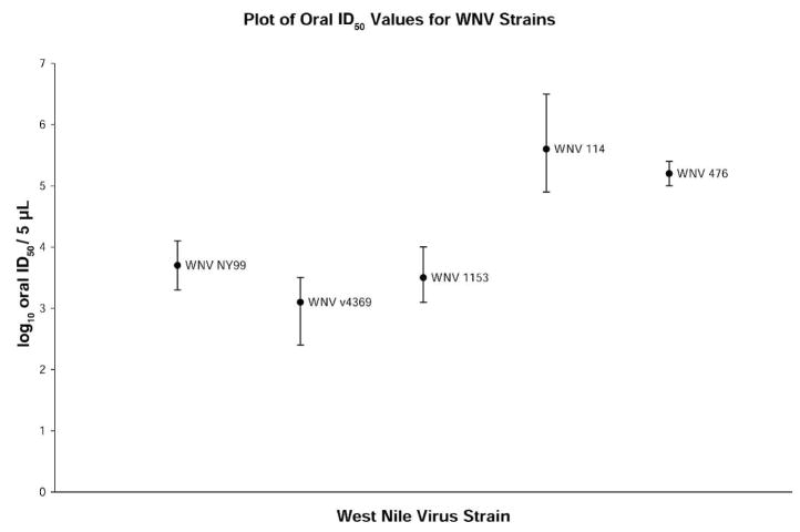
Oral ID50 values for the five viruses were determined in Cx. quinquefasciatus mosquitoes; overlap of the 95% CIs was used as a conservative test of statistical significance (Figure 1). There was no significant difference in the oral ID50 values for WNV NY99 clade virus and the two attenuated genetic variants WNV v4369 and WNV 1153. Similarly, there was no significant difference between the non-attenuated North American clade virus, WNV 114, and the Southeast Coastal Texas clade virus WNV 476. However, at the 95% CI, there was a statistically significantly difference identified between these two groups (Figure 1).
Figure 1.
Oral 50% infectious dose values of five isolates of West Nile virus in Culex quinquefasciatus (Sebring colony). Error bars represent 95% confidence intervals. Titers are reported as log10 50% tissue culture infectious doses0/5 μL, which is the estimated volume of ingested virus.
Plaque assays on Vero cell monolayers were used to determine if the plaque morphology phenotype had changed during replication in the mosquito. On the basis of these plaque assays of whole body homogenates at 14 dpi, the small plaque phenotype of the two attenuated viruses WNV 1153 and WNV v4369 was maintained after replication in mosquitoes.
We compared the phenotype of five isolates of WNV in Cx. quinquefasciatus. The predominant North American clade, represented by the WNV 114 isolate, had a significantly higher oral ID50 in Cx. quinquefasciatus mosquitoes than the NY99 isolate, which it replaced (5.56 and 3.88 log10TCID50/5μL, respectively). This finding suggests that the lower oral dose, which enabled NY99 to infect this mosquito species, did not confer a selective advantage in nature. Previous studies have found that the oral ID50 of WNV 114 presented to field collected Cx. quinquefasciatus F0 mosquitoes was 5.33 log10TCID50/5 μL,10 which is comparable to the oral ID50 value of 5.56 log10TCID50/5 μL for WNV 114 virus in the Cx. quinquefasciatus colony used in this study.
There was no significant difference in the oral ID50 values of the North American clade (WNV 114) and Southeast Coastal Texas clade (WNV 476) viruses in Cx. quinquefasciatus mosquitoes (5.56 and 5.21 log10TCID50/5 μL, respectively), although the Southeast Coastal Texas clade did not persist in nature beyond 2002. Interestingly, the two isolates that have naturally attenuated phenotypes in mice (WNV 1153 and WNV v4369) had oral ID50 values similar to that of to NY99 rather than to the North American clade virus from which they are genetically derived (Figure 1). In nature, these isolates did not persist beyond 2003, which indicated that a lower oral infectious dose for mosquitoes apparently conferred no selective advantage to these isolates.
Previous studies have attempted to identify the source of the genetic variability and the mechanism of selection of one virus genotype over another. A study examining the source of the genetic variability of WNV found that WNV sequences in mosquitoes were more variable than WNV sequences isolated from birds.15 Jerzak and others speculated that mosquitoes in nature may be responsible for the genetic variation.15 Studies examining a possible mechanism of selection of WNV isolates in mosquitoes suggested that the rate of transmission may be important. In one study, 2% of orally infected Cx. tarsalis mosquitoes transmitted a North American clade isolate (WN02) four days earlier than mosquitoes infected with NY99.16 A similar transmission study in Cx. pipiens found that the WN02 genotype virus was transmitted 2–4 days earlier than the NY99 genotype virus.17 In both of these studies, the infection and dissemination rates of WNV in Cx. tarsalis and Cx. pipiens mosquitoes were substantially lower than the infection and dissemination found in colonized and F0 Cx. quinquefasciatus mosquitoes orally infected for this and a previous study.10 These differences in infection and dissemination between the mosquito species and time points examined prohibit a direct comparison of these data.
Overall, our findings indicate that although there are phenotypic differences between the viruses in vitro and in mice, these differences are unrelated to the mosquito infectivity phenotype in terms of the oral ID50 values of these viruses. The selective advantage of a lower oral ID50 in Cx. quinquefasciatus mosquitoes does not appear to be sufficient to influence the selection of this particular viral phenotype. Therefore, we conclude that the replacement of the Eastern U.S. clade with the North American clade has occurred by a mechanism not directly related to viral infectivity for Cx. quinquefasciatus mosquitoes.
Acknowledgments
We thank Jing Haung for expert technical assistance with this project.
Financial support: This study was supported in part by Centers for Disease Control and Prevention grant U50/CCU620539 to Stephen Higgs and National Institutes of Health (NIH) grant RO1 AI 67847 to Alan D. T. Barrett. Dana L. Vanlandingham was supported in part by NIH grant T32 A107536. Charles E. McGee was supported by the Centers for Disease Control and Prevention Fellowship Training Program in Vector-Borne Infectious Diseases (TOI/CCT622892) and NIH grant T32 AI 7526.
References
- 1.Apperson CS, Hassan HK, Harrison BA, Savage HM, Aspen SE, Farajollahi A, Crans W, Daniels TJ, Falco RC, Benedict M, Anderson M, McMillen L, Unnasch TR. Host feeding patterns of established and potential mosquito vectors of West Nile virus in the eastern United States. Vector Borne Zoonotic Dis. 2004;4:71–82. doi: 10.1089/153036604773083013. [DOI] [PMC free article] [PubMed] [Google Scholar]
- 2.Lillibridge KM, Parsons R, Randle Y, Travassos da Rosa AP, Guzman H, Siirin M, Wuithiranyagool T, Hailey C, Higgs S, Bala AA, Pascua R, Meyer T, Vanlandingham DL, Tesh RB. The 2002 introduction of West Nile virus into Harris County, Texas, an area historically endemic for St. Louis encephalitis. Am J Trop Med Hyg. 2004;70:676–681. [PubMed] [Google Scholar]
- 3.Reisen W, Lothrop H, Chiles R, Madon M, Cossen C, Woods L, Husted S, Kramer V, Edman J. West Nile virus in California. Emerging Infect Dis. 2004;10:1369–1378. doi: 10.3201/eid1008.040077. [DOI] [PMC free article] [PubMed] [Google Scholar]
- 4.Davis CT, Ebel GD, Lanciotti RS, Brault AC, Guzman H, Siirin M, Lambert A, Parsons RE, Beasley DW, Novak RJ, Elizondo-Quiroga D, Green EN, Young DS, Stark LM, Drebot MA, Artsob H, Tesh RB, Kramer LD, Barrett AD. Phylogenetic analysis of North American West Nile virus isolates, 2001–2004: evidence for the emergence of a dominant genotype. Virology. 2005;342:252–265. doi: 10.1016/j.virol.2005.07.022. [DOI] [PubMed] [Google Scholar]
- 5.Lanciotti RS, Roehirig JT, Deubel V, Smith J, Parker M, Steele K, Crise B, Volpe KE, Crabtree MB, Scherret JH, Hall RA, MacKenzie JS, Cropp CB, Panigrahy B, Ostlund E, Schmitt B, Malkinson M, Banet C, Weissman J, Komar N, Savage HM, Stone W, McNamara T, Gubler DJ. Origin of the West Nile virus responsible for an outbreak of encephalitis in the northeastern United States. Science. 1999;286:2333–2337. doi: 10.1126/science.286.5448.2333. [DOI] [PubMed] [Google Scholar]
- 6.Snapinn KW, Holmes EC, Young DS, Bernard KA, Kramer LD, Ebel GD. Declining growth rate of West Nile virus in North America. J Virol. 2007;81:2531–2534. doi: 10.1128/JVI.02169-06. [DOI] [PMC free article] [PubMed] [Google Scholar]
- 7.Davis CT, Beasley DW, Guzman H, Siirin M, Parsons RE, Tesh RB, Barrett AD. Emergence of attenuated West Nile virus variants in Texas, 2003. Virology. 2004;330:342–350. doi: 10.1016/j.virol.2004.09.016. [DOI] [PubMed] [Google Scholar]
- 8.Beasley DW, Davis CT, Guzman H, Vanlandingham DL, Travassos da Rosa AP, Parsons RE, Higgs S, Tesh RB, Barrett AD. Limited evolution of West Nile virus has occurred during its southwesterly spread in the United States. Virology. 2003;309:190–195. doi: 10.1016/s0042-6822(03)00150-8. [DOI] [PubMed] [Google Scholar]
- 9.Davis CT, Galbraith SE, Zhang S, Whiteman MC, Li L, Kinney RM, Barrett AD. A combination of naturally occurring mutations in North American West Nile virus nonstructural protein genes and in the 3′ untranslated region alters virus phenotype. J Virol. 2007;81:6111–6116. doi: 10.1128/JVI.02387-06. [DOI] [PMC free article] [PubMed] [Google Scholar]
- 10.Vanlandingham DL, McGee CE, Klinger KA, Vessey N, Fredregillo C, Higgs S. Relative susceptibilties of South Texas mosquitoes to infection with West Nile virus. Am J Trop Med Hyg. 2007;77:925–928. [PubMed] [Google Scholar]
- 11.Vanlandingham DL, Schneider BS, Klingler K, Fair J, Beasley D, Huang J, Hamilton P, Higgs S. Real-time reverse transcriptase-polymerase chain reaction quantification of West Nile virus transmitted by Culex pipiens quinquefasciatus. Am J Trop Med Hyg. 2004;71:120–123. [PubMed] [Google Scholar]
- 12.Higgs S, Beaty BJ. Rearing and containment of arthropod vectors. In: Marquardt WC, Beaty BJ, editors. Biology of Disease Vectors. University Press; Colorado: 1996. pp. 595–605. [Google Scholar]
- 13.Higgs S, Olson KE, Kamrud KI, Powers AM, Beaty BJ. Viral expression systems and viral infections in insects. In: Crampton JM, Beard CB, Louis C, editors. The Molecular Biology of Disease Vectors: A Methods Manual. London: Chapman and Hall; 1997. pp. 457–483. [Google Scholar]
- 14.Vanlandingham DL, Hong C, Klingler K, Tsetsarkin K, McElroy KL, Powers AM, Lehane MJ, Higgs S. Differential infectivities of o’nyong-nyong and chikungunya virus isolates in Anopheles gambiae and Aedes aegypti mosquitoes. Am J Trop Med Hyg. 2005;72:616–621. [PubMed] [Google Scholar]
- 15.Jerzak G, Bernard KA, Kramer LD, Ebel GD. Genetic variation in West Nile virus from naturally infected mosquitoes and birds suggests quasispecies structure and strong purifying selection. J Gen Virol. 2005;86:2175–2183. doi: 10.1099/vir.0.81015-0. [DOI] [PMC free article] [PubMed] [Google Scholar]
- 16.Moudy RM, Meola MA, Morin LL, Ebel GD, Kramer LD. A newly emergent genotype of West Nile virus is transmitted earlier and more efficiently by Culex mosquitoes. Am J Trop Med Hyg. 2007;77:365–370. [PubMed] [Google Scholar]
- 17.Ebel GD, Carricaburu J, Young D, Bernard KA, Kramer LD. Genetic and phenotypic variation of West Nile virus in New York, 2000–2003. Am J Trop Med Hyg. 2004;71:493–500. [PubMed] [Google Scholar]


