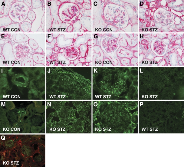
FIG. 2.
Light microscopic features of the glomerular lesions and expression of type VIII collagen. Representative glomerulus of an untreated wild-type mouse (A and E) and a Col8a1−/Col8a2− mouse (C and G) stained against type IV collagen (A and C) and laminin (E and G). Representative glomerulus of a wild-type mouse (B and F) and a Col8a1−/Col8a2− mouse (D and H) treated with STZ and stained against type IV collagen (B and D) and laminin (F and H). Diabetes was associated with an increase in type IV collagen and laminin protein expression. Lack of collagen VIII reduced the accumulation of type IV collagen (C and D) and laminin (G and H) in the glomeruli. Immunofluorescence staining against collagen VIII in wild-type mice (I, J, and K) or EGFP in Col8a1−/EGFP “knock-in” mice (M, N, and O). Strong staining for collagen VIII (I) and EGFP (M) was seen in healthy mice within the adventitia and the endothelium of arteries, whereas no staining was apparent in tubuli or glomeruli. In diabetic mice strong staining within the tubular interstitium, within the glomeruli, and around arterioles (anti–type VIII collagen [J and K] and anti-EGFP [N and O]). No staining against type VIII collagen was seen in Col8a1−/Col8a2− mice (L) or EGFP in wild-type mice (P). Double immunofluorescence revealed that EGFP (red) only partly colocalized with CD31/PECAM-1 (green), a marker for vascular endothelial cells (Q). Original magnification: ×200 or ×400. CON, control; KO, knockout; WT, wild-type. (A high-quality digital representation of this figure is available in the online issue.)

