Abstract
The Pathobiological Determinants of Atherosclerosis in Youth (PDAY) study of autopsied 15-34 year old young people developed a risk score using the coronary heart disease (CHD) risk factors (sex, age, serum lipoprotein concentrations, smoking, hypertension, obesity, and hyperglycemia) to estimate the probability of advanced atherosclerotic lesions in the coronary arteries. The Cardiovascular Risk in Young Finns Study measured CHD risk factors in a population-based cohort in 1986 and 2001 and measured carotid artery intima-media thickness (IMT) with ultrasound in 2001. We computed the PDAY risk score from risk factors measured in 1279 subjects who were 12-24 years old in 1986 and 27-39 years in 2001. The PDAY risk score early in life (1986) and the change in risk score over the following 15 years (between 1986 and 2001) were independent predictors of carotid artery IMT; the multiplicative effect of 1 point in the 1986 risk score was 1.008 (95% CI 1.005-1.012) and the multiplicative effect of a 1 point increase between 1986 and 2001 risk scores was 1.003 (95% CI 1.001-1.006) (multiplicative effect 0.997 for 1 point decrease). In conclusion, the change over time (either a decrease or an increase) in the risk score during adolescence and young adulthood as well as the risk score early in life are important predictors of atherosclerosis.
Keywords: Prevention, Atherosclerosis, Risk factors, Coronary disease
Longitudinal observational studies have shown that coronary heart disease (CHD) risk factors measured in adolescence predict markers of atherosclerosis measured by noninvasive methods. Both carotid artery intima-media thickness (IMT) and coronary artery calcification were associated with concurrently measured risk factors in the Muscatine Study.1 The Muscatine Study,2 the Bogalusa Heart Study,3 and the Cardiovascular Risk in Young Finns Study4 showed that the CHD risk factors predicted carotid artery IMT measured 15 to 20 years later. The Cardiovascular Risk in Young Finns Study also showed that the CHD risk factors measured in childhood and adolescence predicted decreased carotid elasticity in adulthood.5 The Pathological Determinant of Atherosclerosis in Youth (PDAY) study of autopsied young people developed a risk score that provides a weighted summary of the effects of the CHD risk factors on atherosclerotic lesions in the coronary arteries.6 When tested in living young persons in the CARDIA study, this risk score predicted calcium in their coronary arteries up to 15 years after the risk factors were measured.7
Methods
Study Subjects
The Cardiovascular Risk in Young Finns Study is an on-going 5-center follow-up study of atherosclerosis precursors in Finnish children and adolescents. In 1980, 4,320 children and adolescents aged 3-18 years (born in years 1962, 1965, 1968, 1971, 1974 or 1977) were randomly chosen from the Finnish Social Insurance Institution's population register of study areas. Of those invited, 3,596 participated in the cross-sectional study in 1980.8 In 1986, 2,977 of these subjects were re-examined at age 9-24 years, and in 2001, 2,265 were re-examined at age 24-39 years.4 The study was approved by local ethics committees and all subjects gave their written informed consent.
Risk factors measured at ages younger than 12 did not predict carotid IMT.4 We analyzed data from 1279 subjects age 12 and older in 1986 for whom risk factors were measured in 1986 and 2001 and IMT measured in 2001. A subject that was pregnant at either examination was excluded. There were no significant differences in risk factors measured in 1980 between the 1279 selected subjects and other subjects age 6 and older in 1980.
Risk Factor Measurements
Details of methods have been presented elsewhere.9 Smoking, diabetes and medication were assessed by questionnaires. Height and weight were measured and body mass index (BMI) was calculated. Obesity was defined as BMI >30 kg/m2. Lipid and lipoprotein concentrations were determined using enzymatic methods. Non-HDL cholesterol was calculated by subtracting HDL cholesterol from total cholesterol. Blood pressure was measured using a random-zero sphygmomanometer with the subject in a sitting position after 5 minutes rest. Korotkoff's fifth phase was used as the sign of diastolic blood pressure and the first phase as the sign of systolic blood pressure. Readings to the nearest even number of mmHg were performed at least 3 times on each subject; the average of these measurements was used in the statistical analysis. Because of the young age of the cohort, hypertension was defined as systolic blood pressure ≥130 mmHg, or diastolic blood pressure ≥85 mmHg10,11 or taking antihypertensive medication. In 2001, fasting glucose concentrations were measured enzymatically. Hyperglycemia/diabetes was defined as glucose ≥126 mg/dL (6.99 mmol/L) or a self-reported diagnosis of diabetes.
Ultrasound Imaging
Carotid ultrasound studies were performed in 2001 using Sequoia 512 ultrasound mainframes (Acuson, Mountain View, CA, USA) with 13.0 MHz linear array transducer.4 In brief, the image was focused on the posterior (far) wall of the left common carotid artery. A magnified image was recorded from the angle showing the greatest distance between the lumen-intima interface and the media-adventitia interface. At least 4 measurements of the common carotid far wall were taken 10 mm proximal to the bifurcation to derive mean carotid IMT. The ultrasound studies were performed simultaneously in 5 centers and each center had 1 operator. The images were analyzed by a single operator who was blinded to the subjects' details. The between-visit (2 visits 3 months apart) coefficient of variation of IMT measurements was 6.4%; interobserver and intraobserver coefficients of variation derived from another study by the same research team were 5.2% and 4.0% respectively.12 The far and near walls of the left common carotid artery and carotid bulb area were scanned for the presence of atherosclerotic plaque, defined as a distinct area of the vessel wall protruding into the lumen >50% of the adjacent intima-media layer.13
PDAY Risk Score
The PDAY risk score was derived from the associations of CHD risk factors measured postmortem with atherosclerotic lesions in the coronary arteries of 1117 autopsied individuals.6 The target lesions were American Heart Association grade IV or V lesions in the left anterior descending coronary artery14,15 or >9% of the intimal surface area of the right coronary artery involved with gross raised lesions, or both. Risk factors included HDL and non-HDL cholesterol concentrations and thiocyanate concentration (as a marker of smoking) in postmortem serum,16 BMI at autopsy >30 kg/m2 to define obesity,17 hyperglycemia assessed by a red blood cell glycohemoglobin ≥8%,18 and hypertension assessed by the intimal thickness of the small renal arteries.19 The normalization for the PDAY risk score was such that 1 point was equivalent to 1 year of age. Point values for the risk factors are presented in Table 1.
Table 1.
Pathobiological Determinants of Atherosclerosis in Youth (PDAY) risk score for predicting advanced atherosclerotic lesions.
| Risk factor | Category | PDAY coronary artery risk score |
|---|---|---|
| Age, years | 15-19* | 0 |
| 20-24 | 5 | |
| 25-29 | 10 | |
| 30-34 | 15 | |
| Sex | Male* | 0 |
| Female | -1 | |
| Non-HDL cholesterol, mg/dL (mmol/L) | < 130* (3.37) | 0 |
| 130-159 (3.37-4.13) | 2 | |
| 160-189 (4.14-4.91) | 4 | |
| 190-219 (4.92-5.69) | 6 | |
| ≥ 220 (5.70) | 8 | |
| HDL cholesterol, mg/dL (mmol/L) | < 40 (1.04) | 1 |
| 40-59* (1.04-1.54) | 0 | |
| ≥ 60 (1.55) | -1 | |
| Smoker | Nonsmoker* | 0 |
| Smoker | 1 | |
| Blood pressure | Normotensive* | 0 |
| Hypertensive | 4 | |
| Body mass index, kg/m2Men | ≤ 30* | 0 |
| > 30 | 6 | |
| Women | ≤ 30 | 0 |
| > 30 | 0 | |
| Hyperglycemia / Diabetes | Normoglycemic/nondiabetic* | 0 |
| Hyperglycemic/diabetic | 5 |
Reference category
Statistical Analysis
Carotid IMT considered as a continuous variable was analyzed using multiple linear regression.20 The IMT measurements were positively skewed and the logarithm of the IMT measurements was analyzed. The association of risk score with high IMT (defined as IMT> 90th percentile or a plaque or both) was analyzed using binary logistic regression.21
The ability of the 1986 risk score and the difference in risk scores measured in 2001 and 1986 used jointly to predict IMT was examined using linear regression and logistic regression. Predictor variables were sex, age in 2001, risk score due to the modifiable risk factors measured in 1986, and the difference in risk scores computed from the modifiable risk factors measured in 2001 and 1986. For graphical presentation, we grouped 1986 risk scores into low, medium, and high categories; and also grouped the changes in risk score between 1986 and 2001 into low, medium, and high categories. Low risk was defined as -1 or 0 (37%); intermediate risk as 1 to 4 (48%); and high risk as ≥5 (15%). The low risk definition conformed to our a priori idea of low risk. Risk score change was grouped as ≤-2 to represent those subjects that improved their risk score, -1 to 1 to represent those subjects with little or no change, and ≥2 to represent subjects whose risk worsened. The category ≤-2 cannot occur for a subject with 1986 score of -1 or 0.
PDAY risk scores computed from the modifiable risk factors measured in 1986 and 2001 were separately investigated as predictors of carotid IMT. To determine whether the 1986 score was a better predictor of IMT than the 2001 risk score, we compared the regression coefficients using the bootstrap22 with 1000 samples.
Results
PDAY risk score and carotid artery IMT by sex and age
Descriptive statistics are given in Table 2. Geometric mean IMT was higher in men than in women (P=0.0004) and higher in older ages (P=0.0001). Seventeen of the 1279 subjects had a plaque (all in the bulbous region). The prevalence of IMT>90th percentile or a plaque or both was higher in men than women (P=0.0034) and higher in older ages (P=0.0001).
Table 2.
Pathobiological Determinants of Atherosclerosis in Youth (PDAY) risk score and carotid artery intima-media thickness (IMT) by sex and age
| Sex | Age, years (2001) | Number of subjects | PDAY risk scores | Carotid IMT | ||||||
|---|---|---|---|---|---|---|---|---|---|---|
| 1986 | 2001 | 2001 | ||||||||
| Median | 25 th-75th percentiles | Median | 25th-75th percentiles | Median, mm | 25th-75th percentiles, mm | Subjects above 90th percentile and/or plaque | ||||
| Number | Prevalence (%) | |||||||||
| Men | 27 | 122 | 0.5 | 0-3 | 4 | 2-7 | 0.5675 | 0.5175-0.6175 | 10 | 8.20 |
| 30-33 | 254 | 1 | 0-4 | 4 | 2-8 | 0.5825 | 0.5325-0.6400 | 28 | 11.02 | |
| 36-39 | 210 | 4 | 1-6 | 6 | 3-9 | 0.6150 | 0.5450-0.6850 | 40 | 19.05 | |
| Overall | 586 | 2 | 0-4 | 5 | 2-8 | 0.5913 | 0.5325-0.6500 | 78 | 13.31 | |
| Women | 27 | 158 | 1 | 0-2 | 1 | 0-4 | 0.5450 | 0.4975-0.5925 | 6 | 3.80 |
| 30-33 | 292 | 1 | 0-3 | 2 | 0-4 | 0.5675 | 0.5188-0.6165 | 15 | 5.14 | |
| 36-39 | 243 | 1 | 0-3 | 2 | 1-5 | 0.6050 | 0.5425-0.6775 | 35 | 14.40 | |
| Overall | 693 | 1 | 0-3 | 2 | 0-4 | 0.5700 | 0.5200-0.6375 | 56 | 8.08 | |
| Overall | 1279 | 1 | 0-4 | 3 | 1-6 | 0.5800 | 0.5275-0.6425 | 134 | 10.48 | |
Risk score change and change in individual risk factors
Thirty-nine subjects (3.05%) were taking medication for their risk factors in 2001. Three subjects (0.23%) were taking lipid lowering medication only, 35 subjects (2.74%) were taking medication for hypertension only, and 1 subject (0.08%) was taking both types of medication. Of the 256 subjects who reduced their risk score, only 3 (1.17%) were on medication; all 3 were on lipid-lowering medication including the subject on both medications.
In the 1240 subjects not receiving medication for their risk factors, between 1986 and 2001 the PDAY risk score change was ≤-2 points in 12.2% of subjects, remained essentially unchanged (-1 to 1) in 40.6%, and was ≥2 in 47.2%. Overall, the median change was a 1 point increase. Table 3 shows the fraction (%) of subjects whose risk score decreased, did not change, or increased for each individual risk factor by risk score change (between 1986 and 2001) groups. Some subjects whose risk score did not change experienced changes in the individual risk factors that balanced one another.
Table 3.
Fraction (%) of subjects by changes in risk factors between 1986 and 2001 and Pathobiological Determinants of Atherosclerosis in Youth (PDAY) risk score change in 1240 subjects not taking lipid lowering or anti-hypertensive medication in 2001
| Risk factor | Risk factor change 1986 to 2001 | Fraction (%) of subjects by changes in risk factors 1986 to 2001 | |||
|---|---|---|---|---|---|
| PDAY risk score change between 1986 and 2001 | Overall | ||||
| ≤-2 (n=151) | -1 to 1 (n=504) | ≥2 (n=585) | (n=1240) | ||
| Non-HDL cholesterol | Decreased | 64.9 | 10.5 | 2.9 | 13.6 |
| No change | 23.2 | 79.8 | 15.6 | 42.6 | |
| Increased | 11.9 | 9.7 | 81.5 | 43.9 | |
| HDL cholesterol | Decreased | 19.9 | 42.1 | 50.4 | 43.3 |
| No change | 71.5 | 50.6 | 46.7 | 51.3 | |
| Increased | 8.6 | 7.3 | 2.9 | 5.4 | |
| Smoking | Smoker to nonsmoker | 11.3 | 7.5 | 6.2 | 7.3 |
| No change | 84.1 | 84.7 | 77.6 | 81.3 | |
| Nonsmoker to smoker | 4.6 | 7.7 | 16.2 | 11.4 | |
| Blood pressure | Hypertensive to normotensive | 41.7 | 6.9 | 1.9 | 8.8 |
| No change | 58.3 | 92.3 | 72.7 | 78.9 | |
| Normotensive to hypertensive | 0 | 0.8 | 25.5 | 12.3 | |
| Hyperglycemia/diabetes | Hyperglycemic/diabetic to normoglycemic/nondiabetic | 0 | 0 | 0 | 0 |
| No change | 100.0 | 100.0 | 99.2 | 99.6 | |
| Normoglycemic/nondiabetic to hyperglycemic/diabetic | 0 | 0 | 0.9 | 0.4 | |
| Obesity | Men, | n=57 | n=165 | n=346 | n=568 |
| Obese to non-obese | 0 | 0 | 0 | 0 | |
| No change | 100.0 | 100.0 | 81.2 | 88.6 | |
| Non-obese to obese | 0 | 0 | 18.8 | 11.4 | |
| Women, | n=94 | n=339 | n=239 | n=672 | |
| Obese to non-obese | 0 | 0.3 | 0.4 | 0.3 | |
| No change | 95.7 | 91.5 | 79.5 | 87.8 | |
| Non-obese to obese | 4.3 | 8.3 | 20.1 | 11.9 | |
Carotid IMT, risk score, and risk score change
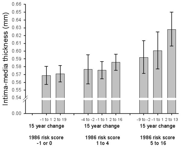
Both the 1986 score and the change in risk score between 1986 and 2001 were significant predictors of carotid IMT (Table 4). The multiplicative effect of 1 year of age was 1.008 (95% CI 1.003-1.010) and the multiplicative effect of female sex was 0.985 (95% CI 0.969-1.003). Figure 1 shows the geometric mean IMT by 1986 risk score and risk score change between 1986 and 2001, adjusted for sex and age.
Table 4.
Multiplicative effecta of a 1 point greater Pathobiological Determinants of Atherosclerosis in Youth (PDAY) risk score due to the modifiable risk factors measured in 1986 on carotid artery intima-media thickness (IMT) and a 1 point increase in risk score due to the modifiable risk factors between 1986 and 2001; and odds ratios for same points for predicting carotid artery intima-media thickness >90th percentile or a plaque. The model includes sex and age in 2001.
| Risk score due to modifiable risk factors | Carotid artery IMT | IMT >90th percentile or a plaque | ||
|---|---|---|---|---|
| Multiplicative effecta (95% CI) | R2 | Odds ratio (95% CI) | c-statisticb | |
| One point increase in 1986 PDAY risk score | 1.008 (1.005-1.012) | 0.097 | 1.118 (1.048-1.194) | 0.674 |
| One point increasec in risk score between 1986 and 2001 | 1.003 (1.001-1.006) | 1.004 (0.994-1.097) | ||
The value of Y (that is, carotid artery IMT) for predictor variable with value X+1, labeled YX+1, is equal to the multiplicative effect eβ (the anti-log of the regression coefficient) multiplied by the Y value at X, that is, YX+1= eβYX.
The c-index is the proportion of all pairs of subjects, one with and one without lesions (>90th percentile or plaque), in which the subject with the lesions has the higher risk score.21
The reciprocal of the multiplicative effect or odds ratio describes the effect of a decrease in the risk score by 1 point between 1986 and 2001
Figure 1.
Geometric mean carotid artery intima-media thickness by 1986 Pathobiological Determinants of Atherosclerosis in Youth (PDAY) risk score and risk score change between 1986 and 2001, adjusted for sex and age. Error bars represent 95% confidence intervals. There was a statistically significant effect of 1986 risk score and a significant effect of risk score change.
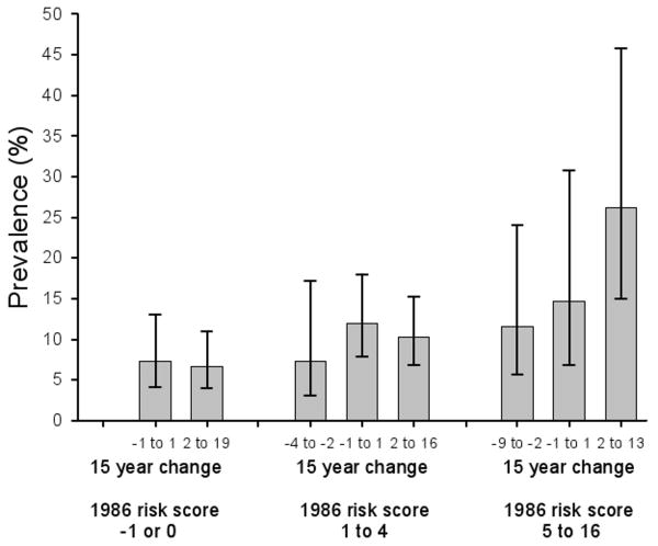
Table 4 also gives odds ratios for predicting IMT> 90th percentile or a plaque or both. The 1986 risk score was a significant predictor but the change in risk score was not a significant predictor. The odds ratio for 1 year of age was 1.106 (95% CI 1.056-1.158). Figure 2 shows the prevalence of IMT> 90th percentile of IMT or a plaque or both by the 1986 risk score and risk score change between 1986 and 2001, adjusted for sex and age.
Figure 2.
Prevalence of carotid artery intima-media thickness >90th percentile of intima-media thickness or a plaque or both by the1986 Pathobiological Determinants of Atherosclerosis in Youth (PDAY) risk score and risk score change between 1986 and 2001, adjusted for sex and age. Error bars represent 95% confidence intervals. There was a statistically significant effect of 1986 risk score.
Carotid IMT and early measurement of risk score
We also examined the 1986 and 2001 risk scores separately as predictors of IMT. The multiplicative effect of the risk score in 1986 [1.007 (95% CI 1.004-1.010)1] was higher, but not significantly higher (P=0.095), than the effect of the 2001 risk score [1.005 (95% CI 1.003-1.007)] although both were significant predictors of IMT. Similarly, the odds ratio for predicting IMT >90th percentile or a plaque or both for the risk score measured in 1986 [1.100 (95% CI 1.033-1.170)1] was higher, but not significantly higher (P=0.290), than the odds ratio for the 2001 risk score [1.066 (95% CI 1.020-1.113)].
Discussion
The PDAY coronary artery risk score, developed to predict coronary artery lesions from CHD risk factors in autopsied individuals, also predicted carotid artery IMT in living young adults. Both the risk score computed from the modifiable risk factors measured 15 years prior to IMT measurement and the change in risk score during the following 15 years were significant predictors of IMT.
Thickness of carotid artery intima and media combined is associated with coronary heart disease risk factors23 and predicts cardiovascular events.24 The intima is more directly involved in atherosclerosis than the media, and the thickness of the intima alone measured by higher resolution ultrasound shows a stronger association with other manifestations of atherosclerosis.25
Linking the results of the present study with previous evidence concerning the significance of carotid IMT suggests that CHD risk is established early in life. The PDAY risk score measured at younger ages was a better predictor of carotid artery IMT (although not significantly better) than the risk score measured concurrently with the ultrasound measurement of carotid lesions. This finding is consistent with the results from analyses of the individual risk factors in the Young Finns cohort4 and in the Bogalusa Heart Study.3 This finding also is consistent with the results from applying the PDAY risk score to the prediction of coronary artery calcification in CARDIA subjects.7
The PDAY risk score provides a simple way to calculate a weighted summary of the effects of the major established risk factors on both carotid and coronary artery atherosclerosis. The PDAY risk score computed from only the modifiable risk factors can be used to identify young people at high relative risk. Controlling their risk factors should prevent their becoming subject to high absolute risk at older ages.
Another potential use of the risk score is to select those young persons who should be screened for markers of precocious atherosclerosis by a noninvasive method. The noninvasive assessment could be used to separate those subjects with risk factors into those without and those with advanced atherosclerosis. Selection of individuals to be screened must consider absolute risk, that is, risk that includes the contributions of sex and age.
Acknowledgments
The Cardiovascular Risk in Young Finns study was supported by the Academy of Finland (grants 117941, 77841, 210283, 53392, 34316), the Social Insurance Institution of Finland, the Turku University Foundation, Federal Grants for the Turku University Central Hospital, Tampere University Hospital Medical Fund, the Juho Vainio Foundation, the Finnish Foundation of Cardiovascular Research, and the Finnish Cultural Foundation. Dr. Jokinen was supported in part by the Finnish Cardiac Foundation. Dr. Gidding was supported in part by grant 1 P20 RR020173-01 from the National Center for Research Resources.
Footnotes
The multiplicative effect or odds ratio of the 1986 risk score is different from that reported in Table 4 because in Table 4 an additional variable, the difference in 2001 and 1986 risk score, was included in the statistical model. Estimates of regression coefficients and odds ratios usually depend on which variables are included in the model.20,21
Publisher's Disclaimer: This is a PDF file of an unedited manuscript that has been accepted for publication. As a service to our customers we are providing this early version of the manuscript. The manuscript will undergo copyediting, typesetting, and review of the resulting proof before it is published in its final citable form. Please note that during the production process errors may be discovered which could affect the content, and all legal disclaimers that apply to the journal pertain.
References
- 1.Davis PH, Dawson JD, Mahoney LT, Lauer RM. Increased carotid intimal-medial thickness and coronary calcification are related in young and middle-aged adults: The Muscatine Study. Circulation. 1999;100:838–842. doi: 10.1161/01.cir.100.8.838. [DOI] [PubMed] [Google Scholar]
- 2.Davis PH, Dawson JD, Riley WA, Lauer RM. Carotid intimal-medial thickness is related to cardiovascular risk factors measured from childhood through middle age: The Muscatine Study. Circulation. 2001;104:2815–9. doi: 10.1161/hc4601.099486. [DOI] [PubMed] [Google Scholar]
- 3.Li S, Chen W, Srinivasan SR, Bond MG, Tang R, Urbina EM, Berenson GS. Childhood cardiovascular risk factors and carotid vascular changes in adulthood: the Bogalusa Heart Study. JAMA. 2003;290:2271–6. doi: 10.1001/jama.290.17.2271. [DOI] [PubMed] [Google Scholar]
- 4.Raitakari O, Juonala M, Kahonen M, Taittonen L, Laitinen T, Maki-Torkko N, Jarvisalo NJ, Uhari M, Jokinen E, Ronnemaa T, Akerblom HK, Viikari JSA. Cardiovascular risk factors in childhood as predictors of carotid artery intima-media thickness in adulthood. The Cardiovascular Risk in Young Finns Study. JAMA. 2003;290:2277–2283. doi: 10.1001/jama.290.17.2277. [DOI] [PubMed] [Google Scholar]
- 5.Juonala M, Jarvisalo MJ, Maki-Torkko N, Kahonen M, Viikari JS, Raitakari OT. Risk factors identified in childhood and decreased carotid artery elasticity in adulthood: the Cardiovascular Risk in Young Finns Study. Circulation. 2005;112:1486–93. doi: 10.1161/CIRCULATIONAHA.104.502161. [DOI] [PubMed] [Google Scholar]
- 6.McMahan CA, Gidding SS, Fayad ZA, Zieske AW, Malcom GT, Tracy RE, Strong JP, McGill HC, Jr, Pathobiological Determinants of Atherosclerosis in Youth Research Group Risk scores predict atherosclerotic lesions in young people. Arch Intern Med. 2005;165:883–90. doi: 10.1001/archinte.165.8.883. [DOI] [PubMed] [Google Scholar]
- 7.Gidding SS, McMahan CA, McGill HC, Colangelo LA, Schreiner PJ, Williams OD, Liu K. Prediction of coronary artery calcium in young adults using the Pathobiological Determinants of Atherosclerosis in Youth (PDAY) risk score: The CARDIA Study. Arch Intern Med. 2006;166:2341–2347. doi: 10.1001/archinte.166.21.2341. [DOI] [PubMed] [Google Scholar]
- 8.Akerblom HK, Viikari J, Uhari M, Rasanen L, Byckling T, Louhivuori K, Pesonen E, Suoninen P, Pietikainen M, Lahde PL. Atherosclerosis precursors in Finnish children and adolescents. I. General description of the cross-sectional study of 1980, and an account of the children's and families' state of health. Acta Paediatrica Scandinavica. 1985;318:49–63. doi: 10.1111/j.1651-2227.1985.tb10082.x. [DOI] [PubMed] [Google Scholar]
- 9.Juonala M, Viikari JS, Hutri-Kahonen N, Pietikainen M, Jokinen E, Taittonen L, Marniemi J, Ronnemaa T, Raitakari OT. The 21-year follow-up of the Cardiovascular Risk in Young Finns Study: risk factor levels, secular trends and east-west difference. Journal Internal Medicine. 2004;255:457–68. doi: 10.1111/j.1365-2796.2004.01308.x. [DOI] [PubMed] [Google Scholar]
- 10.National Cholesterol Education Program. Third Report of the National Cholesterol Education Program (NCEP) Expert Panel on Detection, Evaluation, and Treatment of High Blood Cholesterol in Adults (Adult Treatment Panel III) final report. Circulation. 2002;106:3143–421. [PubMed] [Google Scholar]
- 11.Chobanian AV, Bakris GL, Black HR, Cushman WC, Green LA, Izzo JL, Jr, Jones DW, Materson BJ, Oparil S, Wright JT, Jr, Roccella EJ. The Seventh Report of the Joint National Committee on Prevention, Detection, Evaluation, and Treatment of High Blood Pressure: the JNC 7 report. JAMA. 2003;289:2560–72. doi: 10.1001/jama.289.19.2560. [DOI] [PubMed] [Google Scholar]
- 12.Toikka JO, Laine H, Ahotupa M, Haapanen A, Viikari JS, Hartiala JJ, Raitakari OT. Increased arterial intima-media thickness and in vivo LDL oxidation in young men with borderline hypertension. Hypertension. 2000;36:929–33. doi: 10.1161/01.hyp.36.6.929. [DOI] [PubMed] [Google Scholar]
- 13.Tonstad S, Joakimsen O, Stensland-Bugge E, Leren TP, Ose L, Russell D, Bonaa KH. Risk factors related to carotid intima-media thickness and plaque in children with familial hypercholesterolemia and control subjects. Arterioscler Thromb Vasc Biol. 1996;16:984–991. doi: 10.1161/01.atv.16.8.984. [DOI] [PubMed] [Google Scholar]
- 14.Stary HC, Chandler AB, Dinsmore RE, Fuster V, Glagov S, Insull W, Jr, Rosenfeld ME, Schwartz CJ, Wagner WD, Wissler RW. A definition of advanced types of atherosclerotic lesions and a histological classification of atherosclerosis: a report from the Committee on Vascular Lesions of the Council on Arteriosclerosis, American Heart Association. Arterioscler Thromb Vasc Biol. 1995;15:1512–1531. doi: 10.1161/01.atv.15.9.1512. [DOI] [PubMed] [Google Scholar]
- 15.McGill HC, Jr, McMahan CA, Zieske AW, Tracy RE, Malcom GT, Herderick EE, Strong JP, Pathobiological Determinants of Atherosclerosis in Youth (PDAY) Research Group Association of coronary heart disease risk factors with microscopic qualities of coronary atherosclerosis in youth. Circulation. 2000;102:374–379. doi: 10.1161/01.cir.102.4.374. [DOI] [PubMed] [Google Scholar]
- 16.McGill HC, Jr, McMahan CA, Malcom GT, Oalmann MC, Strong JP, PDAY Research Group Effects of serum lipoproteins and smoking on atherosclerosis in young men and women. Arterioscler Thromb Vasc Biol. 1997;17:95–106. doi: 10.1161/01.atv.17.1.95. [DOI] [PubMed] [Google Scholar]
- 17.McGill HC, Jr, McMahan CA, Herderick EE, Zieske AW, Malcom GT, Tracy RE, Strong JP, Pathobiological Determinants of Atherosclerosis in Youth (PDAY) Research Group Obesity accelerates the progression of coronary atherosclerosis in young men. Circulation. 2002;105:2712–2718. doi: 10.1161/01.cir.0000018121.67607.ce. [DOI] [PubMed] [Google Scholar]
- 18.McGill HC, Jr, McMahan CA, Malcom GT, Oalmann MC, Strong JP, Pathobiological Determinants of Atherosclerosis in Youth (PDAY) Research Group Relation of glycohemoglobin and adiposity to atherosclerosis in youth. Arterioscler Thromb Vasc Biol. 1995;15:431–440. doi: 10.1161/01.atv.15.4.431. [DOI] [PubMed] [Google Scholar]
- 19.McGill HC, Jr, McMahan CA, Tracy RE, Oalmann MC, Cornhill JF, Herderick EE, Strong JP, Pathobiological Determinants of Atherosclerosis in Youth (PDAY) Research Group Relation of a postmortem renal index of hypertension to atherosclerosis and coronary artery size in young men and women. Arterioscler Thromb Vasc Biol. 1998;18:1108–1118. doi: 10.1161/01.atv.18.7.1108. [DOI] [PubMed] [Google Scholar]
- 20.Draper NR, Smith H. Applied Regression Analysis. New York: John Wiley & Sons; 1998. [Google Scholar]
- 21.Hosmer DW, Lemeshow S. Applied Logistic Regression. 2nd. New York: Wiley; 2000. p. 373. [Google Scholar]
- 22.Efron B, Tibshirani R. An Introduction to the Bootstrap. Boca Raton, FL: Chapman & Hall/CRC; 1993. [Google Scholar]
- 23.Poli A, Tremoli E, Colombo A, Sirtori M, Pignoli P, Paoletti R. Ultrasonographic measurement of the common carotid artery wall thickness in hypercholesterolemic patients. A new model for the quantitation and follow-up of preclinical atherosclerosis in living human subjects. Atherosclerosis. 1988;70:253–61. doi: 10.1016/0021-9150(88)90176-1. [DOI] [PubMed] [Google Scholar]
- 24.Hodis HN, Mack WJ, LaBree L, Selzer RH, Liu CR, Liu CH, Azen SP. The role of carotid arterial intima-media thickness in predicting clinical coronary events. Ann Intern Med. 1998;128:262–9. doi: 10.7326/0003-4819-128-4-199802150-00002. [DOI] [PubMed] [Google Scholar]
- 25.Osika W, Dangardt F, Gronros J, Lundstam U, Myredal A, Johansson M, Volkmann R, Gustavsson T, Gan LM, Friberg P. Increasing peripheral artery intima thickness from childhood to seniority. Arterioscler Thromb Vasc Biol. 2007;27:671–6. doi: 10.1161/01.ATV.0000256468.95403.6f. [DOI] [PubMed] [Google Scholar]


