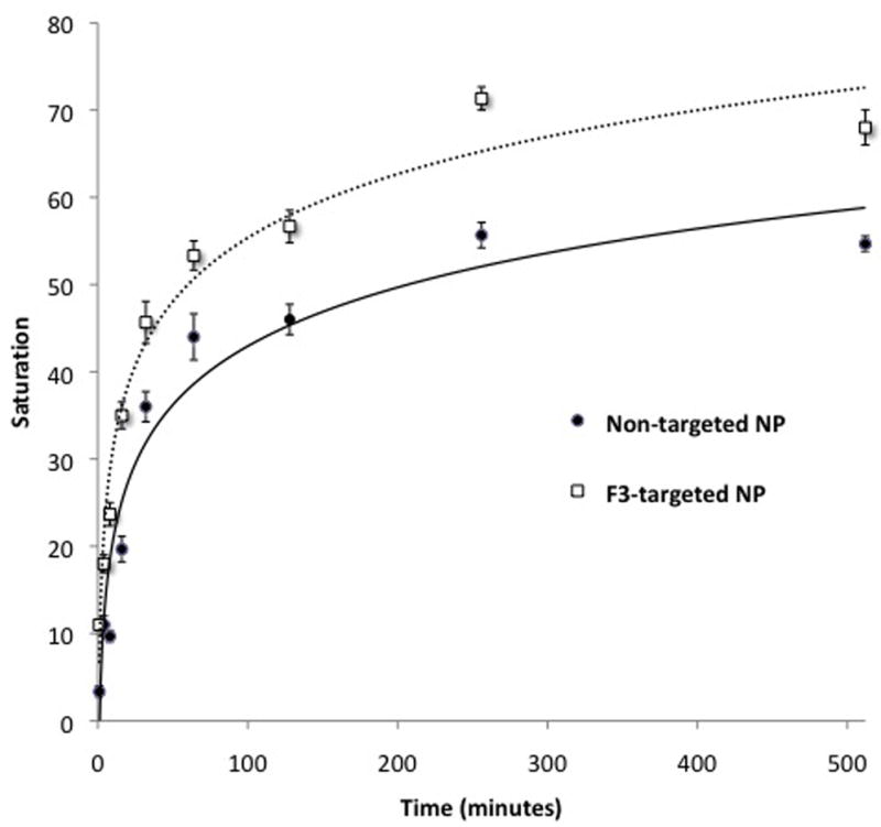
Figure 4.

Effect of treatment duration on gliosarcoma cell tagging by CB-loaded NPs. 9L cells were incubated for varying duration with non-targeted or F3-targeted CB-loaded NPs at time points shown. The extent of tagging is significantly greater for F3-targeted compared to non-targeted NP at all time points (P < 0.04). Lines represent best-fit logarithmic curves for non-targeted (R2=0.94) and F3-targeted (R2=0.97) NP treatments. Error bars indicate standard error of mean.
