Abstract
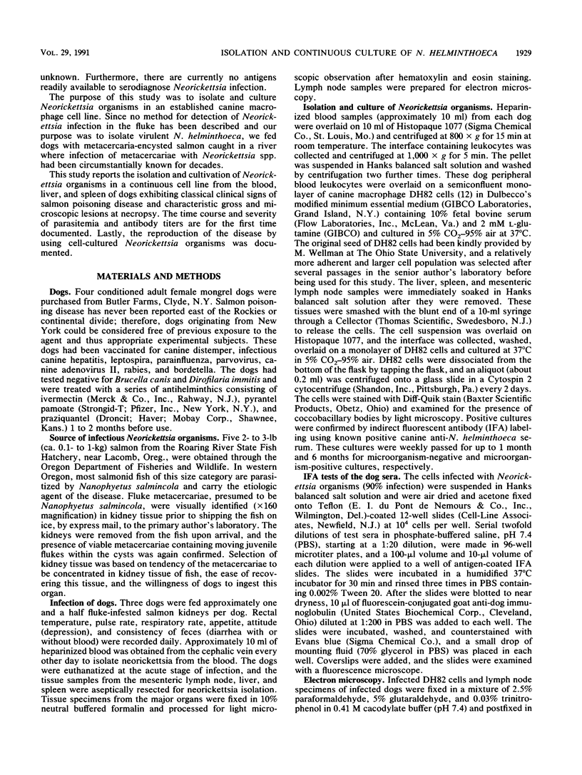
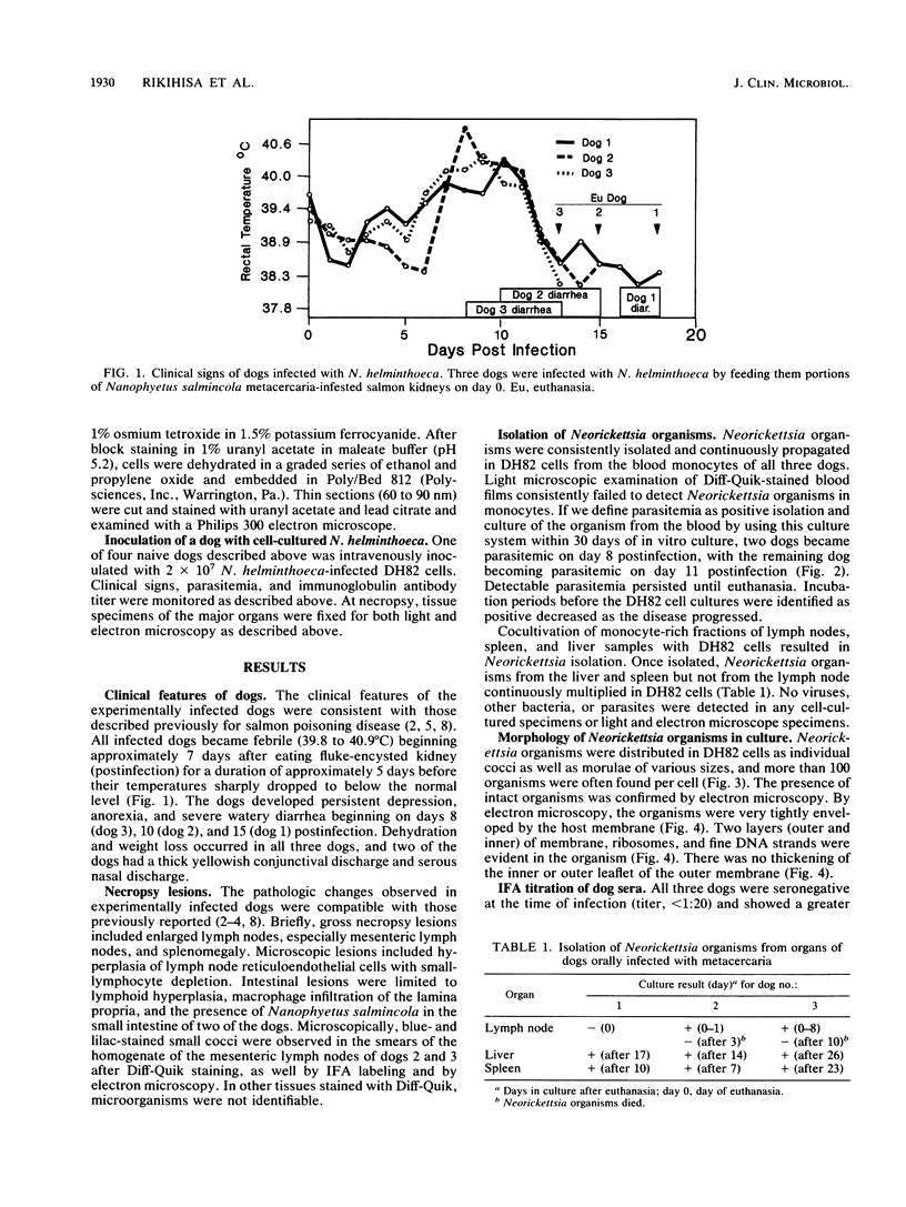
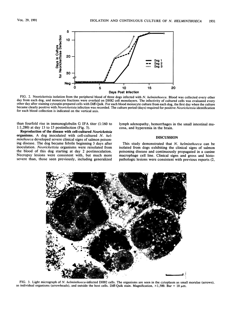
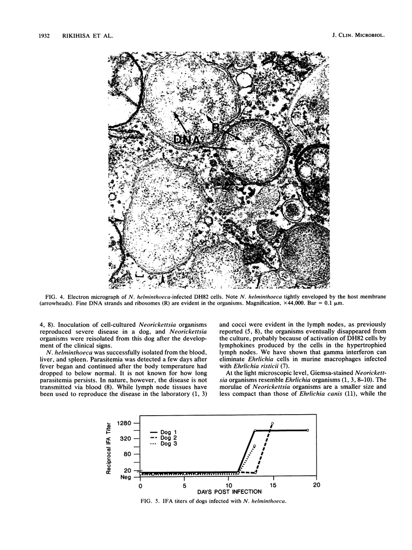
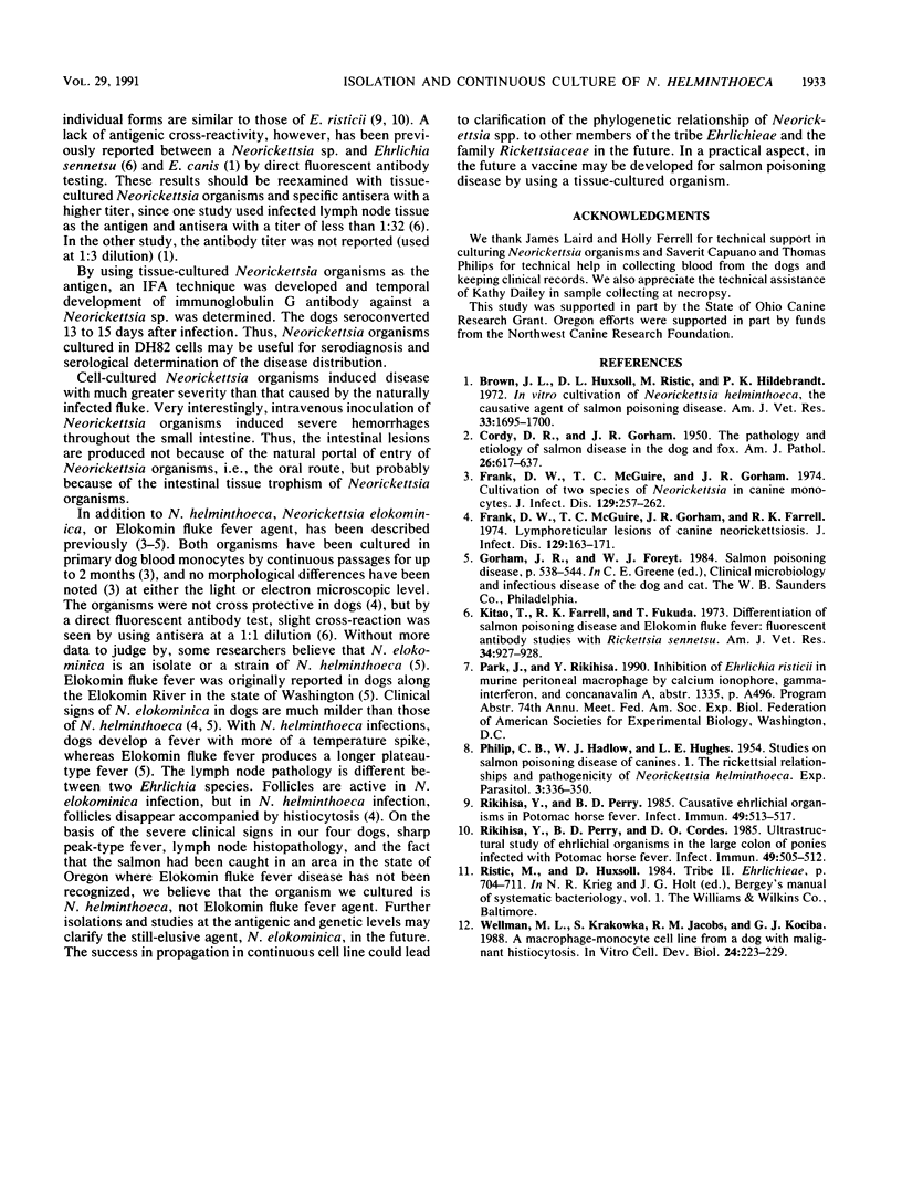
Experimental evidence is presented supporting the development of a system for the isolation and propagation of a Neorickettsia sp. in a continuous canine macrophage cell line (DH82). To isolate a Neorickettsia sp. pathogenic to the canine species, three naive dogs were fed metacercaria-encysted kidneys of salmon caught in a river where infection of metacercariae with Neorickettsia helminthoeca has been circumstantially known for decades. Clinically, the classic course of salmon poisoning disease developed in all of the dogs. Parasitemia began on day 8 to 11 postinfection, when the dogs developed a febrile peak, and continued until euthanasia. At necropsy, characteristic gross and microscopic lesions of the disease were present. A Neorickettsia sp. was also isolated from liver and spleen samples of these animals. The isolates have been continuously propagated and passed in DH82 cells for more than 6 months. Electron microscopic examination confirmed that the rickettsial organisms multiplied in the membrane-bound compartment of DH82 cells and that they morphologically closely resembled rickettsia belonging to the genus Ehrlichia. An indirect fluorescent antibody test using Neorickettsia organisms cultured in DH82 cells showed that all dogs seroconverted 13 to 15 days postinfection. Finally, inoculation of the cell-cultured Neorickettsia organisms into a naive dog reproduced clinically typical salmon poisoning disease which was of greater severity and had a more rapid time course than that in the dogs from which the original isolation was made. On the basis of the clinical and pathologic responses of the dogs in our study, we believe that virulent N. helminthoeca was isolated and cultured in a continuous cell line.
Full text
PDF
Images in this article
Selected References
These references are in PubMed. This may not be the complete list of references from this article.
- Brown J. L., Huxsoll D. L., Ristic M., Hildebrandt P. K. In vitro cultivation of Neorickettsia helminthoeca, the causative agent of salmon poisoning disease. Am J Vet Res. 1972 Aug;33(8):1695–1700. [PubMed] [Google Scholar]
- CORDY D. R., GORHAM J. R. The pathology and etiology of salmon disease. Am J Pathol. 1950 Jul;26(4):617–637. [PMC free article] [PubMed] [Google Scholar]
- Frank D. W., McGuire T. C., Gorham J. R., Davis W. C. Cultivation of two species of Neorickettsia in canine monocytes. J Infect Dis. 1974 Mar;129(3):257–262. doi: 10.1093/infdis/129.3.257. [DOI] [PubMed] [Google Scholar]
- Frank D. W., McGuire T. C., Gorham J. R., Farrell R. K. Lymphoreticular lesions of canine neorickettsiosis. J Infect Dis. 1974 Feb;129(2):163–171. doi: 10.1093/infdis/129.2.163. [DOI] [PubMed] [Google Scholar]
- Kitao T., Farrell R. K., Fukuda T. Differentiation of salmon poisoning disease and Elokomin fluke fever: fluorescent antibody studies with Rickettsia sennetsu? Am J Vet Res. 1973 Jul;34(7):927–928. [PubMed] [Google Scholar]
- PHILIP C. B., HADLOW W. J., HUGHES L. E. Studies on salmon poisoning disease of canines. I. The rickettsial relationships and pathogenicity of Neorickettsia helmintheca. Exp Parasitol. 1954 Jul;3(4):336–350. doi: 10.1016/0014-4894(54)90032-6. [DOI] [PubMed] [Google Scholar]
- Rikihisa Y., Perry B. D. Causative ehrlichial organisms in Potomac horse fever. Infect Immun. 1985 Sep;49(3):513–517. doi: 10.1128/iai.49.3.513-517.1985. [DOI] [PMC free article] [PubMed] [Google Scholar]
- Rikihisa Y., Perry B. D., Cordes D. O. Ultrastructural study of ehrlichial organisms in the large colons of ponies infected with Potomac horse fever. Infect Immun. 1985 Sep;49(3):505–512. doi: 10.1128/iai.49.3.505-512.1985. [DOI] [PMC free article] [PubMed] [Google Scholar]
- Wellman M. L., Krakowka S., Jacobs R. M., Kociba G. J. A macrophage-monocyte cell line from a dog with malignant histiocytosis. In Vitro Cell Dev Biol. 1988 Mar;24(3):223–229. doi: 10.1007/BF02623551. [DOI] [PubMed] [Google Scholar]


