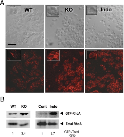
Figure 2.
Loss of PG signaling enhances RhoA activation and actin polymerization. A, F-actin staining of the oviductal cumuli of WT, Ptger2−/− (KO) and indomethacin-pretreated WT mice (Indo). The ampulla of oviducts containing COCs was excised at 14 h, and the frozen tissue was transversely sectioned. F-actin staining with Texas Red-X phalloidin (bottom panels) and bright-field images of the same section (top panels) are shown. Inset, Magnified view of typical cell morphology and F-actin staining of the same field. Note that cumulus cell rounding in KO or Indo was associated with prominent ring-like F-actin signals. Scale bar, 20 μm. B, Increased RhoA activation in PG signal-depleted COCs. COCs isolated from WT and Ptger2−/− (KO) mice at 14 h were subjected to the RhoA pull-down assay (16 mice for each). Similar analysis was also performed between COCs from vehicle- (Cont) and indomethacin-treated mice (Indo). The total RhoA level in each lysate used for the pull-down assay is shown as an input. Note that the levels of total RhoA were relatively reduced in the KO and Indo COCs due to reduced ovulation, but the levels of GTP-RhoA was nevertheless increased in these COCs. The ratio of GTP-RhoA to total RhoA for each sample is also shown. Representative results of two independent experiments are shown.

