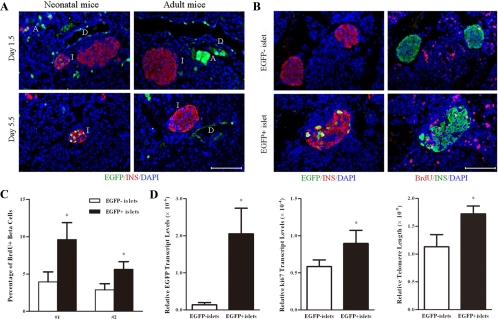
Figure 5.
Identification of newly generated islets by retrograde pancreatic duct labeling. Neonatal and adult mice were injected with adenoviruses carrying an EGFP expression cassette. BrdU was injected at 36, 24, and 12 h before the mice were killed. Panel A, Pancreases were harvested at 1.5 (upper panel) or 5.5 d (lower panel) after the operation. Paraffin sections of the pancreases were immunostained for insulin (red), EGFP (green), and DAPI (blue). D, Duct; I, islet; A, acinus. (n = 6). Scale bar, 100 μm. Panel B, Consecutive paraffin sections of the pancreases were stained for insulin/EGFP/DAPI (left panel) or insulin/BrdU/DAPI (right panel). An EGFP-labeled islet was shown on the lower panel, whereas two EGFP unlabeled islets were shown on the upper panel. Scale bar, 100 μm. Panel C, The percentage of BrdU incorporation was determined in EGFP+ and EGFP− islets isolated from two mice (no. 1, no. 2). Panel D, EGFP+ and EGFP− islets were hand picked under a fluorescence microscope. The ratios of EGFP, Ki67 expression levels, and telomere length between EGFP+ and EGFP− islets were determined by real-time RT-PCR or PCR. *, P < 0.05 (n = 3).

