Abstract
Implantation requires communication between a receptive endometrium and a healthy blastocyst. This maternal-embryonic crosstalk involves local mediators within the uterine microenvironment. We demonstrate that a secreted protein, prokineticin 1 (PROK1), is expressed in the receptive endometrium and during early pregnancy. PROK1 induces expression of leukemia inhibitory factor (LIF) in endometrial epithelial cells and first trimester decidua via a Gq-Ca2+-cSrc-mitogen-activated protein kinase kinase-mediated pathway. We show that human embryonic chorionic gonadotropin (hCG) induces sequential mRNA expression of PROK1 and LIF in an in vivo baboon model, in human endometrial epithelial cells, and in first-trimester decidua. We have used micro RNA constructs targeted to PROK1 to demonstrate that hCG-mediated LIF expression in the endometrium is dependent on prior induction of PROK1. Dual immunohistochemical analysis colocalized expression of the luteinizing hormone/chorionic gonadotropin receptor, PROK1, PROKR1, and LIF to the glandular epithelial cells of the first trimester decidual tissue. PROK1 enhances adhesion of trophoblast cells to fibronectin and laminin matrices, which are mediated predominantly via LIF induction. These data describe a novel signaling pathway mediating maternal-embryonic crosstalk, in which embryonic hCG via endometrial PROK1 may play a pivotal role in enhancing receptivity and maintaining early pregnancy.—Evans, J., Catalano, R. D., Brown, P., Sherwin, R., Critchley, H. O. D., Fazleabas, A. T., Jabbour, H. N. Prokineticin 1 mediates fetal-maternal dialogue regulating endometrial leukemia inhibitory factor.
Keywords: human chorionic gonadotropin, implantation, decidua, adhesion, laminin, fibronectin
Differentiation of the endometrium is essential for successful embryo implantation. The secretory phase encompasses a finite period of time or “window” during which the endometrium is optimally receptive for implantation of a blastocyst. After this period, the endometrium becomes refractory to conceptus implantation. The implantation window in humans is predicted to last for ∼4 d and commences ∼6 d after the luteinizing hormone (LH) peak in a normal menstrual cycle (1, 2). The endometrial expression of a number of cytokines, chemokines, growth factors, and adhesive molecules is associated with the onset of receptivity. In the event of pregnancy, embryonic human chorionic gonadotropin (hCG) maintains corpus luteum-mediated progesterone production, which is essential for the maintenance of early pregnancy. Production of hCG by the blastocyst may also directly regulate expression of endometrial factors that may prolong the period during which the endometrium is receptive (3,4,5).
Recently, the expression of a pleiotropic protein, prokineticin 1 (PROK1), and its receptor, prokineticin receptor 1 (PROKR1), has been characterized in the nonpregnant and early pregnant human endometrium (6, 7). Endometrial PROK1 expression peaks during the secretory phase of the human menstrual cycle (6, 7) and is further elevated during the first trimester of pregnancy (7). PROK1 and PROKR1 are also expressed in human early pregnancy tissues, with elevation in expression between wk 8–9 of pregnancy and localization to the syncytiotrophoblast and cytotrophoblast layers (8). In addition, PROK1 has been demonstrated by gene array analysis to regulate endometrial expression of a host of genes known to be important for implantation, including cyclooxygenase-2, heparin-binding epidermal growth factor (EGF), Dickkopf-1, interleukin (IL)-11, and leukemia inhibitory factor (LIF) (7). LIF is a cytokine that is critical for murine implantation (9). LIF expression in mice is initiated on d 4 of pregnancy before implantation, after the nidatory estrogen peak (10) and in humans is elevated during the receptive phase of the menstrual cycle (11, 12). However, the exact mechanisms regulating LIF expression during this phase of the menstrual cycle in humans are not understood. In models of early pregnancy, it has been established that hCG mediates endometrial LIF expression in humans and baboons (13, 14).
In this study we investigated the role of PROK1 in the hCG-mediated regulation of LIF during embryonic to maternal crosstalk of early pregnancy. We sought to identify the intracellular signaling pathways leading to enhancement of LIF expression and evidence for a functional role of endometrial LIF in early pregnancy events.
MATERIALS AND METHODS
Reagents
DMEM-F12 GlutaMAX culture medium and RPMI 1640 culture medium were purchased from Invitrogen (Paisley, UK). LIF and chorionic gonadotropin (CG) receptor antibodies were purchased from Santa Cruz Biotechnology/Autogen Bioclear (Calne, UK). PROK1 antibodies were purchased from Phoenix Pharmaceuticals (Belmont, CA, USA). PROKR1 antibody was purchased from Life Span Biosciences (Atlanta, GA, USA). SuperFect transfection reagent was purchased from Qiagen (Crawley, UK). Intracellular calcium chelator [1,2-bis(2-aminophenoxy)ethane-N,N,N′,N′-tetraacetic acid-acetoxymethyl ester (BAPTA-AM)] used at a final concentration of 50 μM, protein kinase C (PKC) inhibitor (Ro-318220) used at a final concentration of 1 μM, cSrc inhibitor [4-amino-5-(4-chlorophenyl)-7-(t-butyl)pyrazolo[3,4-d]pyrimidine (PP2)] used at a final concentration of 10 μM, EGF receptor (EGFR) inhibitor (AG1478) used at a final concentration of 200 nM, MEK inhibitor (PD98059) used at a final concentration of 50 μM, and protein synthesis inhibitor (cycloheximide) used at a final concentration of 10 μg/ml were purchased from Calbiochem (Nottingham, UK). Gq inhibitor (YM254890) used at a final concentration of 1 μM was kindly supplied by Dr. Jun Takasaki (Molecular Medicine Laboratories, Yamanouchi Pharmaceutical Co. Ltd., Tokyo, Japan). LIF ELISAs were purchased from R&D Systems (Minneapolis, MN, USA). Recombinant LIF used at a final concentration of 100 ng/ml and adhesion assays were purchased from Chemicon (Chandlers Ford, UK). hCG was purchased from Sigma (Dorset, UK). Recombinant hCG was obtained from Dr. A. F. Parlow (National Hormone and Pituitary Program, Harbor-UCLA Medical Center, Torrance, CA, USA). Recombinant PROK1, used at a final concentration of 40 nM, was purchased from Promokine (Heidelberg, Germany).
Patients and tissue collection
Endometrial biopsies were obtained from women with regular menstrual cycles who had not received hormonal preparations in the 3 mo preceding biopsy collection and were dated according to Noyes criteria by a pathologist. Circulating estradiol and progesterone concentrations were measured and were consistent with the histological assessment. First trimester decidua (7–12 wk of gestation) was collected from women undergoing elective first-trimester surgical termination of pregnancy with gestation confirmed by ultrasound scan. Ethical approval was obtained from Lothian research ethics committee, and written informed patient consent was obtained before tissue collection.
Baboon tissue collection
Baboon tissues were treated as described previously and subjected to microarray analysis (14). Briefly, uterine tissue was obtained from 6 control and 5 hCG-treated adult female baboons (Papio annubis). Tissue was collected by endometrectomy or hysterectomy on d 10 postovulation (PO). Ovulation was detected in cycling female baboons by measuring peripheral serum levels of estradiol beginning on d 7 after the first day of menses. The day of estradiol surge was designated as d −1, with d 0 as the day of ovulatory LH surge and d 1 as the day of ovulation. On d 6 PO, an oviductal cannula was attached to an Alzet osmotic minipump, and recombinant hCG was infused at the rate of 1.25 IU/h for 5 d. The infusion was terminated on d 10 PO (d 10 corresponds to the expected day of implantation in pregnant baboons). All experimental procedures were approved by the animal care committee of the University of Illinois (Chicago, IL, USA).
Cell/tissue culture and treatment
Ishikawa endometrial epithelial cells and JEG-3 epithelial choriocarcinoma cells obtained from the European Collection of Cell Culture (Porton Down, UK) were routinely maintained in DMEM-F12 GlutaMAX culture medium with 10% FBS, 100 IU of penicillin, and 100 μg of streptomycin at 37°C and 5% CO2. PROKR1 stably transfected Ishikawa cells were produced and maintained as described previously (7). First trimester decidual tissue was chopped finely and maintained in DMEM. Tissue was divided into equal portions for experimental procedures. Transient transfections of Ishikawa cells were performed using PROK1 targeting microRNA (miRNA) constructs. Tissue was infected with lentivirus expressing PROK1 miRNA constructs for 72 h. Oligonucleotides encoding human PROK1 miRNA constructs were obtained from Invitrogen and inserted into the pcDNA6.2-GW/EmGFP-miR vector and used for transient transfections. These were recombined to create plenti6/V5-EmGFP-miR-neg control and pLenti6/V5-EmGFP-hum-PROK1-72 and -287. Lentivirus was produced with a Block-iT lentiviral Pol II miR RNAi expression system (Invitrogen) using the manufacturer’s protocols and used to infect tissue for 72 h. Cells and tissue were incubated in serum-free medium overnight before treatment with PROK1 or hCG alone or pretreated for 1 h with inhibitors at the concentrations indicated above. Cells, tissue, and medium were harvested, and RNA or protein was extracted for PCR or ELISA analysis.
Immunohistochemistry and immunofluorescent microscopy
Colocalization of PROK1 with CG receptor or PROKR1 with LIF was performed by dual immunofluorescence histochemistry. Five-micrometer paraffin-embedded sections were dewaxed and rehydrated in graded ethanol. Antigen retrieval was performed by boiling in 0.01 M citrate buffer for 5 min. Endogenous peroxidase activity was quenched by incubation in 3% H2O2/MeOH solution. Sections were blocked using 5% normal horse serum. Sections were incubated with goat anti-CG receptor (K-15, sc-26341) (1:10) or goat anti-LIF (1:10) overnight at 4°C. Sections were then incubated with horse anti-goat biotinylated antibodies, followed by incubation with fluorochrome streptavidin 546 (1:200 in PBS). Sections were reblocked with 5% normal goat serum (PROKR1/LIF localizations) or 5% normal swine serum (PROK1/CG receptor localizations) and incubated with rabbit anti-human PROK1 (1:1500) or rabbit anti-human PROKR1 (1:500) overnight at 4°C. Sections were incubated with peroxidase-labeled goat or swine anti-rabbit antibodies (1:200 in PBS) followed by fluorochrome trichostatin A plus fluorescein (1:50 in substrate). Sections were washed and incubated with nuclear counterstain ToPro (1:2000 in PBS), mounted in PermaFluor, and photographed using a laser scanning microscope (LSM 510; Carl Zeiss, Jena, Germany).
TaqMan quantitative RT-PCR
RNA was extracted with TRI reagent (Thermo Scientific, Cramlington, UK) following the manufacturer’s guidelines using phase lock tubes (Eppendorf, Cambridge, UK). RNA samples were reverse transcribed as described previously (6). PCR reactions were carried out using an ABI Prism 7900 system (Applied Biosystems, Foster City, CA, USA). Primer and FAM (6-carboxyfluorescein)-labeled probe sequences are listed in Table 1. Gene expression was normalized to 18S ribosomal RNA (Applied Biosystems) as an internal standard. Results are expressed as relative to a standard (endometrial tissue cDNA) included in all reactions.
TABLE 1.
TaqMan primer and probe sequences for PROK1, LIF, LH/CG receptor, and 18S ribosomal RNA
| Gene | Primers and probe (5′-3′) |
|---|---|
| PROK1 forward | GTGCCACCCGGGCAG |
| PROK1 reverse | AGCAAGGACAGGTGTGGTGC |
| PROK1 probe (FAM) | ACAAGGTCCCCTTGTTCAGGAAACGCA |
| LIF forward | TGGTGGAGCTGTACCGCATA |
| LIF reverse | TGGTCCCGGGTGATGTTG |
| LIF probe (FAM) | TCGTGTACCTTGGCACCTCCCTGG |
| CG receptor forward | TGGCTGGGACTATGAATATGGTT |
| CG receptor reverse | AGGGATTAAAAGCATCTGGTTCAG |
| CG receptor probe (FAM) | CTGCTTACCCAAGACACCCCGATGTG |
| 18S forward | CGGCTACCACATCCAAGGAA |
| 18S reverse | GCTGGAATTACCGCGGCT |
| 18S probe (VIC) | TGCTGGCACCAGACTTGCCCTC |
LIF measurement
PROKR1 Ishikawa cells and decidual tissue were treated with 40 nM PROK1 over a period of 24 h. Medium was removed and assayed by ELISA for LIF following the manufacturer’s instructions (R&D Systems). Briefly, medium or standards were applied to the microplate coated with mouse anti-LIF monoclonal antibody for 2 h at room temperature. Subsequently, polyclonal LIF antibody conjugated to horseradish peroxidase was applied followed by the color development solution (a 50:50 mix of stabilized hydrogen peroxidase and stabilized tetramethylbezidine) for 20 min. Color development was terminated by addition of sulfuric acid, and the optical density was determined at 450 nm with correction at 540 nm. LIF ELISA values were normalized against total protein content.
Protein extraction
Tissue was harvested in Nonidet P-40 lysis buffer, and protein content was quantified according to the manufacturer’s instructions (Bio-Rad Laboratories, Hemel Hempstead, UK).
Adhesion assay
Conditioned medium was prepared by incubating PROKR1 Ishikawa cells with vehicle (PBS) or 40 nM PROK1 for 12 h. PROK1- and vehicle-conditioned medium were immunoneutralized by incubation overnight at 4°C in the presence of 1 μg/ml LIF antibody. JEG-3 cells were seeded to a density of 5 × 10⁵ cells and treated overnight with conditioned medium, immunoneutralized conditioned medium, and 100 ng/ml recombinant LIF or vehicle (PBS). Cells were subsequently trypsinized and subjected to an adhesion assay according to the manufacturer’s guidelines (Chemicon). Briefly, 5 × 10⁴ cells/well were applied to wells coated with fibronectin or laminin for 1 h at 37°C and 5% CO2. Wells were subsequently washed with PBS (with Ca2+ and Mg2+) followed by addition of 10% crystal violet. Wells were again washed, and cells were solubilized by addition of solubilization buffer. Absorbance was determined at 540 nm.
Statistics
Data were subjected to statistical analysis with ANOVA and Fisher’s protected least-significant differences tests (StatView 5.0; Abacus Concepts Inc., Berkeley, CA, USA) or the Wilcoxon-Mann-Whitney test (GraphPad Prism, GraphPad Software, Inc, LA Jolla, CA, USA) where appropriate.
RESULTS
PROK1 induces expression of LIF via a Ca2+-PKC-cSrc-EGFR-MEK-mediated pathway
Previous microarray analysis has revealed that LIF is a target of transcriptional regulation by PROK1 via PROKR1 (7). In this study, we examined the temporal regulation and mechanism by which PROK1 mediates LIF expression. Treatment of PROKR1 Ishikawa cells with 40 nM PROK1 resulted in a time-dependent increase in LIF mRNA, peaking at 6 h (17.88±3.8-fold above vehicle-treated control, P<0.001) (Fig. 1A) and protein at 12 h (27.8±3.97 pg/ml, P<0.05) (Fig. 1B). No increase in LIF mRNA expression was observed in the wild-type (WT) Ishikawa cells at any time point (Fig. 1A). Cotreatment of PROKR1 Ishikawa cells with 40 nM PROK1 and inhibitors of Gq (YM254890), Ca2+ (BAPTA-AM), PKC (Ro-318220), cSrc (PP2), EGFR (AG1478), or MEK (PD98059) for 6 or 12 h significantly inhibited PROK1-mediated LIF mRNA and protein expression (P<0.001) (Fig. 1C, D).
Figure 1.
PROK1 induces LIF mRNA and protein expression. LIF mRNA (A) and protein (B) expression in WT and PROKR1 Ishikawa cells after treatment with 40 nM PROK1. LIF mRNA and protein expression peaked at 6 and 12 h, respectively, in PROKR1 Ishikawa cells. No elevation in LIF mRNA expression was observed in WT Ishikawa cells. Expression of LIF mRNA (C) and protein (D) was measured after treatment with 40 nM PROK1 (lane 1) in the absence or presence of YM254890 (Gq inhibitor, lane 2), BAPTA-AM (calcium inhibitor, lane 3), PKC inhibitor (Ro-318220, lane 4), cSrc inhibitor (PP2, lane 5), EGFR inhibitor (AG1478, lane 6), or MEK inhibitor (PD98059, lane 7). PROK1-induced LIF mRNA and protein expression was significantly inhibited in the presence of these inhibitors. Bars represent means ± se of at least 3 independent experiments. *P < 0.05. ns, not significant; −, absence of agent; +, presence of agent.
hCG mediates expression of PROK1 and LIF
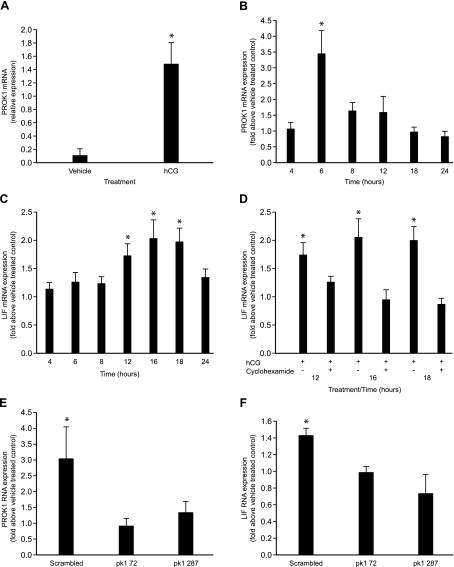
Previous studies have highlighted the fact that the expression of LIF can be induced by hCG in a number of models including primary cell cultures (13), human endometrial infusion (15), and, most recently, a baboon endometrial infusion model (14). Using RNA extracted from baboon endometrium after treatment with hCG for 5 d commencing on d 6 PO, we have shown that hCG increases mRNA expression of PROK1 (5.2±0.3-fold above vehicle-treated control, P<0.05) (Fig. 2A).
Figure 2.
hCG mediates expression of LIF via induction of PROK1. Expression of PROK1 in baboon endometrium was elevated after treatment with 1.25 IU of hCG/h for 5 d (A). Treatment of Ishikawa PROKR1 cells with 1 IU of hCG results in increased expression of PROK1 which peaks at 6 h (B). Similarly, treatment of Ishikawa PROKR1 cells with 1 IU of hCG results in increased expression of LIF, which peaks at 16 h (C). LIF mRNA expression in response to treatment with hCG is inhibited when PROKR1 Ishikawa cells are treated with 1 IU of hCG in the presence of cycloheximide (protein synthesis inhibitor) (D). PROKR1 Ishikawa cells were transiently transfected with miRNA constructs targeting PROK1 or scrambled sequences and subsequently treated with 1 IU of hCG. hCG-mediated PROK1 (E) and LIF (F) expression were significantly inhibited by PROK1 miRNA constructs. Bars represent means ± se of at least 3 independent experiments. *P < 0.05.
To demonstrate the temporal regulation of PROK1 expression mediated by hCG, PROKR1 Ishikawa cells were stimulated with 1 IU of hCG for 4, 6, 8, 12, 18, and 24 h. PROK1 mRNA expression was elevated by hCG with a peak in expression at 6 h (3.44±0.73-fold above vehicle-treated control, P<0.01) (Fig. 2B). LIF mRNA expression was also regulated by hCG in PROKR1 Ishikawa cells, with a peak in LIF expression at 16 h after treatment with hCG (2.03±0.3-fold above vehicle-treated control, P<0.05) (Fig. 2C). The peak in hCG-mediated LIF mRNA expression occurred ∼10 h after the peak in hCG-mediated PROK1 mRNA. Cotreatment of PROKR1 Ishikawa cells with 10 μg/ml cycloheximide, an inhibitor of protein synthesis, and 1 IU of hCG significantly inhibited hCG-mediated LIF expression at 12, 16, and 18 h (P<0.01) (Fig. 2D), suggesting that an intermediary protein is involved in hCG-mediated LIF expression. To investigate whether PROK1 is required for hCG-mediated LIF expression, we generated miRNA constructs expressing sequences targeting PROK1. Transfection of PROKR1 Ishikawa cells with pcDNA6.2-GW/EmGFP-PROK1-72 or PROK1-287 miRNA constructs significantly inhibited hCG-mediated PROK1 expression at 6 h compared with that in PROKR1 Ishikawa cells transfected with scrambled control construct alone (P<0.05) (Fig. 2E). Moreover, transfection of pcDNA6.2-GW/EmGFP-PROK1-72 or PROK1-287 into PROKR1 Ishikawa cells significantly inhibited hCG-mediated LIF expression at 18 h (P<0.05) (Fig. 2F), suggesting that regulation of LIF expression by hCG is mediated via the expression of PROK1.
Nonpregnant endometrium and first trimester decidua tissue express LH/CG receptor, PROK1, and LIF
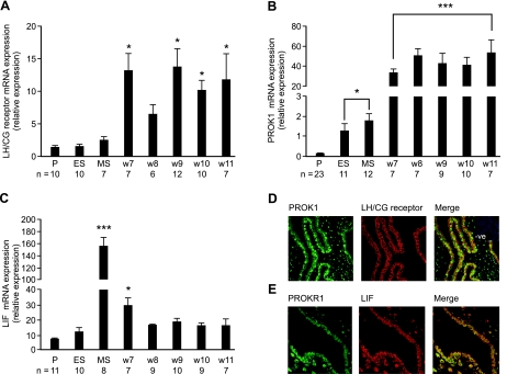
Using primers and probe directed against exon 11 of the LH/CG receptor, we demonstrated elevated expression of the LH/CG receptor in first trimester decidua samples obtained from wk 7–11 of pregnancy compared with that in samples taken throughout the normal menstrual cycle (P<0.05) (Fig. 3A). PROK1 mRNA expression is significantly elevated in the secretory phase compared with that in the proliferative phase of the menstrual cycle (P<0.05) (Fig. 3B) and is further elevated in all samples collected during the first trimester of pregnancy (P<0.001) (Fig. 3B). Expression of LIF mRNA is elevated in the midsecretory phase of the menstrual cycle and in wk 7 of pregnancy compared with that in nonpregnant proliferative or early secretory endometrium and later stages of gestation (P<0.05) (Fig. 3C). To identify the site of expression of the LH/CG receptor, PROK1, PROKR1, and LIF in human first trimester decidua, immunofluorescent histochemistry was used. Immunofluorescent histochemical analysis colocalized the site of expression of PROK1 (green) and the LH/CG receptor (red) to the glandular epithelial cells (yellow, merge) (Fig. 3D) of first trimester decidua. By using an alternate antibody to the LH/CG receptor (H-15, sc-25828; Santa Cruz Biotechnology) known to detect truncated variants of the receptor, staining was also present in the stroma, data not shown. In addition, expression of PROKR1 (green) and LIF (red) colocalized to the glandular epithelial cells (yellow, merge) of the decidua (Fig. 3E).
Figure 3.
Temporal expression and colocalization of signaling components. LH/CG receptor (A), PROK1 (B) and LIF (C) mRNA in human endometrium across the menstrual cycle and first trimester decidua. P, proliferative; ES, early secretory; MS, mid secretory, w7, w8, w9, w10, and w11 represent weeks of gestation (n=sample size from each stage of the cycle and early pregnancy). Immunofluorescent histochemical analysis of the LH/CG receptor (red panel) and PROK1 (green panel) show colocalization to the glandular epithelial cells of first trimester decidua (yellow panel, merge) (D). PROKR1 (green panel) and LIF (red panel) colocalize to glandular epithelial cells and a subset of stromal cells of first trimester decidua (yellow panel, merge) (E). Data are means ± se. *P < 0.05; ***P < 0.001.
hCG mediates expression of PROK1 and LIF in first trimester decidua tissue
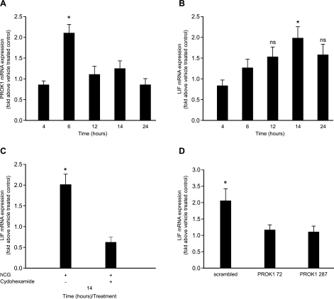
To investigate the regulation of PROK1 by hCG in first trimester decidua, decidual tissue was stimulated with 1 IU of hCG. Temporal regulation of PROK1 mRNA expression by hCG was demonstrated with a peak in expression of PROK1 at 6 h (2.1±0.2-fold above control, P<0.05) (Fig. 4A). Investigation of hCG-mediated LIF mRNA expression in first trimester decidua revealed a peak in LIF expression at 14 h (1.98±0.27-fold above control, P<0.05) (Fig. 4B). Incubation of first trimester decidual tissue explants with cycloheximide for 1 h before treatment with 1 IU of hCG for 14 h significantly inhibited hCG-mediated LIF mRNA expression (P<0.05) (Fig. 4C), suggesting the involvement of an intermediate protein in hCG-mediated LIF expression. Infection of first trimester decidua explants with lentivirus expressing PROK1-72 or PROK1-287 miRNA constructs significantly inhibited hCG-mediated LIF mRNA expression compared with decidual explants infected with lentivirus expressing miRNA negative control constructs (P<0.05) (Fig. 4D).
Figure 4.
hCG mediates expression of LIF via induction of PROK1 in first trimester decidua. Treatment of first trimester decidua with 1 IU of hCG results in increased expression of PROK1 that peaks at 6 h (A). Similarly, treatment of first trimester decidua with 1 IU of hCG results in increased expression of LIF that peaks at 14 h (B). LIF mRNA expression in response to treatment with hCG is inhibited when first trimester decidua is treated with 1 IU of hCG for 14 h in the presence cycloheximide (protein synthesis inhibitor) (C). First trimester decidua tissue was infected with lentivirus encoding miRNA constructs targeting PROK1 or a scrambled sequence. Infected tissue was subjected to stimulation with 1 IU of hCG and subsequently analyzed for LIF mRNA expression. hCG-mediated LIF expression was significantly inhibited by PROK1 miRNA constructs compared with scrambled sequences (D). Bars represent means ± se of at least 3 independent experiments. *P < 0.05.
PROK1 mediates expression of LIF in first trimester decidual explants
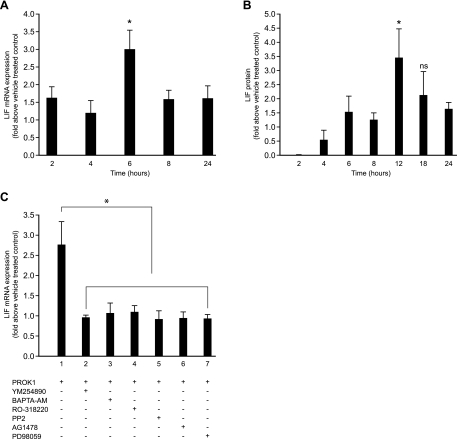
Stimulation of first trimester decidual explants with 40 nM PROK1 demonstrated an elevation in LIF mRNA expression at 6 h (2.99±0.53-fold above vehicle-treated control, P<0.05) (Fig. 5A) followed by a peak in LIF protein production at 12 h (3.44±1.01-fold above vehicle-treated control, P<0.05) (Fig. 5B). These data are consistent with those observed in the PROKR1 Ishikawa cell line, demonstrating that PROK1 regulates LIF expression in decidual tissue. We further investigated the signaling pathway inducing LIF expression in response to PROK1. Cotreatment of first trimester decidual explants with inhibitors of Gq (YM254890), Ca2+ (BAPTA-AM), PKC (Ro-318220), cSrc (PP2), EGFR (AG1478), or MEK (PD98059) significantly inhibited PROK1-mediated LIF mRNA expression (P<0.05) (Fig. 5C).
Figure 5.
PROK1 induces LIF expression in first trimester decidua. Treatment of first trimester decidua with 40 nM PROK1 induced LIF mRNA (A) and protein (B) expression that peaked at 6 and 12 h, respectively. Expression of LIF mRNA in first trimester decidua was measured after treatment with 40 nM PROK1 (lane 1) in the absence or presence of YM254890 (Gq inhibitor, lane 2), BAPTA-AM (calcium inhibitor, lane 3), PKC inhibitor (Ro-318220, lane 4), cSrc inhibitor (PP2, lane 5), EGFR inhibitor (AG1478, lane 6), or MEK inhibitor (PD98059, lane 7) (C). PROK1-induced LIF mRNA expression was significantly inhibited in the presence of these inhibitors. Bars represent means ± se of at least 3 independent experiments. *P < 0.05.
PROK1-mediated LIF expression enhances trophoblast adhesion
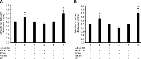
LIF has previously been described to enhance trophoblast adhesion to extracellular matrices (16). We therefore sought to investigate whether PROK1-mediated epithelial LIF expression could induce trophoblast adhesion and which components of the extracellular matrix were involved. PROKR1 Ishikawa cells were treated with vehicle or 40 nM PROK1 for 12 h, and conditioned medium was collected. JEG-3 human trophoblast cells were subsequently treated overnight with conditioned medium from vehicle- or PROK1-treated cells or conditioned medium (vehicle- and PROK1-treated) that had been immunoneutralized using 1 μg/ml LIF antibody, or treatment of JEG-3 cells with 100 ng/ml recombinant LIF alone was used as a positive control. Treatment of JEG-3 cells with PROK1-conditioned medium enhanced adhesion to fibronectin (Fig. 6A, lane 2) and laminin (Fig. 6B, lane 2) matrices to a level comparable with that observed after treatment with recombinant LIF protein alone (Fig. 6A, B; lane 6). The adhesion mediated by PROK1-conditioned medium was abrogated after immunoneutralization of LIF from the medium (P<0.05) (Fig. 6A, B; lane 4). These data suggest that PROK1-mediated LIF expression may enhance trophoblast-endometrium adhesion.
Figure 6.
PROK1 via LIF increases adhesion of trophoblast cells to fibronectin and laminin. Treatment of JEG-3 trophoblast cells with LIF increased adhesion to fibronectin (A, lane 6) and laminin (B, lane 6) compared with vehicle-treated cells (A, B; lane 5). Treatment of JEG-3 cells with conditioned medium collected from PROKR1 Ishikawa cells treated with 40 nM PROK1 for 24 h (PROK1 CM) also increased adhesion to fibronectin (A, lane 2) and laminin (B, lane 2) compared with conditioned medium collected from PROKR1 Ishikawa cells treated with vehicle for 24 h (vehicle CM; A, B; lane 1). This PROK1-mediated adhesion was abolished when LIF was immunoneutralized from the PROK1-conditioned medium (A, B; lane 4). Bars represent means ± se of at least 3 independent experiments. *P < 0.05. Ab, immunoneutralization.
DISCUSSION
Preimplantation signaling between the developing blastocyst and the endometrium is essential for successful implantation and maintenance of early pregnancy. Endometrial cytokines prepare the endometrium to become receptive to the blastocyst, and increasing evidence suggests that paracrine and juxtacrine signals from the blastocyst can influence the endometrial cytokine milieu and enhance receptivity. Blastocysts are known to secrete various factors (reviewed in ref. 17) that initiate early embryonic communication with the endometrium. There is evidence that embryonic hCG can influence implantation by regulating the expression of endometrial factors that are important in receptivity, such as endometrial LIF (13,14,15, 18, 19).
LIF has been shown to be critical in murine implantation (9, 20) and has been suggested to have a role in human fertility (21, 22). We have previously demonstrated regulation of a host of implantation-related genes including LIF by PROK1 using gene array analysis (7). We demonstrate that the temporal expression of LIF mRNA and protein is regulated by PROK1 in an endometrial epithelial cell line and in first trimester decidual tissue explants. LIF is known to be regulated by cytokines including TNF, IL-1, and TGF-β via activation of a signaling cascade involving intracellular calcium, PKC, and MEK (23,24,25,26,27). To examine whether PROK1 activates LIF expression through a similar pathway, chemical inhibitors of signaling pathway molecules were used. We demonstrate that PROKR1, a Gq-coupled receptor that mediates extracellular signal-regulated kinase 1/2 phosphorylation (7), induces LIF expression via a cSrc-EGFR-MEK signaling pathway, and is also dependent on intracellular calcium release in both PROKR1 Ishikawa cells and first trimester decidual explants.
This study demonstrates that hCG mediates both PROK1 and LIF expression with a delay in the peak of LIF expression after the peak in PROK1 expression. By using an inhibitor of protein synthesis, hCG induction of LIF was abolished suggesting the involvement of PROK1 as an intermediate in hCG-mediated LIF expression. Using miRNA constructs targeting PROK1, we confirmed that hCG-stimulated LIF expression is dependent on expression of PROK1. These data provide the first evidence that an intermediate protein, PROK1, is required in hCG-mediated endometrial LIF expression.
To demonstrate that the data represent an active signaling pathway in vivo LH/CG receptor is required to be present and colocalized in the endometrium and decidua to permit up-regulation of PROK1 via hCG. We therefore investigated the expression of the LH/CG receptor in our decidua samples using quantitative PCR. The results demonstrated the expression of the receptor in the normal cycling endometrium, with an elevation in expression in first trimester decidua. To demonstrate LH/CG receptor mRNA translation and localization, immunohistochemical analysis of first trimester decidua was performed with an antibody recognizing the full-length receptor. This confirmed protein expression of the receptor with localization to the glandular epithelial cells. Coexpression of PROK1 and LH/CG receptor was demonstrated in first trimester decidual tissue, suggesting potential regulation of PROK1 by hCG in vivo. Furthermore, the colocalization of PROKR1 and LIF in first trimester decidual tissue suggests that the pathway we have identified, hCG-LH/CG receptor binding activating PROK1-PROKR1 signaling, is responsible for regulation of LIF expression in human early pregnancy.
LIF expression was elevated in midsecretory endometrium and during early pregnancy up to wk 7 in first trimester decidua. LIF expression significantly decreased thereafter, in agreement with previous reports that decidual LIF production is decreased in the second trimester compared with that in the first trimester (28). Week 8 of pregnancy shows a shift in the phenotype of the trophoblasts from predominantly an anchoring villi to an increase in invasive interstitial trophoblasts (29, 30). By 8 wk of pregnancy the interstitial trophoblasts have invaded to the decidual-myometrial border and during the second trimester, there is a further invasion into the inner myometrium (29, 30). In first trimester trophoblast populations, LIF has been suggested to increase fibronectin production, inhibit gelatinase activity, increase spreading and adhesion, and induce differentiation toward syncytialization (31,32,33). Our data confirm a role for LIF in enhancing adhesion of trophoblast cells to fibronectin and laminin mediated via PROK1-PROKR1 signaling. At the interface between the chorionic villi and the decidua at the implantation site, a predominantly acellular zone develops, known as Nitabuch’s fibrinoid (34). This zone is rich in laminin, suggesting that PROK1-induced LIF promotes adhesion of the anchoring trophoblast cells to this layer (35). Down-regulation of LIF in decidua after wk 7 of pregnancy may be controlled by the trophoblast as a requirement to reduce adhesion and allow controlled invasion through the decidua. Therefore, PROK1 enhancement of LIF may only be required during early pregnancy before wk 8 of gestation during the establishment of the anchoring villi. In addition, decidualizing stromal cells also produce a pericellular laminin basement membrane that may regulate this process (36). The increase in PROK1 and LIF in the midsecretory phase of the menstrual cycle and enhancement before implantation may also regulate trophoblast adhesion. The basal lamina associated with the epithelium increases in thickness during the secretory phase, reaching maximum thickness in the midsecretory phase. The basal lamina consists of collagen type IV with proteoglycan molecules such as laminin and fibronectin (37). The thickened basal lamina may provide an important structural scaffold for epithelial and trophoblast attachment. A number of studies suggest that LIF production in fertile women is elevated in the secretory phase of the menstrual cycle compared with that in the proliferative phase of the cycle but is dysregulated in infertile women (22, 38,39,40). Therefore, disrupted regulation of PROK1 expression and its downstream target LIF may be associated with infertility, spontaneous abortion, and placental abnormalities.
In conclusion, this study demonstrates that embryonic hCG regulates endometrial PROK1-PROKR1 signaling, which induces expression of LIF via induction of intracellular calcium and activation of intracellular phosphorylation cascades. These data support a role for PROK1 in initial blastocyst adhesion to basement membrane matrices and subsequent trophoblast invasion. Dysregulation in the expression of PROK1 and consequently LIF may lead to suboptimal fertility and trophoblast invasion.
Acknowledgments
We thank Sharon Donaldson and Catherine Murray for consenting patients and tissue collection.
References
- Psychoyos A. Hormonal control of ovoimplantation. Vitam Horm. 1973;31:201–256. doi: 10.1016/s0083-6729(08)60999-1. [DOI] [PubMed] [Google Scholar]
- Tabibzadeh S. Molecular control of the implantation window. Hum Reprod Update. 1998;4:465–471. doi: 10.1093/humupd/4.5.465. [DOI] [PubMed] [Google Scholar]
- Goto S, Kadowaki T, Hashimoto H, Kokeguchi S, Shiotani M. Stimulation of endometrium embryo transfer: injection of embryo culture supernatant into the uterine cavity before blastocyst transfer can improve implantation and pregnancy rates. Fertil Steril. 2007;88:1339–1343. doi: 10.1016/j.fertnstert.2007.01.011. [DOI] [PubMed] [Google Scholar]
- Filicori M, Fazleabas A T, Huhtaniemi I, Licht P, Rao C H V, Tesarik J, Zygmunt M. Novel concepts of human chorionic gonadotropin: reproductive system interactions and potential in the management of infertility. Fertil Steril. 2005;84:275–284. doi: 10.1016/j.fertnstert.2005.02.033. [DOI] [PubMed] [Google Scholar]
- De Ziegler D, Mattenberger C, Schwarz C, Ibecheole V, Fournet N, Bianchi-Demicheli F. New tools for optimizing endometrial receptivity in controlled ovarian hyperstimulation: aromatase inhibitors and LH/(mini)hCG. Ann N Y Acad Sci. 2004;1034:262–277. doi: 10.1196/annals.1335.028. [DOI] [PubMed] [Google Scholar]
- Battersby S, Critchley H O, Morgan K, Millar R P, Jabbour H N. Expression and regulation of the prokineticins (endocrine gland-derived vascular endothelial growth factor and Bv8) and their receptors in the human endometrium across the menstrual cycle. J Clin Endocrinol Metab. 2004;89:2463–2469. doi: 10.1210/jc.2003-032012. [DOI] [PubMed] [Google Scholar]
- Evans J, Catalano R D, Morgan K, Critchley H O, Millar R P, Jabbour H N. Prokineticin 1 signaling and gene regulation in early human pregnancy. Endocrinology. 2008;149:2877–2887. doi: 10.1210/en.2007-1633. [DOI] [PMC free article] [PubMed] [Google Scholar]
- Hoffmann P, Feige J J, Alfaidy N. Expression and oxygen regulation of endocrine gland-derived vascular endothelial growth factor/prokineticin-1 and its receptors in human placenta during early pregnancy. Endocrinology. 2006;147:1675–1684. doi: 10.1210/en.2005-0912. [DOI] [PubMed] [Google Scholar]
- Stewart C L, Kaspar P, Brunet L J, Bhatt H, Gadi I, Kontgen F, Abbondanzo S J. Blastocyst implantation depends on maternal expression of leukaemia inhibitory factor. Nature. 1992;359:76–79. doi: 10.1038/359076a0. [DOI] [PubMed] [Google Scholar]
- Bhatt H, Brunet L J, Stewart C L. Uterine expression of leukaemia inhibitory factor coincides with the onset of blastocyst implantation. Proc Natl Acad Sci U S A. 1991;88:11408–11412. doi: 10.1073/pnas.88.24.11408. [DOI] [PMC free article] [PubMed] [Google Scholar]
- Arici A, Engin O, Attar E, Olive D L. Modulation of leukemia inhibitory factor gene expression and protein biosynthesis in human endometrium. J Clin Endocrinol Metab. 1995;80:1908–1915. doi: 10.1210/jcem.80.6.7775640. [DOI] [PubMed] [Google Scholar]
- Vogiagis D, Marsh M M, Fry R C, Salamonsen L A. Leukaemia inhibitory factor in human endometrium throughout the menstrual cycle. J Endocrinol. 1996;148:95–102. doi: 10.1677/joe.0.1480095. [DOI] [PubMed] [Google Scholar]
- Perrier d'Hauterive S, Charlet-Renard C, Berndt S, Dubois M, Munaut C, Goffin F, Hagelstein M T, Noel A, Hazout A, Foidart J M, Geenen V. Human chorionic gonadotropin and growth factors at the embryonic-endometrial interface control leukaemia inhibitory factor (LIF) and interleukin 6 (IL-6) secretion by human endometrial epithelium. Hum Reprod. 2004;19:2633–2643. doi: 10.1093/humrep/deh450. [DOI] [PubMed] [Google Scholar]
- Sherwin J R, Sharkey A M, Cameo P, Mavrogianis P M, Catalano R D, Edassery S, Fazleabas A T. Identification of novel genes regulated by chorionic gonadotropin in baboon endometrium during the window of implantation. Endocrinology. 2007;148:618–626. doi: 10.1210/en.2006-0832. [DOI] [PubMed] [Google Scholar]
- Licht P, Russu V, Lehmeyer S, Wildt L. Molecular aspects of direct LH/hCG effects on human endometrium—lessons from intrauterine microdialysis in the human female in vivo. Reprod Biol. 2001;1:10–19. [PubMed] [Google Scholar]
- Tapia A, Salamonsen L A, Manuelpillai U, Dimitriadis E. Leukemia inhibitory factor promotes human first trimester extravillous trophoblast adhesion to extracellular matrix and secretion of tissue inhibitor of metalloproteinases-1. Hum Reprod. 2008;23:1724–1732. doi: 10.1093/humrep/den121. [DOI] [PMC free article] [PubMed] [Google Scholar]
- Norwitz E R, Schust D J, Fisher S J. Implantation and the survival of early pregnancy. N Engl J Med. 2001;345:1400–1408. doi: 10.1056/NEJMra000763. [DOI] [PubMed] [Google Scholar]
- Srisuparp S, Strakovam Z, Fazleabas A T. The role of chorionic gonadotropin (CG) in blastocyst implantation. Arch Med Res. 2001;32:627–634. doi: 10.1016/s0188-4409(01)00330-7. [DOI] [PubMed] [Google Scholar]
- Licht P, Fluhr H, Neuwinger J, Wallwiener D, Wildt L. Is human chorionic gonadotropin directly involved in the regulation of human implantation? Mol Cell Endocrinol. 2007;269:85–92. doi: 10.1016/j.mce.2006.09.016. [DOI] [PubMed] [Google Scholar]
- Fouladi-Nashta A A, Jones C J, Nijjar N, Mohamet L, Smith A, Chambers I, Kimber S J. Characterization of the uterine phenotype during the peri-implantation period for LIF-null, MF1 strain mice. Dev Biol. 2005;281:1–21. doi: 10.1016/j.ydbio.2005.01.033. [DOI] [PubMed] [Google Scholar]
- Steck T, Giess R, Suetterlin M W, Bolland M, Wiest S, Poehls U G, Dietl J. Leukaemia inhibitory factor (LIF) gene mutations in women with unexplained infertility and recurrent failure of implantation after IVF and embryo transfer. Eur J Obstet Gynecol Reprod Biol. 2004;112:69–73. doi: 10.1016/s0301-2115(03)00315-4. [DOI] [PubMed] [Google Scholar]
- Hambartsoumian E. Endometrial leukaemia inhibitory factor (LIF) as a possible cause of unexplained infertility and multiple failures of implantation. Am J Reprod Immunol. 1998;39:137–143. doi: 10.1111/j.1600-0897.1998.tb00345.x. [DOI] [PubMed] [Google Scholar]
- Carlson C D, Bai Y, Jonakait G M, Hart R P. Interleukin-1β increases leukaemia inhibitory factor mRNA levels through transient stimulation of transcription rate. Glia. 1996;18:141–151. doi: 10.1002/(SICI)1098-1136(199610)18:2<141::AID-GLIA6>3.0.CO;2-3. [DOI] [PubMed] [Google Scholar]
- Elias J A, Zheng T, Whiting N L, Marcovici A, Trow T K. Cytokine-cytokine synergy and protein kinase C in the regulation of lung fibroblast leukemia inhibitory factor. Am J Physiol. 1994;266:426–435. doi: 10.1152/ajplung.1994.266.4.L426. [DOI] [PubMed] [Google Scholar]
- Fan Z, Bau B, Yang H, Aigner T. IL-1β induction of IL-6 and LIF in normal articular human chondrocytes involves the ERK, p38 and NFκB signalling pathways. Cytokine. 2004;28:17–24. doi: 10.1016/j.cyto.2004.06.003. [DOI] [PubMed] [Google Scholar]
- Bamberger A M, Erdmann I, Jenatschke S, Bamberger C M, Schulte H M. Regulation of the human leukaemia inhibitory factor (LIF) promoter in HEC-1B endometrial adenocarcinoma cells. Mol Hum Reprod. 1997;3:789–793. doi: 10.1093/molehr/3.9.789. [DOI] [PubMed] [Google Scholar]
- Sawai K, Matsuzaki N, Okada T, Shimoya K, Koyama M, Azuma C, Saji F, Murata Y. Human decidual cell biosynthesis of leukemia inhibitory factor: regulation by decidual cytokines and steroid hormones. Biol Reprod. 1997;56:1274–1280. doi: 10.1095/biolreprod56.5.1274. [DOI] [PubMed] [Google Scholar]
- Ren S G, Melmed S, Braunstein G D. Decidual leukemia inhibitory factor production and action on human chorionic gonadotropin secretion at different stages of gestation in vitro. Early Pregnancy. 1997;3:102–108. [PubMed] [Google Scholar]
- Red-Horse K, Zhou Y, Genbacev O, Prakobphol A, Foulk R, McMaster M, Fisher S J. Trophoblast differentiation during embryo implantation and formation of the maternal-fetal interface. J Clin Invest. 2004;114:744–754. doi: 10.1172/JCI22991. [DOI] [PMC free article] [PubMed] [Google Scholar]
- Lunghi L, Ferretti M E, Merci S, Bondi C, Vesce F. Control of human trophoblast function. Reprod Biol Endocrinol. 2007;5:6. doi: 10.1186/1477-7827-5-6. [DOI] [PMC free article] [PubMed] [Google Scholar]
- Sawai K, Matsuzaki N, Kameda T, Hashimoto K, Okada T, Shimoya K, Nobunaga T, Taga T, Kishimoto T, Saji F. Leukemia inhibitory factor produced at the fetomaternal interface stimulates chorionic gonadotropin production: its possible implication during pregnancy, including implantation period. J Clin Endocrinol Metab. 1995;80:1449–1456. doi: 10.1210/jcem.80.4.7714123. [DOI] [PubMed] [Google Scholar]
- Nachtigall M J, Kliman H J, Feinberg R F, Olive D L, Engin O, Arici A. The effect of leukemia inhibitory factor (LIF) on trophoblast differentiation: a potential role in human implantation. J Clin Endocrinol Metab. 1996;81:801–806. doi: 10.1210/jcem.81.2.8636307. [DOI] [PubMed] [Google Scholar]
- Bischof P, Haenggeli L, Campana A. Effect of leukemia inhibitory factor on human cytotrophoblast differentiation along the invasive pathway. Am J Reprod Immunol. 1995;34:225–230. doi: 10.1111/j.1600-0897.1995.tb00945.x. [DOI] [PubMed] [Google Scholar]
- Ferenczy A. Ultrastructure of the normal menstrual cycle: a review. Microsc Res Tech. 1993;25:91–105. doi: 10.1002/jemt.1070250203. [DOI] [PubMed] [Google Scholar]
- Fernandez P L, Merino M J, Nogales F F, Charonis A S, Stetler-Stevenson W, Liotta L. Immunohistochemical profile of basement membrane proteins and 72 kilodalton type IV collagenase in the implantation placental site: an integrated view. Lab Invest. 1992;66:572–579. [PubMed] [Google Scholar]
- Faber M, Wewer U M, Berthelsen J G, Liotta L A, Albrechtsen R. Laminin production by human endometrial stromal cells relates to the cyclic and pathologic state of the endometrium. Am J Pathol. 1986;124:384–391. [PMC free article] [PubMed] [Google Scholar]
- Dockery P, Khalid J, Sarani S A, Bulut H E, Warren M A, Li T C, Cooke I D. Changes in basement membrane thickness in the human endometrium during the luteal phase of the menstrual cycle. Hum Reprod Update. 1998;4:486–495. doi: 10.1093/humupd/4.5.486. [DOI] [PubMed] [Google Scholar]
- Delage G, Moreau J F, Taupin J L, Freitas S, Hambartsoumian E, Olivennes F, Fanchin R, Letur-Konirsch H, Frydman R, Chaouat G. In-vitro endometrial secretion of human interleukin for DA cells/leukaemia inhibitory factor by explant cultures from fertile and infertile women. Hum Reprod. 1995;10:2483–2488. doi: 10.1093/oxfordjournals.humrep.a136328. [DOI] [PubMed] [Google Scholar]
- Lédée-Bataille N, Laprée-Delage G, Taupin J L, Dubanchet S, Frydman R, Chaouat G. Concentration of leukaemia inhibitory factor (LIF) in uterine flushing fluid is highly predictive of embryo implantation. Hum Reprod. 2002;17:213–218. doi: 10.1093/humrep/17.1.213. [DOI] [PubMed] [Google Scholar]
- Mikolajczyk M, Skrzypczak J, Szymanowski K, Wirstlein P. The assessment of LIF in uterine flushing—a possible new diagnostic tool in states of impaired fertility. Reprod Biol. 2003;3:259–270. [PubMed] [Google Scholar]


