Abstract
Bacillus thuringiensis subsp. israelensis produces a potent mosquitocidal protein, Cry4A. We have identified a 15-bp catabolite responsive element (cre), overlapping the −35 element of the cry4A promoter. Changing a guanine to adenine at position −49 in the promoter abolished glucose catabolite repression of cry4A and enhanced promoter activity two- to threefold. This cis regulatory element is essential for controlled toxin synthesis, vital to evolutionary success of B. thuringiensis subsp. israelensis.
Bacillus thuringiensis produces insecticidal crystal proteins highly toxic to insects (6, 14). Synthesis of insecticidal proteins by B. thuringiensis is transcriptionally linked to sporulation through common transcription factors (3). Transcription of many stationary-phase genes, including cry genes, is held in check until effectors regulating the developmental process are induced (1). The developmental process in B. thuringiensis is temporally controlled at transcription level by recruitment of successive sigma subunits of the RNA polymerase (RNAP) (13). The following five σ factors are involved in the developmental process of B. thuringiensis: σH, which functions in the stationary phase in the undivided mother cell; σ35 and σ28, which control early and late sporulation genes and most of the crystal protein genes in the mother cell; and σF and σG, which are active in the forespore compartment of the sporulating bacterium (25).
Toxin production in B. thuringiensis is subject to a variety of sophisticated controls at various levels to maximize protein yields (1, 6). B. thuringiensis subsp. israelensis produces an ovoid inclusion toxic to dipteran insects: e.g., mosquitoes and blackflies (10). The parasporal crystals are composed of a number of polypeptides, namely, Cry4A, Cry4B, Cry11A, and CytA (15). The cry4A gene encodes a 130-kDa protoxin protein, expressed weakly in the transition phase through σH-specific RNAP (27), while major amount of the protein is synthesized in the stationary phase under the control of σ35- and σ28-specific RNAP (33, 34).
Our earlier investigations probing the role of environmental factors regulating Cry4A expression in B. thuringiensis subsp. israelensis (5) demonstrated that glucose and its metabolite fructose 6-phosphate repressed cry4A gene expression through catabolite repression (CR) (16), a global, negative regulatory mechanism of gene transcription in gram-positive bacteria (11). CR establishes priorities in the utilization of carbon and energy sources according to their selective advantage (11) for competitive success of the bacteria in the natural environment. In gram-positive bacteria, the essential components of CR are catabolite control protein CcpA, a member of LacI/GalR family of bacterial regulatory proteins (31); an allosteric effector protein, HPr (4); and a cis-acting, partially palindromic binding sequence, the catabolite responsive element (cre) in the gene (9, 30).
Glucose repression of cry4A transcription in B. thuringiensis subsp. israelensis (5) and its derepression in an HPr-serine phosphorylation (Hpr-Ser-P)-defective mutant (16) suggested catabolite repression of the δ-endotoxin gene. To get an insight into the mechanism of cry4A repression, here we analyzed the cry4A gene and its upstream regulatory sequence for potential regulatory elements. We present data characterizing interaction of different components of the catabolite repression network with the cry4A gene leading to identification of a cre.
Search for cre-like sequence in the cry4A gene and upstream promoter region.
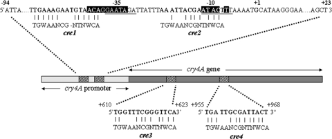
Based on the 14-nucleotide consensus sequence TGWAANCGNTNWCA, proposed by Weickert and Chambliss (30), a pattern-matching search was performed in the cry4A gene and its flanking regions of B. thuringiensis subsp. israelensis strain ATCC 522. Four sequences matching the consensus cre sequence were identified in the latter (Fig. 1). Two sequences, cre1 and cre2 were present in the promoter region, 50 and 21 bases upstream of the transcriptional start site, showing 85.7% and 57.14% sequence similarity, respectively. cre3 and cre4 were present in the coding region of the gene and showed 71.43% and 78.57% similarity, respectively, to the consensus sequence.
FIG. 1.
Identification of cre sequences in the cry4A gene. Shown is the cry4A gene, with four cre-like sequences (cre1, cre2, cre3, and cre4) homologous to consensus sequence TGWAANCGNTNWCA. +1 represents the cry4A transcription initiation nucleotide, σ35-recognized −35 and −10 elements are underlined, and the SigH promoter is shaded.
Interaction of CcpA and HPr-Ser-P with identified cre sequences.
The bacterial strains and plasmids used in this study are listed in Table 1, and the oligonucleotide primers are shown in a table in the supplemental material.
TABLE 1.
Strains and plasmids used in this study
| Plasmid or strain | Relevant characteristic(s) | Source or reference |
|---|---|---|
| Plasmids | ||
| pGEM-T Easy | Ampr, E. coli cloning vector | Promega |
| pHT370 | Ampr Eryr, shuttle vector | Pasteur Institute |
| pSK1 | pGEM-T Easy containing cry4A coding sequence with native promoter | This work |
| pSK2 | pHT370 containing cry4A coding sequence with native promoter | This work |
| pSK3 | pGEM-T Easy containing 117-bp cre1-2 | This work |
| pSK4 | pGEM-T Easy containing cre1-2 with mutation (G49→A) in cre1 | This work |
| pSK5 | pGEM-T Easy containing 458-bp cry4A promoter | This work |
| pSK6 | pGEM-T Easy containing 3,067-bp lacZ | This work |
| pSK7 | pGEM-T Easy containing cry4Apro-lacZ fusion | This work |
| pSK8 | pHT370 containing cry4Apro-lacZ fusion | This work |
| pSK9 | pHT370 with cry4A coding sequence with G49→A in cre1 | This work |
| Strains | ||
| 4Q7 | B. thuringiensis subsp. israelensis plasmidless mutant | BGSCa |
| SK2 | 4Q7 containing pSK2 | This work |
| SK9 | 4Q7 containing pSK9 [(G49→A) mutation in cre1 in pSK2] | This work |
| SK10 | 4Q7 containing pSK8 | This work |
| SK11 | 4Q7 containing pSK11 [(A36→C) in pSK8]b | This work |
| SK12 | 4Q7 containing pSK12 [(C37→A) in pSK8]b | This work |
| SK13 | 4Q7 containing pSK13 [(T40→C) in pSK8]b | This work |
| SK14 | 4Q7 containing pSK14 [(A43→G) in pSK8]b | This work |
| SK15 | 4Q7 containing pSK15 [(A44→C) in pSK8]b | This work |
| SK16 | 4Q7 containing pSK16 [(A47→C) in pSK8]b | This work |
| SK17 | 4Q7 containing pSK17 [(A48→C) in pSK8]b | This work |
| SK18 | 4Q7 containing pSK18 [(G49→A) in pSK8]b | This work |
| SK19 | 4Q7 containing pSK19 [(G49→T) in pSK8]b | This work |
| SK20 | 4Q7 containing pSK20 [(G49→C) in pSK8]b | This work |
BGSC, Bacillus Genetic Stock Center (Ohio State University, Columbus).
Point mutations are done in the cre1 region of the promoter.
Protein DNA interactions were evaluated by mobility shift assays. A 117-bp probe DNA (−94 to +23) containing cre1 and cre2 (Fig. 1) was amplified by PCR with primers 3 and 4 using a B. thuringiensis subsp. israelensis megaplasmid as a template and cloned in pGEM-T Easy vector, producing pSK3. The G49→A mutation was obtained using G49→A forward and reverse primers with pSK3 as a template, producing plasmid pSK4. The plasmids pSK3 and pSK4 were digested with SpeI and EcoRI, and the 117-bp inserts were end labeled in the presence of [α-32P]dCTP (Perkin-Elmer), deoxynucleotide triphosphates (dNTPs), and the Klenow fragment of DNA polymerase for use as a probe. The probe DNA was purified on a Sephadex G-25 column (Pharmacia).
Double-stranded oligonucleotide fragments encompassing cre3 (+594 to +651) and cre4 (+931 to +986) were generated by annealing long complementary primer sequences 5 with 6 and 7 with 8 in annealing buffer (1× Tris-EDTA buffer [pH 8.0] and 50 mM NaCl). The reaction mixture was heated to 80°C and allowed to cool slowly to 25°C, and the 3′ overhangs were radiolabeled by end-filling reaction.
Recombinant CcpA and HPr kinase were prepared as described earlier (7, 12). HPr from B. thuringiensis subsp. israelensis was phosphorylated at Ser-45 using HPr kinase and purified as described earlier (17). The binding reaction mixture contained labeled probe (20,000 cpm/μl) with 0.6 nM HPr and 0.78 nM CcpA, 2 μg poly(dI-dC) (Pharmacia), 15 mM HEPES (pH 7.5), 35 mM KCl, 1 mM EDTA (pH 7.5), 1 mM dithiothreitol, 6% glycerol, and 1 mM MgCl2 in a total volume of 45 μl. The proteins were incubated with poly(dI-dC) in the reaction buffer for 5 min at room temperature, followed by addition of 1.5 μl of probe DNA. The reaction mixture was incubated at 37°C for 20 min and loaded on a 5% nondenaturing polyacrylamide gel in 0.5× Tris-borate-EDTA (TBE) buffer. Electrophoresis was conducted at 4°C for 1 h. The gel was dried and exposed to a PhosphorImager screen for 2 h, and the autoradiograms were analyzed with a Typhoon 9210 PhosphorImager (Amersham).
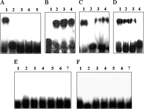
Binding of the proteins CcpA and HPr-Ser-P was tested with all four putative cre sequences. HPr or HPr-Ser-P alone showed no shift with any of the selected cre sequences (Fig. 2A, E, and F). CcpA alone retarded mobility of the DNA containing cre1 and cre2 (cre1-2) (Fig. 2A, lane 1), but large excess (400-fold) of cold, specific DNA (Fig. 2B) or 4 μg of nonspecific poly(dI-dC) DNA could not inhibit binding competitively, indicating the nonspecific nature of the interaction. Miwa et al. have also reported such nonspecific CcpA interaction earlier (24). Incubation of cre1-2 with HPr-Ser-P and CcpA together reduced the mobility of DNA in a concentration-dependent manner (Fig. 2C), although addition of HPr-Ser-P to CcpA produced only a marginal increase in mobility shift over CcpA alone. This could be due to the smaller size of HPr (runs like a 10-kDa protein in sodium dodecyl sulfate-polyacrylamide gel electrophoresis), compounded with an additional negative charge on HPr-Ser-P, pulling the complex more toward anode. The mobility shift of cre1-2 with CcpA-HPr-Ser-P complex was specific, as a 200-fold excess of cold DNA abolished the binding completely (Fig. 2D). These results indicate that formation of CcpA-HPr-Ser-P complex in solution is conducive to DNA binding. These observations are supported by the structural study (29) showing significant local structural changes in the DNA binding domain of CcpA induced directly by binding of HPr-Ser-P. No shift in mobility of cre3 or cre4 DNA was observed when CcpA, HPr, and HPr-Ser-P alone or as complex were tested (Fig. 2E and F), excluding their involvement in regulation of the cry4A gene.
FIG. 2.
(A) Binding of CcpA, HPr, and HPr-Ser-P to 117-bp probe containing cre1-2 by electrophoretic mobility shift assay. Lane 1, 0.78 nM CcpA; lane 2, free probe; lane 3, 0.6 nM HPr; lanes 4 and 5, 0.6 nM and 1.2 nM HPr-Ser-P, respectively. (B) Binding of CcpA to 117-bp probe and competition with cold DNA. Lane 1, free probe; lane 2, 0.78 nM CcpA; lanes 3 and 4, 0.78 nM CcpA plus 200- and 400-fold excess cold probe DNA, respectively. (C) Binding of CcpA and HPr-Ser-P to 117-bp probe. Lane 1, 0.78 nM CcpA; lane 2, free probe; lanes 3 and 4, 0.78 nM CcpA plus 0.6 nM and 1.2 nM HPr-Ser-P, respectively. (D) Competition of CcpA-HPr-Ser-P complex binding to 117-bp probe with cold probe DNA. Lane 1, 0.78 nM CcpA plus 0.6 nM HPr-Ser-P; lanes 2, 3, and 4, 0.78 nM CcpA plus 0.6 nM HPr-Ser-P with 50-, 100-, and 200-fold excess cold competitor DNA, respectively. (E) Binding of CcpA, HPr, and HPr-Ser-P to probe DNA containing cre3. Lane 1, free probe; lane 2, 0.78 nM CcpA; lane 3, 0.6 nM HPr; lanes 4 and 5, 0.6 nM and 1.2 nM HPr-Ser-P, respectively; lanes 6 and 7, 0.78 nM CcpA plus 0.6 nM and 1.2 nM HPr-Ser-P, respectively. (F) Binding of CcpA, HPr, and HPr-Ser-P to probe DNA containing cre4. The lanes are as in panel E.
Identification of CcpA-HPr-P complex binding site on cre1-2 by DNase I footprinting assay.
To identify the precise binding sequence of cre1-2, DNase I footprinting was performed as described by Schmitz and Galas (28) with minor modification. The 117-bp fragment containing cre1 and cre2 was end labeled with [α-32P]dCTP at the SpeI end of the coding strand. The wild-type DNA or the G2→A altered variant (20,000 cpm/μl) was incubated with CcpA and HPr-Ser-P proteins under conditions described above for gel retardation, except no EDTA was added to the buffer and each reaction mixture contained 2 μg poly(dI-dC) in a total volume of 45 μl. One unit of DNase I (Sigma) was added to the reaction mixture and incubated at room temperature for 2 min. The DNase I digestion was stopped by adding phenol-chloroform-isoamyl alcohol (25:24:1), and the DNA was precipitated with ethanol in the presence of 6 μg glycogen. The DNA was resolved in a 10% urea-polyacrylamide sequencing gel. A base-specific chemical cleavage sequencing reaction (21) was also performed in parallel on the same gel. The gels were dried and exposed to a PhosphorImager screen overnight.
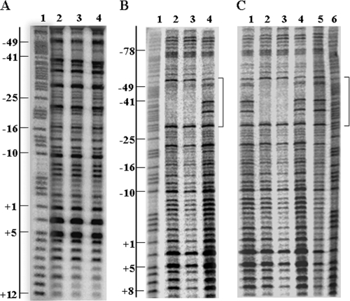
No protection was observed when CcpA or HPr-Ser-P alone was used (Fig. 3A). However, binding of the CcpA-HPr-Ser-P complex protected the cre1 region extending from position −50 to −29 (23 bp) from DNase I digestion (Fig. 3B, lanes 2 and 3). The protected region overlapped with the −35 element of the σ35-driven promoter of cry4A and showed 86% similarity to the 14-bp consensus sequence proposed by Weickert and Chambliss (30).
FIG. 3.
DNase I footprinting analysis of CcpA and HPr-Ser-P binding to the cre1 in the 117-bp fragment of the cry4A promoter. (A) Lane 1, G+A marker; lane 2, no-protein control; lane 3, 0.78 nM CcpA; lane 4, 0.6 nM HPr-Ser-P. Nucleotide positions (with +1 representing the transcription initiation nucleotide) are given on the left. (B) Protection of 117-bp wild-type DNA with CcpA-HPr-Ser-P complex. Lane 1, G+A marker; lanes 2 and 3, 0.78 nM CcpA plus 0.6 nM and 1.2 nM HPr-Ser-P, respectively; lane 4, no-protein control. The protected region is indicated on the right by a bracket in panels B and C. (C) DNase I protection of 117-bp wild-type and G49→A variant promoter DNA. Lane 1, no-protein control; lanes 2 and 3, 117-bp wild-type DNA with 0.78 nM CcpA plus 0.6 nM and 1.2 nM HPr-Ser-P, respectively; lanes 4 and 5, 117-bp G49→A DNA with 0.78 nM CcpA plus 0.6 nM and 1.2 nM HPr-Ser-P, respectively; lane 6, G+A marker.
Mutational analysis of the DNase I-protected sequence identified G49 as the critical nucleotide base (results described in the following section), which was subsequently verified by footprinting also. The 117-bp variant with G49→A mutation in the cre1 region showed no protection from DNase I in the presence of CcpA-HP-Ser-P complex (Fig. 3C, lanes 2, 3, 4, and 5), corroborating the promoter activity results.
Evaluation of promoter activity in cry4Apro-lacZ transcriptional fusions.
The cry4A promoter (cry4Apro) (458 bp upstream of the start codon) was amplified by PCR using primers 9 and 10 with KpnI and BamHI sites, respectively, with B. thuringiensis subsp. israelensis plasmid DNA. The coding region of the lacZ gene (3,067 bp) was obtained from Escherichia coli K-12 genomic DNA using primers 11 and 12 with BamHI and HindIII sites, respectively. The amplified DNA fragments were cloned into pGEM-T Easy vector, giving pSK5 and pSK6, respectively. The cry4A-lacZ fusion (pSK7) was produced by excising cry4Apro from pSK5 with NcoI/BamHI and ligating it to lacZ sequence in pSK6 at complementary sites. The promoter-lacZ fusion was excised from pSK7 with KpnI/HindIII and ligated in pHT370, an E. coli-Bacillus shuttle vector (Pasteur Institute), resulting in reporter construct pSK8.
To map the cre1 region revealed by footprinting, we performed base exchange mutagenesis using the Quik Change site-directed mutagenesis kit (Stratagene). Plasmids pSK8 (wild-type cry4Apro-lacZ gene fusion) and pSK2 (wild-type cry4A gene with promoter) were used as template for mutations with primers listed in the table in the supplemental material. The resulting plasmids with designated mutations in the cre1 region are listed in Table 1. A crystal-negative B. thuringiensis subsp. israelensis strain, 4Q7, obtained from the Bacillus Genetic Stock Center (Columbus, OH) was electroporated with the plasmids by the protocol described by Macaluso and Mettus (19). Two hundred microliters of competent cells was electroporated with 1 to 5 μl of plasmid DNA at 2.5 kV, 200 Ω, and 25-μF capacitance. The transformants were selected on Luria-Bertani plates containing 25 μg of erythromycin/ml.
Cells of strain 4Q7 containing the promoter variants were grown in 50 ml G medium (2) containing G salts, 0.2% glucose, and 0.15% yeast extract with erythromycin for 10 h (optical density at 595 nm [OD595] of 1.5), 14 h (OD595 of 1.8) or 18 h (OD595 of 1.75). To induce Cry4A synthesis, the cells were harvested, washed, and suspended in a resuspension medium (RM) [50 mM Tris-HCl [pH 7.5], 0.008% CaCl2·2H2O, 0.05% K2HPO4, 0.2% (NH4)2SO4, 0.00005% FeSO4·7H2O, 0.0005% CuSO4·5H2O, 0.005% MnSO4·H2O, 0.02% MgSO4], at 25 ml each in the presence of 0.6% glucose or no glucose. The cultures were incubated at 29°C with shaking, 1-ml aliquots were withdrawn every 2 h up to 8 h, and OD595 was measured. The cells were centrifuged, washed with ice-cold 25 mM Tris-HCl (pH 7.5), and suspended in 0.8 ml buffer Z (100 mM sodium phosphate buffer [pH 7.5], 10 mM KCl, 1 mM MgSO4, 50 mM β-mercaptoethanol) containing 0.4 mg lysozyme for permeabilization. The specific activity of β-galactosidase was determined using O-nitrophenyl-β-d-galactopyranoside (ONPG) (Sigma) as described earlier (22). One unit of β-galactosidase is the amount of enzyme producing 1 nmol of O-nitrophenol per min at 30°C. The average of triplicate samples was determined. The experiment was performed more than three times, and the data from one representative experiment are presented.
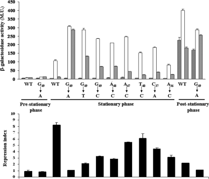
The mutations were selected to substitute most conserved bases in the consensus sequence proposed by Weickert and Chambliss (30). The basic principle was to introduced transversions from AT to CG and vice versa. A progressive decrease in glucose repression occurred as the position of the mutation moved across the cre from 3′ to 5′ and was abolished at G49→A, the putative σ factor contact site (18). The substitutions G49→T and G49→C also reduced the repression ratio, but not as dramatically as G49→A (Fig. 4). Mutations of A44→C and A43→G were performed to create the conserved CG at the center. Contrary to expectations, the mutations resulted in partial relief from glucose repression instead of enhancing it (data not shown). A second possibility of replacement of A43 and T42 with CG was not attempted, so its positive effect on repression cannot be ruled out. The centrally placed CG is considered highly conserved, and mutations of these bases are reported to result in loss in activity of the cre (9, 30, 32). However, like the cry4A gene, in several instances their absence is of little or no consequence to the activity (23). The substitution A48→C close to G49 also caused substantial relief from glucose repression. Although mutation of the other conserved positions at the 3′ end of the cre, namely A36→C, resulted in low repression ratio, the promoter activity was also reduced below the wild-type level (Fig. 4). The above results establish the presence of a 15-bp cre element in the cry4A gene which although 1 base longer shows strong similarity to the commonly observed 14-bp consensus sequence, although a longer (16 bp) cre, associated with the lev operon, has also been reported earlier in B. subtilis (20). The identified cre sequence overlaps the −35 element partially and extends up to the −50 position on the promoter. Apparently the CcpA-HPr-Ser-P complex blocks the RNAP recognition site on the promoter, inhibiting transcription (26).
FIG. 4.
Effect of glucose on cry4A promoter activity in vivo during different growth phases. cry4A promoter-lacZ fusion variants were grown in G medium (8 h for pre-stationary phase, 14 h for stationary phase, and 18 h for post-stationary phase) and resuspended in RM (upper panel) with glucose (gray bars) or without glucose (white bars). β-Galactosidase activities were measured at 8 h after resuspension. The hatched bars (T0) show β-galactosidase activities at the time of resuspension in RM. WT, wild type. The repression index (lower panel) was calculated as the ratio of β-galactosidase activity obtained in the absence of glucose to that in the presence of glucose. Subscripts show nucleotide positions relative to +1, and substitutions are shown with arrows. Values are the average of three independent experiments, each assayed in duplicate. Error bars represent the standard deviation. M.U., Miller units.
Promoter activity was low in cells containing the wild-type promoter grown for 10 h, and no glucose repression occurred. The promoter activity increased in both stationary-phase (14 h) and post-stationary-phase (18 h) cells and was highest in the latter, perhaps due to transcription from two different σ35 and σ28 promoters, known to occur commonly in other cry genes (6, 33). Parallel to the increase in transcriptional activity, glucose repression was also observed in the cells grown for 14 h (88%) and 18 h (55%) (Fig. 4). The repression was relieved significantly by G49→A mutation in both cells grown for 14 h (93%) and 18 h (89%). The results reveal that glucose repression of cry4A lasts until late in the post-stationary phase (18-h grown), when most of the glucose is consumed; hence, a glucose metabolite such as fructose-1,6-bisphosphate or one whose identity is not known at the moment might be mediating the glucose effect in the later phase.
Interestingly, the promoter activity also increased approximately threefold by G49→A mutation, (Fig. 4), suggesting more efficient binding/transcription of the promoter by RNAP. An explanation that needs to be verified experimentally could be generation of a sequence resembling the UP element, the A+T-rich component of bacterial promoters that stimulates transcription by interacting with the α subunit of RNAP (8). The RNAP holoenzyme in its extended form covers 70 to 80 bp from −55 to +20 of DNA, while the σ factor, which initiates recognition, covers the region from −55 to −35 (18). In the light of the above facts, a more efficient recognition/initiation of the promoter by the σ or α subunit of RNAP may result in elevated promoter activity. Thus, the vital role of guanine at −49 in orchestrating toxin gene regulation through twin effects on promoter activity is the key finding of this study and could be useful in developing a high-toxin-yielding strain.
Evaluation of cry4A gene expression by semiquantitative RT-PCR and Western blotting.
The cry4A gene with the promoter region (4001 bp) was amplified from B. thuringiensis subsp. israelensis plasmid, with forward primer 1 and backward primer 2. The amplified DNA fragment was cloned in the pGEM-T Easy vector (Promega), producing pSK1. The gene was ligated in the pHT370 vector, and pSK2 was obtained; G49→A mutation was performed in pSK2, producing pSK9. Both plasmids were transformed in strain 4Q7 (Table 1), producing SK2 and SK9.
The SK2 (cry4A with wild type promoter) and SK9 (cry4A with G49→A variant promoter) strains were grown in G medium for ∼14 h and resuspended in RM as described above, with or without 0.6% glucose. Total RNA was isolated from 5 ml culture using the RNeasy minikit (Qiagen) and treated with DNase I. About 0.3 μg of DNA-free total RNA was used as the template in reverse transcription-PCR (RT-PCR) using the One Step RT-PCR kit (Qiagen). Primer 14 and internal reverse primer 15 were used to amplify a 414-bp fragment of the cry4A gene. A 16S RNA gene fragment was amplified using primers 16 and 17 as a loading control. The PCR conditions were 94°C denaturation for 45 s, 54°C annealing for 45 s, and 72°C extension for 30 s for cry4A and 1.5 min for 16S RNA.
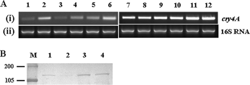
Glucose repressed cry4A transcripts in cells containing the wild-type construct at 1, 2, and 3 h after resuspension (Fig. 5A, lanes 1 to 6), while no effect was observed in the G49→A variant (Fig. 5A, lanes 7 to 12). At all time points, concentrations of cry4A transcripts were two- to threefold higher in the G49 mutant than the wild-type strain, implying increased promoter activity.
FIG. 5.
(A) Evaluation of glucose effect on cry4A transcription. Cells of strain 4Q7 were grown in G medium for 14 h and resuspended in RM with or without glucose. Samples were collected at different time points after resuspension in RM. (i) cry4A transcripts in the SK2 cells, lanes 1 to 6. Lanes 1, 3, and 5, with glucose after 1, 2, and 3 h, respectively; lanes 2, 4, and 6, no glucose after 1, 2, and 3 h, respectively. Lanes 7 to 12 contained cry4A transcripts in SK9 cells. Lanes 7, 9, and 11, with glucose after 1, 2, and 3 h, respectively; lanes 8, 10, and 12, no glucose after 1, 2, and 3 h, respectively (ii) 16S RNA loading control of respective test samples. (B) Western blot of Cry4A expression in 4Q7 strains. The SK2 and SK9 strains were grown in G medium and resuspended in RM, and samples were taken at 24 h. Lanes 1 and 2, total protein from SK2 cells without and with glucose, respectively; lanes 3 and 4, total protein from the SK9 (G49→A) variant without and with glucose, respectively.
Total proteins were isolated from SK2 and SK9 cells grown as described above for RNA isolation. Two-milliliter samples were removed at different intervals, and the cell pellets were solubilized in sodium dodecyl sulfate-polyacrylamide gel electrophoresis loading dye. Proteins were resolved by electrophoresis and transferred on nitrocellulose membrane by standard methods, and expression of Cry4A protein was detected using polyclonal antiserum diluted 1:10,000. The antiserum against recombinant domain I of the cry4A gene (762 bp, 35 kDa) was raised in mice. The effect of glucose on Cry4A expression in SK2 and SK9 is in accordance with the effect on mRNA observed earlier. The 130-kDa Cry4A protein was repressed by glucose in the wild-type strain but not in the G49→A variant as expected (Fig. 5B). In addition, the amount of Cry4A synthesis in the variant strain was also higher than that in the wild-type cells.
To allow smooth operation of essential metabolic processes that share common transcription factors in the mother cell, a stringent control of cry genes seems imperative. Since σ subunits play a key role in temporal control of Cry4A synthesis as well as sporulation in Bacillus (16, 25, 34), maintenance of adequate σ35 and σ28 levels is crucial for the cell. A high transcription rate of cry4A is likely to titrate out the cellular σ subunit pool and may lead to disruption of the developmental process. Indeed suppression of sporulation was observed earlier in the ptsH knockout mutant (16) and also in a separate study during overexpression of the recombinant Cry proteins (1, 27).
In conclusion, we demonstrate a cis-acting regulatory sequence, the cre associated with the cry4A promoter, which performs a vital function for the evolutionary success of the cell. The importance of this regulatory element lies in maintaining normal cellular growth, development and synthesis of large amounts of toxin protein in B. thuringiensis subsp. israelensis. Considering the significance of B. thuringiensis as the most successful biopesticide, understanding toxin protein regulation is the key to maximizing protein yield for biotechnological applications.
Supplementary Material
Acknowledgments
This work was supported by the Council of Scientific and Industrial Research, New Delhi, India.
We thank Didier Lereclus for the gift of vector pHT370.
Footnotes
Published ahead of print on 22 May 2009.
Supplemental material for this article may be found at http://jb.asm.org/.
REFERENCES
- 1.Agaisse, H., and D. Lereclus. 1995. How does Bacillus thuringiensis produce so much insecticidal crystal protein? J. Bacteriol. 1776027-6032. [DOI] [PMC free article] [PubMed] [Google Scholar]
- 2.Aronson, A. I., N. Angelo, and S. C. Holt. 1971. Regulation of extracellular protease production in Bacillus cereus T: characterization of mutants producing altered amounts of protease. J. Bacteriol. 1061016-1025. [DOI] [PMC free article] [PubMed] [Google Scholar]
- 3.Aronson, A. I., W. Beckman, and P. Dunn. 1986. Bacillus thuringiensis and related insect pathogens. Microbiol. Rev. 501-24. [DOI] [PMC free article] [PubMed] [Google Scholar]
- 4.Aung-Hilbrich, L. M., G. Seidel, A. Wagner, and W. Hillen. 2002. Quantification of the influence of HPrSer46P on CcpA-cre interaction. J. Mol. Biol. 31977-85. [DOI] [PubMed] [Google Scholar]
- 5.Banerjee-Bhatnagar, N. 1998. Modulation of CryIVA toxin protein expression by glucose in Bacillus thuringiensis israelensis. Biochem. Biophys. Res. Commun. 252402-406. [DOI] [PubMed] [Google Scholar]
- 6.Baum, J. A., and T. Malvar. 1995. Regulation of insecticidal crystal protein production in Bacillus thuringiensis. Mol. Microbiol. 181-12. [DOI] [PubMed] [Google Scholar]
- 7.Dossonnet, V., V. Monedero, M. Zagorec, A. Galinier, G. Pérez-Martinez, and J. Deutscher. 2000. Phosphorylation of HPr by the bifunctional HPr kinase/P-Ser-HPr phosphatase from Lactobacillus casei controls catabolite repression and inducer exclusion but not inducer expulsion. J. Bacteriol. 1822582-2590. [DOI] [PMC free article] [PubMed] [Google Scholar]
- 8.Estrem, S. T., T. Gaal, W. Ross, and R. L. Gourse. 1998. Identification of an UP element consensus sequence for bacterial promoters. Proc. Natl. Acad. Sci. USA 959761-9766. [DOI] [PMC free article] [PubMed] [Google Scholar]
- 9.Fujita, Y., Y. Miwa, A. Galinier, and J. Deutscher. 1995. Specific recognition of the Bacillus subtilis gnt cis-acting catabolite-responsive element by a protein complex formed between CcpA and seryl-phosphorylated HPr. Mol. Microbiol. 17953-960. [DOI] [PubMed] [Google Scholar]
- 10.Goldberg, L., and J. Margalitt. 1977. A bacterial spore demonstrating rapid larvicidal activity against Anopheles sergentii, Uranotaenia unguiculata, Aedes aegypti and Culex pipiens. Mosq. News 37355-358. [Google Scholar]
- 11.Gorke, B., and J. Stulke. 2008. Carbon catabolite repression in bacteria: many ways to make the most out of nutrients. Nat. Rev. Microbiol. 6613-624. [DOI] [PubMed] [Google Scholar]
- 12.Gosseringer, R., E. Kuster, A. Galinier, J. Deutscher, and W. Hillen. 1997. Cooperative and non-cooperative DNA binding modes of catabolite control protein CcpA from Bacillus megaterium result from sensing two different signals. J. Mol. Biol. 266665-676. [DOI] [PubMed] [Google Scholar]
- 13.Helmann, J. D., and M. J. Chamberlin. 1988. Structure and function of bacterial sigma factors. Annu. Rev. Biochem. 57839-872. [DOI] [PubMed] [Google Scholar]
- 14.Höfte, H., and H. R. Whiteley. 1989. Insecticidal crystal proteins of Bacillus thuringiensis. Microbiol. Rev. 53242-255. [DOI] [PMC free article] [PubMed] [Google Scholar]
- 15.Insell, J. P., and P. C. Fitz-James. 1985. Composition and toxicity of the inclusion of Bacillus thuringiensis subsp. israelensis. Appl. Environ. Microbiol. 5056-62. [DOI] [PMC free article] [PubMed] [Google Scholar]
- 16.Khan, S. R., and N. Banerjee-Bhatnagar. 2002. Loss of catabolite repression function of HPr, the phosphocarrier protein of the bacterial phosphotransferase system, affects expression of the cry4A toxin gene in Bacillus thuringiensis subsp. israelensis. J. Bacteriol. 1845410-5417. [DOI] [PMC free article] [PubMed] [Google Scholar]
- 17.Khan, S. R., J. Deutscher, R. A. Vishwakarma, V. Monedero, and N. B. Bhatnagar. 2001. The ptsH gene from Bacillus thuringiensis israelensis. Characterization of a new phosphorylation site on the protein HPr. Eur. J. Biochem. 268521-530. [DOI] [PubMed] [Google Scholar]
- 18.Lewin, B. 2000. Genes VII, p. 241. Oxford University Press, Inc., Oxford, United Kingdom.
- 19.Macaluso, A., and A.-M. Mettus. 1991. Efficient transformation of Bacillus thuringiensis requires nonmethylated plasmid DNA. J. Bacteriol. 1731353-1356. [DOI] [PMC free article] [PubMed] [Google Scholar]
- 20.Martin-Verstraete, I., J. Deutscher, and A. Galinier. 1999. Phosphorylation of HPr and Crh by HprK, early steps in the catabolite repression signalling pathway for the Bacillus subtilis levanase operon. J. Bacteriol. 1812966-2969. [DOI] [PMC free article] [PubMed] [Google Scholar]
- 21.Maxam, A. M., and W. Gilbert. 1980. Sequencing end-labeled DNA with base-specific chemical cleavages. Methods Enzymol. 65499-560. [DOI] [PubMed] [Google Scholar]
- 22.Miller, J. H. 1972. Experiments in molecular genetics. Cold Spring Harbor Laboratory, Cold Spring Harbor, NY.
- 23.Miwa, Y., K. Nagura, S. Eguchi, H. Fukuda, J. Deutscher, and Y. Fujita. 1997. Catabolite repression of the Bacillus subtilis gnt operon exerted by two catabolite-responsive elements. Mol. Microbiol. 231203-1213. [DOI] [PubMed] [Google Scholar]
- 24.Miwa, Y., M. Saikawa, and Y. Fujita. 1994. Possible function and some properties of the CcpA protein of Bacillus subtilis. Microbiology 1402567-2575. [DOI] [PubMed] [Google Scholar]
- 25.Moran, C. P., Jr. 1989. Sigma factors and the regulation of transcription, p. 167-184. In I. Smith, R. A. Slepecky, and P. Setlow (ed.), Regulation of prokaryotic development: structural and functional analysis of bacterial sporulation and germination. American Society for Microbiology, Washington, DC.
- 26.Nicholson, W. L., Y. K. Park, T. M. Henkin, M. Won, M. J. Weickert, J. A. Gaskell, and G. H. Chambliss. 1987. Catabolite repression-resistant mutations of the Bacillus subtilis alpha-amylase promoter affect transcription levels and are in an operator-like sequence. J. Mol. Biol. 198609-618. [DOI] [PubMed] [Google Scholar]
- 27.Poncet, S., E. Dervyn, A. Klier, and G. Rapoport. 1997. Spo0A represses transcription of the cry toxin genes in Bacillus thuringiensis. Microbiology 1432743-2751. [DOI] [PubMed] [Google Scholar]
- 28.Schmitz, A., and D. J. Galas. 1978. DNase I foot printing: a simple method for the detection of protein-DNA specificity. Nucleic Acids Res. 53157-3170. [DOI] [PMC free article] [PubMed] [Google Scholar]
- 29.Schumacher, M. A., G. S. Allen, M. Diel, G. Seidel, W. Hillen, and R. G. Brennan. 2004. Structural basis for allosteric control of the transcription regulator CcpA by the phosphoprotein HPr-Ser46-P. Cell 118731-741. [DOI] [PubMed] [Google Scholar]
- 30.Weickert, M. J., and G. H. Chambliss. 1990. Site-directed mutagenesis of a catabolite repression operator sequence in Bacillus subtilis. Proc. Natl. Acad. Sci. USA 876238-6242. [DOI] [PMC free article] [PubMed] [Google Scholar]
- 31.Weickert, M. J., and S. Adhya. 1992. A family of bacterial regulators homologous to Gal and Lac repressors. J. Biol. Chem. 26715869-15874. [PubMed] [Google Scholar]
- 32.Wray, L. V., Jr., F. K. Pettengill, and S. H. Fisher. 1994. Catabolite repression of the Bacillus subtilis hut operon requires a cis-acting site located downstream of the transcription initiation site. J. Bacteriol. 1761894-1902. [DOI] [PMC free article] [PubMed] [Google Scholar]
- 33.Yoshisue, H., H. Sakai, K. Sen, M. Yamagiwa, and T. Komano. 1997. Identification of a second transcriptional start site for the insecticidal protein gene cryIVA of Bacillus thuringiensis subsp. israelensis. Gene 185251-255. [DOI] [PubMed] [Google Scholar]
- 34.Yoshisue, H., T. Fukada, K.-I. Yoshida, K. Sen, S.-I. Kurosawa, H. Sakai, and T. Komano. 1993. Transcriptional regulation of Bacillus thuringiensis subsp. israelensis mosquito larvicidal crystal protein gene cryIVA. J. Bacteriol. 1752750-2753. [DOI] [PMC free article] [PubMed] [Google Scholar]
Associated Data
This section collects any data citations, data availability statements, or supplementary materials included in this article.


