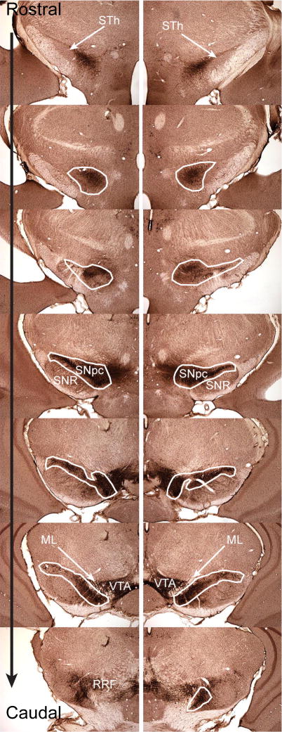
Figure 1.
Outline of Substantia Nigra pars compacta used to determine cell number. Forty micron free-floating cryostat sections were stained for tyrosine hydroxylase expression. The sections shown are spaced every 240 microns and thus represent every 6th section from a single brain. White outlines indicate areas used to define the SNpc for the purposes of stereology. The rostral portion of the SNpc starts with the first TH positive cells near the end of the subthalamic nucleus (STh) and the caudal SNpc is where the retrorubral field (RRF). The outlines used include the substantia nigra lateralis. The mediodorsal boundary of the SNpc is defined by TH expression. The dorsal portion of the SN pars reticulata (SNR) defines the ventrolateral boundary. The anterior medial boundary is defined by the ventral tegmental area (VTA) and by size and orientation of stained cells. DA neurons of the SNpc are larger than ventral tegmental area DA neurons, and SNpc DA neurons orient along the long axis of the SNpc. The posterior medial portion of the SNpc is defined by the medial lemniscus (ML).

