

Abstract
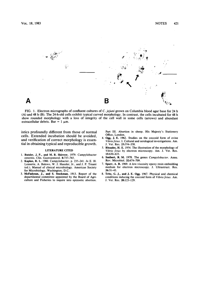
Confluent cultures of Campylobacter jejuni incubated for 24 and 48 h each were examined by electron microscopy. Although the 24-h-old cells exhibited typical curved morphology, the 48-h-old cells showed rounded morphology with a loss of cell integrity. This appeared to be an autolytic process that occurred very rapidly after the culture became mature. These results confirm previous evidence that the coccoid form of this organism is a degenerate state.
Full text
PDF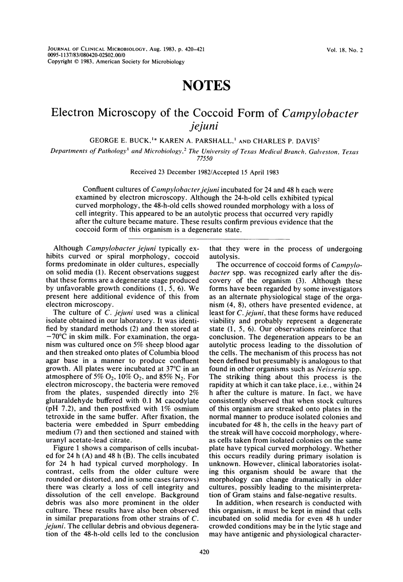
Images in this article
Selected References
These references are in PubMed. This may not be the complete list of references from this article.
- Butzler J. P., Skirrow M. B. Campylobacter enteritis. Clin Gastroenterol. 1979 Sep;8(3):737–765. [PubMed] [Google Scholar]
- OGG J. E. Studies on the coccoid form of ovine Vibrio fetus I. Cultural and serologic investigations. Am J Vet Res. 1962 Mar;23:354–358. [PubMed] [Google Scholar]
- RHOADES H. E. The illustration of the morphology of vibrio fetus by electron microscopy. Am J Vet Res. 1954 Oct;15(57):630–633. [PubMed] [Google Scholar]
- Smibert R. M. The genus Campylobacter. Annu Rev Microbiol. 1978;32:673–709. doi: 10.1146/annurev.mi.32.100178.003325. [DOI] [PubMed] [Google Scholar]
- Spurr A. R. A low-viscosity epoxy resin embedding medium for electron microscopy. J Ultrastruct Res. 1969 Jan;26(1):31–43. doi: 10.1016/s0022-5320(69)90033-1. [DOI] [PubMed] [Google Scholar]