Abstract
The hemagglutinin-neuraminidase (HN) protein of Newcastle disease virus (NDV) is a multifunctional protein that plays a crucial role in virus infectivity. In this study, using the mesogenic strain Beaudette C (BC), we mutated three conserved amino acids thought to be part of the binding/catalytic active site in the HN protein. We also mutated five additional residues near the proposed active site that are nonconserved between BC and the avirulent strain LaSota. The eight recovered NDV HN mutants were assessed for effects on biological activities. While most of the mutations had surprisingly little effect, mutation at conserved residue Y526 reduced the neuraminidase, receptor binding, and fusion activities and attenuated viral virulence in eggs and young birds.
Newcastle disease virus (NDV) is an avian pathogen of the genus Avulavirus in the family Paramyxoviridae (10). The envelope of NDV contains two surface glycoproteins, the fusion (F) protein and the HN (hemagglutinin-neuraminidase [NA]) protein. The F protein mediates viral penetration and requires cleavage-activation by host protease. Cleavability of the F protein is a major determinant of virulence. However, other viral proteins, including HN, also contribute to virulence (5). HN is a multifunctional glycoprotein. It recognizes sialic acid-containing receptors on cell surfaces; promotes the fusion activity of F protein, thereby allowing the virus to penetrate the cell surface; and acts as an NA that removes sialic acid from progeny virus particles to prevent viral self-aggregation (9).
HN is a type II homotetrameric glycoprotein with a monomer length of 577 amino acids for most NDV strains (14). The ectodomain of the HN protein consists of a 95-amino-acid stalk region supporting a 428-amino-acid terminal globular head. Although mutations in the transmembrane and stalk regions of the HN protein can affect the structure and activities of the protein (11, 15), the antigenic, receptor recognition, and NA active sites are all localized in the globular head (12, 16). The X-ray crystal structure of the globular head of the NDV HN protein has identified residues that appear to contribute to receptor recognition, NA, and fusion activities (4). Previous studies have proposed that conserved residues R174, I175, D198, K236, R416, R498, Y526, and E547 are important in receptor recognition and NA activities and that residues R174 and E547 influence the fusion promotion activity of the HN protein (3, 4, 6). Although transfection studies using plasmids expressing HN mutants of NDV have highlighted the importance of these residues in different biological functions of the HN protein, their contribution to NDV biology and pathogenesis in the context of the complete virus was not known.
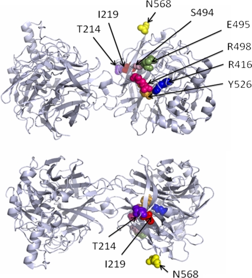
In this study, we examined the roles of three of the above-named conserved residues, R416, R498, and Y526 (all located near the sialic acid binding site), in the biological activities and pathogenesis of the HN protein of NDV in the context of infectious virus. In addition, comparison of the HN protein sequence between the avirulent strain LaSota and the moderately virulent strain Beaudette C (BC) identified 12 amino acid differences in the globular head region of the HN protein (H203, T214, I219, S228, L269, A271, E293, G310, S494, E495, T502, and N568, named according to the BC amino acid assignment). We also examined five of these nonconserved residues, T214, I219, S494, E495, and N568, located in close proximity to residues identified earlier by crystal structure studies, to determine whether these might affect HN function and contribute to the difference in pathogenicity between the LaSota and BC strains (Fig. 1).
FIG. 1.
Three-dimensional structure of the NDV HN protein showing the positions of amino acid residues that were substituted in the present study. The residues are shown in space-filling mode and represented in different colors. The MacPymol (DeLano Scientific) software was used to generate the model of the globular domain of the NDV HN monomer. The structure was derived from the crystal structure of the NDV HN protein reported by Crennell et al. (4).
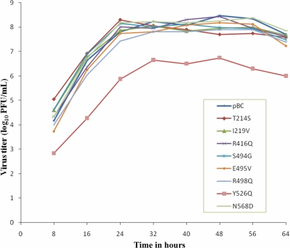
We used site-directed mutagenesis (2) to introduce individual amino acid substitutions into a cDNA of the HN gene of strain BC. For the conserved residues, we changed arginine at positions 416 and 498 and tyrosine at position 526 to polar glutamine. For the nonconserved residues, the assignments T214, I219, S494, E495, and N568 of strain BC were altered to the corresponding assignments of strain LaSota: S214, V219, G494, V495, and D568, respectively. Each mutagenized HN gene was then inserted into a full-length cDNA clone of the BC antigenome. These clones were transfected into HEp2 cells, and mutant viruses were recovered as previously described (8). These viruses were designated according to the substitutions introduced: T214S, I219V, R416Q, S494G, E495V, R498Q, Y526Q, and N568D. The HN genes from recovered viruses were sequenced. This confirmed the presence of each introduced mutation and the lack of adventitious mutations in the HN gene. To determine the stability of each HN mutation, the recovered viruses were passaged five times in 9-day-old embryonated chicken eggs and five times in chicken embryo fibroblast DF-1 cells. Sequence analysis of the HN gene of the mutant viruses at each passage showed that the introduced mutations were unaltered (data not shown). To rule out the possibility that change in the HN protein sequence could be compensated for by a mutation in the F protein, the F gene from each recovered virus was sequenced. No compensatory mutations in the F gene were observed (data not shown). The HN protein content of each mutant virus, determined by sodium dodecyl sulfate-polyacrylamide gel electrophoresis and Coomassie staining, was very similar to that of the parental BC virus (pBC) (Table 1). The multicycle growth kinetics of the recombinant HN mutant viruses in DF-1 cells (Fig. 2) showed that the replication kinetics of all of the HN mutant viruses were similar to those of pBC, with the exception of the Y526Q mutant, which showed delayed growth and had a lower virus yield (1.5 to 2.0 log10 PFU/ml) than the parental and other mutant viruses. In addition, the Y526Q mutant produced syncytia at 72 h, whereas the parental and other mutant viruses initiated syncytia at 24 h postinfection. These studies showed the importance of amino acid residue Y526 at the active site of the HN protein of NDV.
TABLE 1.
Biological activities of HN mutants of NDV
| Virus | Expressiona | Cell surface expressionb | NA activityc | HAd activityc | Fusiond |
|---|---|---|---|---|---|
| pBC | 100.00 | 100.00 | 100.00 | 100.00 | 100.00 |
| T214S mutant | 110.1 ± 15.5 | 102.5 ± 4.9 | 109.1 ± 8.3 | 99.1 ± 8.2 | 101.5 ± 4.2 |
| I219V mutant | 105.8 ± 5.2 | 100.1 ± 2.8 | 112.2 ± 9.2 | 99.3 ± 9.5 | 92.9 ± 5.4 |
| R416Q mutant | 101.2 ± 6.3 | 99.5 ± 2.5 | 106.5 ± 9.1 | 101.0 ± 9.1 | 90.6 ± 4.3 |
| S494G mutant | 110.3 ± 12.5 | 105.7 ± 6.5 | 87.6 ± 6.2 | 103.2 ± 7.5 | 99.1 ± 2.4 |
| E495V mutant | 106.1 ± 12.2 | 101.2 ± 3.2 | 94.4 ± 3.1 | 101.1 ± 7.2 | 89.2 ± 4.5 |
| R498Q mutant | 108.5 ± 13.9 | 106.9 ± 8.1 | 102.8 ± 5.4 | 101.8 ± 8.8 | 102.0 ± 6.2 |
| Y526Q mutant | 112.2 ± 15.6 | 103.9 ± 4.1 | 66.2 ± 4.2 | 70.0 ± 4.1 | 50.4 ± 3.1 |
| N568D mutant | 105.1 ± 7.8 | 98.9 ± 2.1 | 102.5 ± 8.1 | 103.7 ± 7.1 | 87.4 ± 5.2 |
Shown is the HN protein content of purified virus relative to that of the pBC parent determined by sodium dodecyl sulfate-polyacrylamide gel electrophoresis and Coomassie staining. All values are averages ± standard deviations of three independent experiments.
Shown are the cell surface expression levels of HN mutants relative to the level of the pBC parent. Expression of the HN protein was quantitated by Western blot analysis using HN-specific monoclonal antibodies. All values are averages ± standard deviations of three independent experiments.
Shown are the HAd and NA activities of HN mutants expressed as normalized values relative to the amount of HN expressed at the cell surface. Each value is relative to the activity of the pBC parent. All values are averages ± standard deviations of three independent experiments.
Shown are the fusion promotion activity of HN mutants expressed relative to the activity of the pBC parent. Cell fusion was calculated as the ratio of the total number of nuclei in multinuclear cells to the total number of nuclei in the field. The values are averages ± standard deviations of three independent experiments.
FIG. 2.
Multicycle growth kinetics of HN mutants of NDV in chicken embryo fibroblast (DF-1) cells. Cells were infected with the indicated parental or mutant virus at an multiplicity of infection of 0.01. Supernatant samples were collected at 8-h intervals until 64 h postinfection, and virus titers were determined at different time points by plaque assay. Values are averages from three independent experiments.
Next we analyzed whether the mutations in the HN protein modulated the biological activities of NDV in cultured cells (Table 1). Vero cells were infected with pBC or the HN mutant viruses, and cell surface expression was quantitated by Western blot analysis using HN-specific monoclonal antibodies. The amount of HN protein expressed on the cell surface by each mutant virus was similar to that of pBC. The NA activity of the mutant viruses was assayed by a fluorescence-based assay (13). The percent biological activity of each virus is shown relative to that of pBC, whose biological activities were considered to be 100%. The NA activity of the Y526Q mutant was 66% of that of pBC, which was the greatest reduction of all of the mutants, followed by 88% for the S494G virus. Hemadsorption (HAd) activity was assayed at 4°C by incubating the infected Vero cells with guinea pig red blood cells. The HAd activity of the Y526Q mutant was 70% of that of pBC, while the other mutants maintained HAd activity comparable to that of pBC. We also evaluated the fusion activity of each HN mutant virus in Vero cells (Table 1) by calculating the fusion index as described previously (7). The fusion activity of the Y526Q mutant virus was only 50% of that of pBC, followed by 89% for the E495V mutant. The other HN mutants did not have fusion activities different from that of pBC. These studies emphasize the importance of the tyrosine residue present at position 526, found near the sialic acid binding site of the HN protein of NDV, in fusion promotion and NA activities.
To determine whether the differences in the in vitro biological characteristics of the Y526Q mutant virus resulted in decreased virulence in chickens in vivo, two internationally accepted pathogenicity tests were performed. The mean death time (MDT) test with 9-day-old embryonated chicken eggs was performed as described previously (1). The MDT was recorded as the time (in hours) for a minimum lethal dose of virus to kill all of the chicken embryos infected (Table 2). The MDT result showed a significant increase in the time required by the Y526Q HN mutant virus (98 h) to kill 9-day-old chicken embryos compared to that required for pBC (60 h), indicating a reduced virulence of the Y526Q mutant virus. The S494G HN mutant virus, involving a nonconserved residue, also had an MDT (70 h) slightly longer than that of pBC. The intracerebral pathogenicity index (ICPI) test was performed as described previously (1). Each virus was inoculated intracerebrally into groups of 10 1-day-old chicks. The birds were observed for paralysis and death once every 12 h for 8 days, and ICPI values were calculated (1). The ICPI values of both of these mutants were lower than that of pBC (Table 2). In aggregate, these results indicated that mutation of the residues at positions 526 and 494 attenuated the virus.
TABLE 2.
Pathogenicitya of HN mutants of NDV
| Virus | MDT (h)b | ICPI scorec |
|---|---|---|
| pBC | 58 | 1.51 |
| T214S mutant | 59 | NDd |
| I219V mutant | 60 | ND |
| R416Q mutant | 59 | ND |
| S494G mutant | 70 | 1.36 |
| E495V mutant | 58 | ND |
| R498Q mutant | 64 | ND |
| Y526Q mutant | 98 | 1.33 |
| N568D mutant | 57 | ND |
The virulence of the mutant and parental BC viruses was evaluated by MDT in 9-day-old chicken embryos and by ICPI in 1-day-old chickens.
The MDT duration is >90 h for lentogenic strains, 60 to 90 h for mesogenic strains, and <60 h for velogenic strains.
The ICPI values for velogenic strains approach the maximum score of 2.00, whereas lentogenic strains give values close to 0.
ND, not determined.
In summary, we investigated the importance of three conserved residues, namely, R416, R498, and Y526, which appear to be part of the active site of the HN protein (4). In the previous studies, mutation of R416 to Q or L essentially eliminated NA and strongly reduced or eliminated HAd activities in transfected cells, although effects on fusion activity were not evaluated (4, 6). Other substitutions at this position involving A, D, E, or K also strongly reduced both NA and HAd activities but resulted in only a marginal decrease in fusion activity (3). In contrast, in the present study, the R416Q mutation in the context of the complete infectious virus had little or no effect on the HAd, NA, and fusion activities and had no effect on pathogenicity as measured by MDT. In one previous study, mutation of R498 to Q resulted in a moderate reduction in NA activity and little effect on HAd activity when evaluated by cDNA transfection (4), whereas in other studies, mutation of R498 to Q or L had more-severe effects on NA and HAd activities (3, 6) but little effect on fusion activity (3). In contrast, in the present study, the same mutation in the context of infectious virus had little or no effect on HAd, NA, and fusion activities or on the MDT. Finally, when evaluated in previous work with transfected HN cDNA, mutation of Y526 to Q or L strongly reduced or eliminated both NA and HAd activities (4, 6). Fusion promotion was not measured in this previous study for the Y526Q mutant, but mutation to F or H, which also strongly inhibited NA and HAd activities, had no effect on fusion activity (3). In contrast, in the present study, the Y526Q mutation in the complete virus resulted in decreased HAd, NA, and fusion activities, as well as a reduction in pathogenicity. This highlighted the importance of residue Y526 in the biological activities of the HN protein. The various activities of the HN protein were much less sensitive to mutation when evaluated in the context of the complete virus than in the context of transfected cDNA. In addition, while there sometimes was dissociation of the NA, HAd, and fusion promotion activities in the transfected cDNA assay, it was not observed in the context of the complete mutant virus.
Second, we investigated the functional importance of five other residues that differ between the lentogenic LaSota and mesogenic BC strains of NDV and are in close proximity to the above-mentioned conserved residues in the crystal structure. We found that mutations at these positions generally had little or no effect on the NA, HAd, or fusion promotion activity of the HN protein and did not alter the virulence of the virus. The one exception was the S494G mutation, which resulted in a modest reduction in NA activity and virulence. We previously showed that the HN protein of strain BC contributes to viral tropism and virulence, compared to strain LaSota (5). Thus, residue S494 may play a role in the difference between these two strains and may contribute to the tropism and virulence of the BC strain. This study indicates that mutating certain key amino acids in the globular head region of the NDV HN glycoprotein can attenuate the virulence of NDV and may provide a means to produce a live attenuated vaccine virus.
Acknowledgments
We thank Flavia Dias and all our laboratory members for their excellent technical assistance and help. We thank Daniel R. Perez for his help in drawing the three-dimensional structure of the NDV HN protein. We also thank Ireen Dryburgh-Barry for proofreading the manuscript.
This research was supported by NIAID contract N01A060009 (85% support) and the NIAID NIH Intramural Research Program (15% support).
The views expressed herein do not necessarily reflect the official policies of the Department of Health and Human Services, and the mention of trade names, commercial practices, or organizations does not imply endorsement by the U.S. Government.
Footnotes
Published ahead of print on 27 May 2009.
REFERENCES
- 1.Alexander, D. J. 1989. Newcastle disease, p. 114-120. In H. G. Purchase, L. H. Arp, C. H. Domermuth, and J. E. Pearson (ed.), A laboratory manual for the isolation and identification of avian pathogens, 3rd ed. American Association for Avian Pathologists, Inc., Kennett Square, PA.
- 2.Byrappa, S., D. K. Gavin, and K. C. Gupta. 1995. A highly efficient procedure for site-specific mutagenesis of full-length plasmids using Vent DNA polymerase. Genome Res. 5404-407. [DOI] [PubMed] [Google Scholar]
- 3.Connaris, H., T. Takimoto, R. Russell, S. Crennell, I. Moustafa, A. Portner, and G. Taylor. 2002. Probing the sialic acid binding site of the hemagglutinin-neuraminidase of Newcastle disease virus: identification of key amino acids involved in cell binding, catalysis, and fusion. J. Virol. 761816-1824. [DOI] [PMC free article] [PubMed] [Google Scholar]
- 4.Crennell, S., T. Takimoto, A. Portner, and G. Taylor. 2000. Crystal structure of the multi-functional paramyxovirus hemagglutinin-neuraminidase. Nat. Struct. Biol. 71068-1074. [DOI] [PubMed] [Google Scholar]
- 5.Huang, Z., A. Panda, S. Elankumaran, D. Govindarajan, D. D. Rockemann, and S. K. Samal. 2004. The hemagglutinin-neuraminidase protein of Newcastle disease virus determines tropism and virulence. J. Virol. 784176-4184. [DOI] [PMC free article] [PubMed] [Google Scholar]
- 6.Iorio, R. M., G. M. Field, J. M. Sauvron, A. M. Mirza, R. Deng, P. J. Mahon, and J. P. Langedijk. 2001. Structural and functional relationship between the receptor recognition and neuraminidase activities of the Newcastle disease virus hemagglutinin-neuraminidase protein: receptor recognition is dependent on neuraminidase activity. J. Virol. 751918-1927. [DOI] [PMC free article] [PubMed] [Google Scholar]
- 7.Kohn, A. 1965. Polykaryocytosis induced by Newcastle disease virus in monolayers of animal cells. Virology 26228-245. [DOI] [PubMed] [Google Scholar]
- 8.Krishnamurthy, S., Z. Huang, and S. K. Samal. 2000. Recovery of a virulent strain of Newcastle disease virus from cloned cDNA: expression of a foreign gene results in growth retardation and attenuation. Virology 278168-182. [DOI] [PubMed] [Google Scholar]
- 9.Lamb, R. A., and D. Kolakofsky. 2001. Paramyxoviridae: the viruses and their replication, p. 1305-1340. In D. M. Knipe and P. M. Howley (ed.), Fields virology. Lippincott Williams & Wilkins, Philadelphia, PA.
- 10.Lamb, R. A., P. L. Collins, D. Kolakofsky, J. A. Melero, Y. Nagai, M. B. A. Oldstone, C. R. Pringle, and B. K. Rima. 2005. Family Paramyxoviridae, p. 655-668. In C. M. Fauquet (ed.), Virus taxonomy: the classification and nomenclature of viruses. The eighth report of the International Committee on Taxonomy of Viruses. Elsevier Academic Press, San Diego, CA.
- 11.McGinnes, L., T. Sergel, and T. Morrison. 1993. Mutations in the transmembrane domain of the HN protein of Newcastle disease virus affect the structure and activity of the protein. Virology 196101-110. [DOI] [PubMed] [Google Scholar]
- 12.Mirza, A. M., R. T. Deng, and R. M. Iorio. 1994. Site-directed mutagenesis of a conserved hexapeptide in the paramyxovirus hemagglutinin-neuraminidase glycoproteins: effects on antigenic structure and function. J. Virol. 685093-5099. [DOI] [PMC free article] [PubMed] [Google Scholar]
- 13.Potier, M., L. Mameli, M. Belishem, L. Dallaire, and S. B. Melancon. 1979. Fluorometric assay of neuraminidase with a sodium (4-methylumbelliferyl-α-d-N-acetylneuraminate) substrate. Anal. Biochem. 94287-296. [DOI] [PubMed] [Google Scholar]
- 14.Schuy, W., W. Garten, D. Linder, and H. D. Klenk. 1984. The carboxy terminus of the hemagglutinin-neuraminidase of Newcastle disease virus is exposed at the surface of the viral envelope. Virus. Res. 1415-426. [DOI] [PubMed] [Google Scholar]
- 15.Stone-Hulslander, J., and T. G. Morrison. 1999. Mutational analysis of heptad repeats in the membrane-proximal region of Newcastle disease virus HN protein. J. Virol. 733630-3637. [DOI] [PMC free article] [PubMed] [Google Scholar]
- 16.Thompson, S. D., W. G. Laver, K. G. Murti, and A. Portner. 1988. Isolation of a biologically active soluble form of the hemagglutinin-neuraminidase protein of Sendai virus. J. Virol. 624653-4660. [DOI] [PMC free article] [PubMed] [Google Scholar]


