Abstract
While endocytosis can regulate morphogen distribution, its precise role in shaping these gradients is unclear. Even more enigmatic is the role of retromer, a complex that shuttles proteins between endosomes and the Golgi apparatus, in Wnt gradient formation. Here we report that DPY-23, the C. elegans μ subunit of the clathrin adaptor AP-2 that mediates the endocytosis of membrane proteins, regulates Wnt function. dpy-23 mutants display Wnt phenotypes, including defects in neuronal migration, neuronal polarity and asymmetric cell division. DPY-23 acts in Wnt-expressing cells to promote these processes. MIG-14, the C. elegans homolog of the Wnt-secretion factor Wntless, also acts in these cells to control Wnt function. In dpy-23 mutants, MIG-14 accumulates at or near the plasma membrane. By contrast, MIG-14 accumulates in intracellular compartments in retromer mutants. Based on our observations, we propose that intracellular trafficking of MIG-14 by AP-2 and retromer plays an important role in Wnt secretion.
Introduction
During development, morphogens control pattern formation by specifying individual cell fates along their gradients (Tabata and Takei, 2004). These graded signaling molecules can also function as guidance cues by providing instructive information for migrating cells and axon growth cones (Zou and Lyuksyutova, 2007). Over the years, many morphogens and guidance cues have been identified, yet how gradients of these molecules are produced and shaped is often mysterious.
Wnts are a family of secreted glycoproteins that have been shown to act as classical morphogens (Zecca et al., 1996). Wnts can also function as guidance cues to control the migrations of axon growth cones in C. elegans, Drosophila and the vertebrate central nervous system (Liu et al., 2005; Lyuksyutova et al., 2003; Pan et al., 2006; Yoshikawa et al., 2003). In C. elegans, Wnts also act as positional cues to control the asymmetric divisions of the T and EMS blast cells (Goldstein et al., 2006).
Two classes of molecules have recently been shown to function in Wnt-secreting cells to produce a functional Wnt. The first are components of the retromer complex. Retromer retrieves from endosomes receptors that shuttle vacuolar or lysosomal hydrolases, returning the receptors to the Golgi (Seaman, 2005; Seaman et al., 1998). C. elegans homologs of Vps26p, Vps29p and Vps35p, components in the retromer complex, are necessary for several developmental processes that require Wnts, and VPS-35 has been shown to function in cells that produce the Wnt EGL-20 (Coudreuse et al., 2006; Prasad and Clark, 2006).
Three groups have found a new molecule known as Wntless, Evi or Sprinter that is necessary for Wnt function (Banziger et al., 2006; Bartscherer et al., 2006; Goodman et al., 2006). Wntless interacts physically with Wingless, targeting it to the cell surface for secretion (Banziger et al., 2006). C. elegans MIG-14, which is also known as MOM-3, is the homolog of Wntless (Banziger et al., 2006). The mig-14 alleles were originally identified in screens for mutants with defects in QL migration, which the Wnt EGL-20 regulates (Harris et al., 1996; Maloof et al., 1999). A screen for mutants with defects in the asymmetric division of the EMS blast cell identified the original mom-3 allele. MOM-3 acts in P2, the cell that secretes the Wnt MOM-2 and signals to EMS, causing it to divide asymmetrically (Thorpe et al., 1997).
In this paper, we establish a connection between endocytosis, retromer function and MIG-14. The C. elegans gene dpy-23 encodes the μ subunit of the AP-2 clathrin adaptor complex that is necessary for receptor-mediated endocytosis and functions in several Wnt-related processes. Our observations indicate that efficient Wnt secretion requires endocytosis and trafficking of MIG-14 by retromer.
Results
dpy-23 Mutants Display Wnt Phenotypes
During embryogenesis, the two HSN neurons migrate anteriorly from the tail to positions near the middle of the embryo (Sulston et al., 1983). In a screen for mutants with defective HSN migration, we isolated dpy-23(gm17). The HSNs were usually displaced posteriorly in the mutant, migrating only part of the distance from the tail to their normal destinations (Figure S1). The mutant also had a squat body stature that is referred to as the Dumpy (Dpy) phenotype. The mutation mapped to the same region of the X chromosome as dpy-23 and failed to complement dpy-23(e840). Both e840 and gm17 mutants displayed Dpy, HSN and several other phenotypes that are described below. All of these phenotypes were maternally and zygotically rescued: only homozygous mutants of homozygous mothers exhibited the mutant phenotypes.
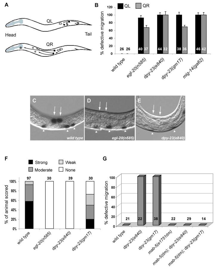
Several Wnts contribute to HSN migration (Pan et al., 2006) (Figure S1), and further phenotypic analysis revealed that dpy-23 mutants exhibited a number of other phenotypes shared by Wnt mutants. In the first larval stage (L1), the left Q neuroblast (QL) and its descendents migrate posteriorly, while the right Q neuroblast (QR) and its descendents migrate anteriorly (Figure 1A) (Sulston and Horvitz, 1977). Loss of the Wnt EGL-20, the Frizzled receptors MIG-1 or LIN-17, the Dishevelled MIG-5, the β-catenin BAR-1 or the TCF transcription factor POP-1 cause the QL descendents to migrate anteriorly (Harris et al., 1996; Herman, 2001; Maloof et al., 1999; Walston et al., 2006). The two mutations in dpy-23 caused a similar phenotype (Figure 1B).
Figure 1. dpy-23 Regulates QL Migration by Transcriptionally Activating Wnt Target Gene mab-5.
(A) Schematic diagram of left and right lateral views of a first larval stage animal showing the migrations of the QL and QR descendents, respectively. Filled squares, QL.a and QR.a; filled circles, QL.p and QR.p; shaded squares, PQR (left side) and AQR (right side); shaded circles, PVM and SDQL (left side), and AVM and SDQR (right side). (B) Graph shows the percentage (with S.E.M.) of QL and QR descendents that are migration defective. In B, F and G, numbers of cells scored for each genotype are provided. (C–E) β-galactosidase activity in wild-type (C), egl-20 (D) and dpy-23 (E) animals with the integrated array muIs3[Pmab-5::lacZ]. Arrows indicate the positions of the QL neuroblast daughters; arrowheads indicate the positions of motor neurons that express β-galactosidase. (F) Graph shows quantification of mab-5 transcriptional activity. Intensity of β-galactosidase activity in the Q lineage was classified into four categories: strong, moderate, weak, or no staining. (G) Graph shows the percentage of defective migrations of the QL descendents for each genotype.
The Wnt EGL-20 and its downstream effectors direct QL migration by activating transcription of the Hox gene mab-5 (Maloof et al., 1999). Animals with compromised EGL-20 signaling display diminished expression of mab-5 in QL and its descendents. Using a Pmab-5::lacZ reporter, we found that dpy-23 mutants also had attenuated β-galactosidase expression in QL and its descendents (Figure 1C–F). The effects of the e840 mutation were more severe than those caused by gm17.
Consistent with the hypothesis that dpy-23 regulates mab-5 expression in QL, the gain-of-function mab-5 mutation e1751dm completely suppressed the QL migration defects of dpy-23 mutants (Figure 1G). This allele causes expression of mab-5 in the absence of Wnt signaling and suppresses the QL defects of Wnt signaling mutants (Maloof et al., 1999). Taken together, these results suggest that DPY-23 regulates the migrations of the QL descendents through mab-5, the target gene for EGL-20/Wnt.
The dpy-23 mutants displayed several other Wnt phenotypes. Loss of either of the two Wnts EGL-20 or CWN-1 cause QR and its descendents to terminate their anterior migrations prematurely (Harris et al., 1996) (C-L.P. and G.G., unpublished observations). The dpy-23 mutations caused a similar QR phenotype that was more severe than either single Wnt mutant but similar to the cwn-1; egl-20 double mutant (Figure 1B and data not shown).
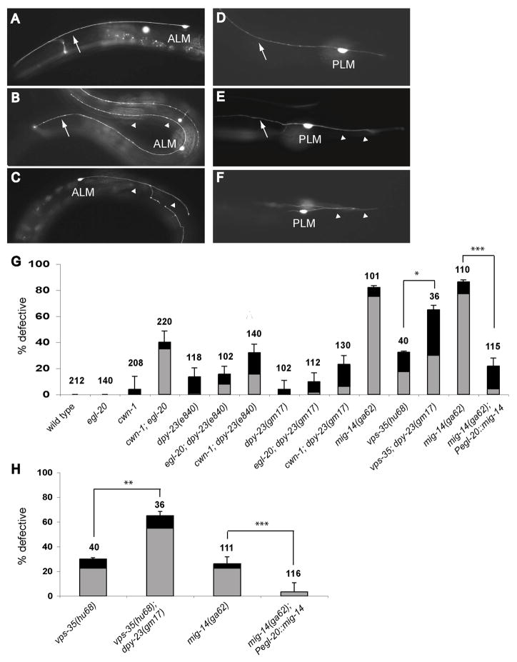
In addition to cell migration, Wnt signaling controls the polarity of neurons and non-neural cells. The mechanosensory neuron ALM extends a single anterior process in wild-type animals (White et al., 1986) (Figure 2A). While ALM neuronal polarity is normal in egl-20, cwn-1 or cwn-2 single mutants, the ALMs of cwn-1; egl-20 and cwn-1; cwn-2 double mutants are often bipolar or have reversed axonal polarity (Figures 2B, C, G) (Hilliard and Bargmann, 2006; Prasad and Clark, 2006). In dpy-23 mutants, the ALMs occasionally extended processes posteriorly, and this phenotype was enhanced by a cwn-1 mutation (Figure 2G). The ALM and QR defects suggest that dpy-23 functions with multiple Wnts.
Figure 2. dpy-23, mig-14, and vps-35 Mutants Display Defective ALM and PLM Neuronal Polarity.
(A–F) Photomicrographs showing ALM (A–C) and PLM (D–F) neuronal morphology. The ALM and PLM cell bodies are labeled. Anterior processes are indicated by arrows and posterior processes by arrowheads. (A) Wild-type ALM with a single, anterior process. (B) Bipolar ALM with a normal anterior process and an ectopic posterior process. (C) ALM with a single posterior process, indicating a reversal of polarity. (D) Wild-type PLM with a long anterior process and a short posterior process. (E) PLM with symmetric polarity. The cell has a normal anterior process and a long posterior process. (F) PLM with reversed polarity. The anterior process is short, and the abnormally long posterior process often folds back on itself at the tip of the tail and projects anteriorly. (G, H) Graph shows the percentage (with S.E.M.) of ALM (G) and PLM (H) neurons with defective polarity for each genotype. Black bars indicate the bipolar phenotype, and gray bars indicate reversed polarity. Numbers of axons scored for each genotype are provided. * p < 0.05; ** p < 0.01; *** p < 0.001, Fisher’s exact test.
The V5 cells normally divide asymmetrically to produce an anterior cell that joins the hypodermal syncytium and a posterior blast cell (Sulston and Horvitz, 1977). In egl-20 mutants, the division is occasionally reversed to produce an anterior blast cell and a posterior syncytial cell (Whangbo et al., 2000), a phenotype that we found was enhanced by a cwn-1 mutation (Figure S2). dpy-23 mutants also displayed this phenotype (Figure S2).
The phenotypic similarities between dpy-23 and Wnt mutants argue that DPY-23 regulates Wnt signaling. Developmental processes controlled by other signaling pathways, such as neuronal and growth cone migrations that are regulated by the guidance cues UNC-6/Netrin and SLT-1/Slit, appeared normal in dpy-23 mutants (data not shown). While DPY-23 regulates the EGF receptor in vulval development (Yoo and Greenwald, 2005), the lack of many signaling phenotypes in dpy-23 mutants suggests that DPY-23 plays a relatively specific role in Wnt signaling.
dpy-23 Encodes the C. elegans Homolog of the μ2 Subunit of the AP-2 Complex
Genetic mapping and transformation rescue of dpy-23 showed that it encodes the C. elegans ortholog of the μ subunit of the AP-2 complex (data not shown). The e840 allele is a large deletion removing the entire dpy-23 gene and flanking sequences. The gm17 allele is a splice donor site mutation in the last intron and is predicted to truncate the C-terminal 40 amino acids of the protein. Since the phenotypes of e840 are generally more severe than gm17 animals, we speculate that either some of the e840 phenotypes are attributed to additional genes removed in the e840 deletion or that the gm17 mutation does not eliminate dpy-23 function. We expressed a genomic dpy-23 fragment tagged with GFP at the C-terminus in e840 mutant animals and found that the mutant phenotypes were completely rescued, suggesting that the adjacent genes deleted in e840 do not contribute significantly to the dpy-23 phenotypes (Figure 3A and data not shown).
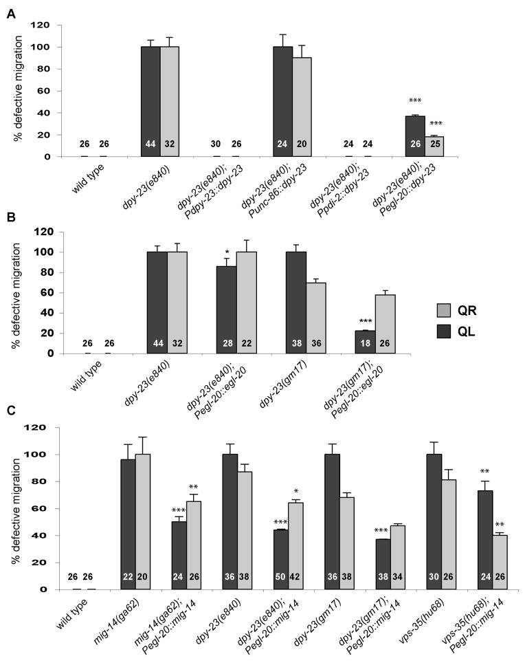
Figure 3. dpy-23 and mig-14 Function in EGL-20/Wnt-Secreting Cells.
(A) Graph shows the percentage (with S.E.M.) of rescue of the neuronal migration defects in dpy-23(e840) mutants by expressing dpy-23 from the endogenous dpy-23, the neuronal unc-86, the hypodermal pdi-2, and the egl-20 promoters. In A, B and C, numbers of cells scored for each genotype are provided. (B) Effects of excess EGL-20 on the migrations of QL and QR descendents in dpy-23 mutants. (C) Effects of MIG-14 expression on the Q migration defects of mig-14(ga62), dpy-23(e840), dpy-23(gm17) and vps-35(hu68) mutants. * p < 0.05; ** p < 0.01; *** p < 0.001, Fisher’s exact test.
dpy-23 Functions in Wnt-Producing Cells
To determine where DPY-23 functions, we expressed a dpy-23 cDNA fused to GFP from tissue-specific promoters and asked whether these transgenes rescued the defects of dpy-23 mutants. Expression of DPY-23::GFP in the HSN and Q descendents from the neuronal specific unc-86 promoter failed to rescue the neuronal migration defects (Figures 3A and S1). We previously used this promoter to express a cDNA for the Frizzled receptor MIG-1 and showed that it could rescue the HSN defects of a mig-1 mutant (Pan et al., 2006). Expression of DPY-23::GFP in hypodermal cells from the pdi-2 promoter, by contrast, completely rescued the Q defects and partially rescued the HSN defects of dpy-23 mutants (Figures 3A and S1). The pdi-2 and dpy-23 promoters are active in the B, F, K, and U hypodermal cells that also produce the Wnt EGL-20 (Figure S3), raising the possibility that dpy-23 could function in EGL-20-producing cells. Consistent with this hypothesis, expression of DPY-23::GFP from the egl-20 promoter partially rescued the Q and HSN defects, suggesting that DPY-23 functions in Wnt-producing cells (Figures 3A and S1). The lack of complete rescue for the HSN and QR phenotypes might be explained by the requirement for multiple Wnts that function in these migrations (Pan et al., 2006) (C.-L.P. and G.G., unpublished results).
Genetic Interactions between dpy-23, vps-35, and mig-14
Several recently identified proteins also regulate the production of functional Wnts. Homologs of Vps35p, Vps26p and Vps29p, subunits of the yeast retromer complex responsible for the trafficking of proteins between the Golgi and endosomes (Seaman et al., 1998), regulate Wnt function in C. elegans (Coudreuse et al., 2006; Prasad and Clark, 2006). C. elegans vps-35 mutants display Wnt phenotypes, including defects in neuronal migrations and polarity, and the Egl-20 phenotypes displayed by vps-35 mutants were rescued by expression of VPS-35 in cells that produce EGL-20.
Wntless, also known as Evi or Sprinter, was identified in Drosophila as a membrane protein required for Wnt secretion (Banziger et al., 2006; Bartscherer et al., 2006; Goodman et al., 2006). Mutations in the C. elegans homolog of Wntless MIG-14, also known as MOM-3, disrupt the asymmetric divisions of EMS and V5 and the migrations of the HSNs and Q neuroblasts, resulting in mutant phenotypes similar to those caused by mutations in the Wnt genes mom-2 or egl-20 (Harris et al., 1996; Thorpe et al., 1997; Whangbo et al., 2000). We will refer to the Drosophila homolog as Wntless and the C. elegans homolog as MIG-14.
We analyzed the phenotypes caused by four mig-14 alleles. Both or78 and gm2 resulted in maternal-effect lethality, whereas ga62 and mu71 were viable as homozygous strains. All four alleles caused HSN and Q migration defects (Figure S1 and data not shown), and our observations suggest that or78 and gm2 alleles severely reduce or eliminate mig-14 activity. ga62 and mu71 alleles only partially disrupt mig-14 function, with mu71 being weaker. We focused on ga62 for the experiments described below.
In addition to neuronal migration defects, we also discovered ALM polarity defects in mig-14(ga62) mutants. mig-14 mutants also had defects in PLM polarity, a phenotype displayed by lin-44 Wnt mutants (Figure 2). Each PLM neuron extends a single anterior process and a very short posterior process (Figure 2D). In lin-44, vps-26, vps-35 or mig-14 mutants, the PLMs extended a short anterior process and a long posterior process, a reversal in polarity (Figures 2E and F) (Hilliard and Bargmann, 2006; Prasad and Clark, 2006). mig-14 mutations also reverse the polarity of the V5 division (Figure S2) (Whangbo et al., 2000). These results confirm and extend previous descriptions of mig-14 mutant phenotypes, and they suggest that MIG-14 regulates Wnt signaling in C. elegans.
To investigate genetic interactions among these genes, we attempted to construct double mutant combinations. mig-14; dpy-23 and vps-35(hu68); dpy-23(e840) double mutants were not viable, precluding us from further analysis. Homozygous vps-35(hu68); dpy-23(gm17) animals from vps-35(hu68)/+; dpy-23(gm17) mothers, however, retain the maternal contribution of VPS-35 and although extremely sick and sterile, they are viable. Both the vps-35 ALM and PLM polarity defects were significantly enhanced in the vps-35(hu68); dpy-23(gm17) double mutant compared to either single mutant (Figures 2G and H). Enhancement of the PLM defects reveals a role for DPY-23 in PLM polarity that was not apparent in dpy-23 single mutants.
The Subcellular Localization of MIG-14 Depends on DPY-23 and Retromer
EGL-20/Wnt is expressed at similar levels in wild-type, dpy-23 and mig-14 animals, indicating that dpy-23 and mig-14 does not regulate Wnt expression (Figure S4). The egl-20 translational GFP reporter used had been shown to fully rescue the neuronal defects of egl-20 mutants (Whangbo and Kenyon, 1999), and we previously showed that it caused excessive HSN migration in sensitized genetic backgrounds, indicating a high level of expression (Pan et al., 2006). This reporter was able to rescue most of the QL defects of the dpy-23(gm17) mutant, and also a small but significant percentage of QL defects of the dpy-23(e840) mutant (Figure 3B). However, it failed to rescue the HSN and QR defects of either allele (Figure 3B and data not shown). The ability of excess EGL-20 to rescue the QL migration defects of dpy-23 mutants may reflect the fact that EGL-20 is the sole Wnt involved in QL migration and hence plays a more prominent role than it does in the QR and HSN migrations.
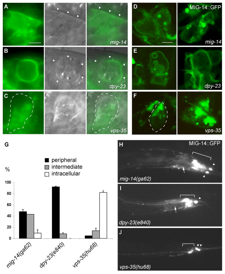
The discovery of Wntless as a component required for Wingless secretion suggested that endocytosis could be necessary for the recycling of the Wntless protein. We examined the subcellular distributions of MIG-14 in EGL-20/Wnt-producing cells (Figure 4). A translational GFP fusion of mig-14 was expressed under the control of the egl-20 promoter. We first observed MIG-14::GFP expression in the 1.5-fold embryo, and it continued through all larval and adult stages, consistent with previous reports on the temporal profile of egl-20 promoter activity (data not shown) (Pan et al., 2006; Whangbo and Kenyon, 1999). The MIG-14::GFP fusion protein was functional: when expressed in EGL-20/Wnt-secreting cells, it partially rescued the HSN and Q neuroblast migration defects and the ALM and PLM neuronal polarity defects of mig-14 mutants, indicating that it acts in Wnt-secreting cells (Figures 2G, 2H, 3C, and S5) (Banziger et al., 2006; Thorpe et al., 1997). Epifluorescence (Figure 4A) and confocal imaging (Figure 5D) of MIG-14::GFP in living animals showed that MIG-14::GFP localized to the cell periphery and intracellular compartments (Figures 4A, D, G).
Figure 4. Subcellular Localization of MIG-14 Requires DPY-23 and VPS-35.
(A–C) Epifluorescence and Nomarski images of a translational MIG-14::GFP fusion protein expressed in EGL-20 secreting cells of living animals. Left, GFP epifluorescence; middle, Nomarski images; right, overlay. Arrowheads (A, B) or dashed lines (C) mark cell boundaries. To eliminate endogenous MIG-14, we expressed MIG-14::GFP in mig-14(ga62) mutants and used this strain as a control. (A) mig-14(ga62): MIG-14::GFP was localized at or near the plasma membrane and in intracellular compartments. (B) dpy-23(e840) mutant: MIG-14::GFP accumulated at or near the plasma membrane. (C) vps-35(hu68) mutant: MIG-14::GFP accumulated in intracellular compartments and was significantly decreased from the cell periphery. (D–F) Representative confocal images of various MIG-14::GFP patterns in living animals. We classified MIG-14::GFP distribution into three categories: (D) intermediate pattern, where MIG-14::GFP is both at the cell periphery and in the intracellular compartments; (E) peripheral pattern, where MIG-14::GFP is predominantly at the cell periphery; and (F) intracellular pattern, where MIG-14::GFP is localized to intracellular compartments with little or no signal at the cell periphery. (G) The distribution of MIG-14::GFP was quantified from single confocal sections through the center of the GFP-expressing cells. Four lines of fluorescence intensity, one horizontal, one vertical and two diagonal, were scanned. A peripheral distribution was defined when the fluorescence intensity on the cell membrane was more than 2 fold the highest intracellular fluorescence intensity in three out of the four measurements (Figure S6A). An intracellular distribution was defined when the peak intracellular fluorescence intensity was more than 2 fold the fluorescence intensity on the cell membrane in three out of the four measurements (Figure S6C). Distributions other than those described above were defined as an intermediate distribution (Figure S6B). Graph shows the percentage (with S.E.M.) of different MIG-14::GFP distributions in mig-14(ga62), dpy-23(e840) and vps-35(hu68) mutants. Total numbers of cells scored: mig-14(ga62), 21; dpy-23(e840), 25; vps-35(hu68), 44. (H–J) Epifluorescence photomicrographs showing the tails of the larvae; anterior is to the left and posterior to the right. In vps-35(hu68) but not in the dpy-23(e840) mutants, the level of MIG-14::GFP often decreased significantly. Camera settings were the same for all three genotypes. Brackets indicate the location of hypodermal cells expressing MIG-14::GFP. Intestinal muscles which are immediately anterior to these cells also expressed this transgene at a much lower level (arrows). Arrowheads indicate PLM neurons marked by the integrated GFP reporter zdIs5[Pmec-4::gfp]. Scale bar is 5 μm.
MIG-14::GFP localization changed in dpy-23 and vps-35 mutants. MIG-14::GFP accumulated at or near the plasma membrane in dpy-23 mutants (Figures 4B, 4G and S6). We often observed MIG-14::GFP to be in large vesicular structures closely associated with the cell membrane (Figure 4E). These vesicles were never seen in retromer mutants, and were only infrequently observed in mig-14 mutants. In vps-35 mutants, by contrast, MIG-14::GFP accumulated in discrete intracellular structures that were absent in control animals, and it rarely localized to the cell periphery (Figures 4C, 4F, 4G and S6). Moreover, we observed a significant decrease in MIG-14 levels in vps-35 mutants (Figures 4H–J), suggesting that in the absence of retromer function, MIG-14 may be targeted for degradation.
These results suggest that DPY-23 and VPS-35 control Wnt secretion by regulating MIG-14 recycling. A prediction of this model is that excess MIG-14 should at least partially bypass the requirement for DPY-23 and VPS-35. Consistent with this model, expression of excess MIG-14 in Wnt-secreting cells rescued the QL, QR and HSN defects of dpy-23 and vps-35 mutants (Figures 3C and S5). By contrast, expression of excess DPY-23 in Wnt-secreting cells failed to rescue the neuronal defects of mig-14 mutants (data not shown).
Discussion
Wnt function requires the C. elegans AP-2 μ subunit DPY-23. Together with retromer, DPY-23 regulates the intracellular distribution of MIG-14, a Wnt-binding factor required for Wnt secretion. We speculate that newly synthesized EGL-20/Wnt binds to MIG-14 in the Golgi, targeting the Wnt to the cell membrane for secretion. In this model, AP-2-mediated endocytosis and retromer retrieval at the sorting endosome would recycle MIG-14 to the Golgi, where it can bind to EGL-20/Wnt for next cycle of secretion.
Studies in Drosophila demonstrated a role for endocytosis in the formation of a Wingless gradient. Models based on a nonautonomous requirement for dynamin in Wnt function implicated endocytosis as part of a relay that transferred Wingless from one cell to the next (Bejsovec and Wieschaus, 1995; Moline et al., 1999). Other investigators proposed that the Wingless gradient was generated by diffusion. These investigators proposed that the effects of dynamin loss on Wnt function reflected a lack of Wingless secretion from cells expressing the morphogen (Strigini and Cohen, 2000). While our results do not directly resolve this controversy, the requirement for DPY-23 in MIG-14 endocytosis supports the hypothesis that endocytosis is necessary for Wnt secretion and provides a mechanism for how endocytosis regulates Wnt secretion.
A previous study argued that retromer was not necessary for Wnt secretion, but instead was necessary for production of a functional Wnt (Coudreuse et al., 2006). These investigators also proposed that retromer was necessary for long-range Wnt signaling, but only played a minor role in short-range signaling. They argued that retromer mutants produced Wnt molecules that could only act on nearby cells but failed to act on more distant cells. Our findings that retromer is required for MIG-14 trafficking and the previous discovery that Wntless, the Drosophila MIG-14 homolog, is necessary for Wingless secretion are at odds with the interpretation that retromer plays a specific role in production of a Wnt that acts in long-range signaling (Banziger et al., 2006; Bartscherer et al., 2006).
An argument for retromer playing a specific role in long-range signaling was based on the observations that retromer mutants have little effect on processes that require MOM-2 and LIN-44, Wnts that are produced near responding cells (Coudreuse et al., 2006). Further support for the long-range hypothesis was based on the higher frequency of V5 defects in egl-20 mutants compared to retromer mutants (Coudreuse et al., 2006). This difference contrasted with the high frequency of QL migration defects in both egl-20 and retromer mutants. The discrepancy between the V5 and QL defects in the two types of mutants was explained by the closer proximity of the V5 cell to the EGL-20 source. The model that retromer plays a specific role in long-range Wnt signaling has led to speculation that the trafficking events regulated by this complex might control the production of a specifically modified form of Wnt (Coudreuse and Korswagen, 2007; Coudreuse et al., 2006; Hausmann et al., 2007), for example, a Wnt that could associate with lipoprotein particles (Panakova et al., 2005).
We favor a simpler hypothesis where retromer is required for MIG-14 recycling and where blocked recycling leads to defects in Wnt secretion. Our observation that excess MIG-14 can ameliorate the Wnt phenotypes of dpy-23 and vps-35 mutants is consistent with the notion that low levels of functional Wnts are still secreted in these mutants. We propose that the phenotypic differences observed between retromer and egl-20 mutants may result from differential sensitivities of various responding cells to lowered Wnt levels, and a similar explanation could account for the phenotypic differences between dpy-23 and Wnt mutants.
While the phenotypes of mig-14 mutants have most of the defects displayed by either single Wnt mutants or Wnt mutant combinations, dpy-23 mutants do not exhibit certain Wnt mutant phenotypes. They do not have the severe ALM polarity defects that are exhibited by cwn-1; egl-20 or cwn-1; cwn-2 double mutants and completely lack the PLM polarity defects of lin-44 mutant. Yet the dpy-23 defects in HSN and QL migration are extremely severe. One explanation for these differences between dpy-23 and mig-14 mutants, as well as the differences between retromer and mig-14 mutants, is that different Wnt-producing cells varying in their dependence on AP-2 or retromer to secrete Wnts. We speculate that endocytosis and retromer recycle MIG-14 for multiple rounds of Wnt secretion. If this hypothesis is correct, phenotypic differences could reflect the ability of some cells to synthesize sufficient MIG-14 resulting in less dependence on recycling. Alternatively, independent mechanisms for trafficking MIG-14 could operate in different Wnt-secreting cells.
Experimental Procedures
Details on C. elegans genetics, molecular biology and β-galactosidase immunohistochemistry and fluorescence confocal microscopy are available in Supplemental Experimental Procedures.
Nomarski Microscopy for HSN Migration, Q Migration and V5 Polarity
Positions of HSN and Q descendents were scored in newly hatched L1 and L1 4–6 hours after hatching, respectively. Positions of HSNs were scored as previously described (Pan et al., 2006). QL migration was scored as defective when PVM and SDQL were positioned anterior to the V4.p cell, which indicates that the QL descendents migrated toward the anterior, the wrong direction. QR migration was scored as defective when AVM and SDQR were positioned posterior to V2.p, which indicates undermigration. The polarity of V cells were identified by Nomarski optics and confirmed with a GFP reporter the jcIs1[ajm-1::gfp] that labels adherens junctions (Koppen et al., 2001).
ALM and PLM Axon Scoring
Neuronal polarity of ALM and PLM was scored using the integrated array zdIs5[Pmec-4::gfp], which is expressed in the six mechanosensory neurons ALMs, PLMs, AVM and PVM. For ALM, the bipolar phenotype was defined as a normal anterior process and a posterior process that is longer than ten ALM cell diameters in length. For PLM, the symmetric phenotype was defined as a normal anterior process and a posterior process that extends to the tip of the tail. Reversed polarity was defined as an abnormally long posterior process that is longer than the anterior process, which is often truncated. In these cases, the posterior process reaches the tip of the tail, and then folds back to project anteriorly.
Supplementary Material
Acknowledgments
We thank Andy Fire, Cynthia Kenyon, Hendrik Korswagen, and Lianna Wong for strains and reagents, and the C. elegans Genetics Center and the C. elegans Knockout Consortium for some of the strains used in this study. This work was supported by National Institutes of Health (NIH) grants NS32057 to G.G., NS034307 to E.M.J. and NS39397 to S.G.C. C.-L.P was supported by a Fellowship for Graduate Study from the Ministry of Education, Taiwan.
Footnotes
Publisher's Disclaimer: This is a PDF file of an unedited manuscript that has been accepted for publication. As a service to our customers we are providing this early version of the manuscript. The manuscript will undergo copyediting, typesetting, and review of the resulting proof before it is published in its final citable form. Please note that during the production process errors may be discovered which could affect the content, and all legal disclaimers that apply to the journal pertain.
Reference List
- Banziger C, Soldini D, Schutt C, Zipperlen P, Hausmann G, Basler K. Wntless, a conserved membrane protein dedicated to the secretion of Wnt proteins from signaling cells. Cell. 2006;125:509–522. doi: 10.1016/j.cell.2006.02.049. [DOI] [PubMed] [Google Scholar]
- Bartscherer K, Pelte N, Ingelfinger D, Boutros M. Secretion of Wnt ligands requires Evi, a conserved transmembrane protein. Cell. 2006;125:523–533. doi: 10.1016/j.cell.2006.04.009. [DOI] [PubMed] [Google Scholar]
- Bejsovec A, Wieschaus E. Signaling activities of the Drosophila wingless gene are separately mutable and appear to be transduced at the cell surface. Genetics. 1995;139:309–320. doi: 10.1093/genetics/139.1.309. [DOI] [PMC free article] [PubMed] [Google Scholar]
- Brenner S. The genetics of Caenorhabditis elegans. Genetics. 1974;77:71–94. doi: 10.1093/genetics/77.1.71. [DOI] [PMC free article] [PubMed] [Google Scholar]
- Coudreuse D, Korswagen HC. The making of Wnt: new insights into Wnt maturation, sorting and secretion. Development. 2007;134:3–12. doi: 10.1242/dev.02699. [DOI] [PubMed] [Google Scholar]
- Coudreuse DY, Roel G, Betist MC, Destree O, Korswagen HC. Wnt gradient formation requires retromer function in Wnt-producing cells. Science. 2006;312:921–924. doi: 10.1126/science.1124856. [DOI] [PubMed] [Google Scholar]
- Goldstein B, Takeshita H, Mizumoto K, Sawa H. Wnt signals can function as positional cues in establishing cell polarity. Dev Cell. 2006;10:391–396. doi: 10.1016/j.devcel.2005.12.016. [DOI] [PMC free article] [PubMed] [Google Scholar]
- Goodman RM, Thombre S, Firtina Z, Gray D, Betts D, Roebuck J, Spana EP, Selva EM. Sprinter: a novel transmembrane protein required for Wg secretion and signaling. Development. 2006;133:4901–4911. doi: 10.1242/dev.02674. [DOI] [PubMed] [Google Scholar]
- Guenther C, Garriga G. Asymmetric distribution of the C. elegans HAM-1 protein in neuroblasts enables daughter cells to adopt distinct fates. Development. 1996;122:3509–3518. doi: 10.1242/dev.122.11.3509. [DOI] [PubMed] [Google Scholar]
- Harris J, Honigberg L, Robinson N, Kenyon C. Neuronal cell migration in C. elegans: regulation of Hox gene expression and cell position. Development. 1996;122:3117–3131. doi: 10.1242/dev.122.10.3117. [DOI] [PubMed] [Google Scholar]
- Hausmann G, Banziger C, Basler K. Helping Wingless take flight: how WNT proteins are secreted. Nat Rev Mol Cell Biol. 2007;8:331–336. doi: 10.1038/nrm2141. [DOI] [PubMed] [Google Scholar]
- Herman M. C. elegans POP-1/TCF functions in a canonical Wnt pathway that controls cell migration and in a noncanonical Wnt pathway that controls cell polarity. Development. 2001;128:581–590. doi: 10.1242/dev.128.4.581. [DOI] [PubMed] [Google Scholar]
- Hilliard MA, Bargmann CI. Wnt signals and frizzled activity orient anterior-posterior axon outgrowth in C. elegans. Dev Cell. 2006;10:379–390. doi: 10.1016/j.devcel.2006.01.013. [DOI] [PubMed] [Google Scholar]
- Koppen M, Simske JS, Sims PA, Firestein BL, Hall DH, Radice AD, Rongo C, Hardin JD. Cooperative regulation of AJM-1 controls junctional integrity in Caenorhabditis elegans epithelia. Nat Cell Biol. 2001;3:983–991. doi: 10.1038/ncb1101-983. [DOI] [PubMed] [Google Scholar]
- Liu Y, Shi J, Lu CC, Wang ZB, Lyuksyutova AI, Song XJ, Zou Y. Ryk-mediated Wnt repulsion regulates posterior-directed growth of corticospinal tract. Nat Neurosci. 2005;8:1151–1159. doi: 10.1038/nn1520. [DOI] [PubMed] [Google Scholar]
- Lyuksyutova AI, Lu CC, Milanesio N, King LA, Guo N, Wang Y, Nathans J, Tessier-Lavigne M, Zou Y. Anterior-posterior guidance of commissural axons by Wnt-frizzled signaling. Science. 2003;302:1984–1988. doi: 10.1126/science.1089610. [DOI] [PubMed] [Google Scholar]
- Maloof JN, Whangbo J, Harris JM, Jongeward GD, Kenyon C. A Wnt signaling pathway controls hox gene expression and neuroblast migration in C. elegans. Development. 1999;126:37–49. doi: 10.1242/dev.126.1.37. [DOI] [PubMed] [Google Scholar]
- Mello CC, Kramer JM, Stinchcomb D, Ambros V. Efficient gene transfer in C. elegans: extrachromosomal maintenance and integration of transforming sequences. EMBO J. 1991;10:3959–3970. doi: 10.1002/j.1460-2075.1991.tb04966.x. [DOI] [PMC free article] [PubMed] [Google Scholar]
- Moline MM, Southern C, Bejsovec A. Directionality of wingless protein transport influences epidermal patterning in the Drosophila embryo. Development. 1999;126:4375–4384. doi: 10.1242/dev.126.19.4375. [DOI] [PubMed] [Google Scholar]
- Pan CL, Howell JE, Clark SG, Hilliard M, Cordes S, Bargmann CI, Garriga G. Multiple Wnts and frizzled receptors regulate anteriorly directed cell and growth cone migrations in Caenorhabditis elegans. Dev Cell. 2006;10:367–377. doi: 10.1016/j.devcel.2006.02.010. [DOI] [PubMed] [Google Scholar]
- Panakova D, Sprong H, Marois E, Thiele C, Eaton S. Lipoprotein particles are required for Hedgehog and Wingless signalling. Nature. 2005;435:58–65. doi: 10.1038/nature03504. [DOI] [PubMed] [Google Scholar]
- Prasad BC, Clark SG. Wnt signaling establishes anteroposterior neuronal polarity and requires retromer in C. elegans. Development. 2006;133:1757–1766. doi: 10.1242/dev.02357. [DOI] [PubMed] [Google Scholar]
- Seaman MN. Recycle your receptors with retromer. Trends Cell Biol. 2005;15:68–75. doi: 10.1016/j.tcb.2004.12.004. [DOI] [PubMed] [Google Scholar]
- Seaman MN, McCaffery JM, Emr SD. A membrane coat complex essential for endosome-to-Golgi retrograde transport in yeast. The Journal of cell biology. 1998;142:665–681. doi: 10.1083/jcb.142.3.665. [DOI] [PMC free article] [PubMed] [Google Scholar]
- Strigini M, Cohen SM. Wingless gradient formation in the Drosophila wing. Curr Biol. 2000;10:293–300. doi: 10.1016/s0960-9822(00)00378-x. [DOI] [PubMed] [Google Scholar]
- Sulston JE, Horvitz HR. Post-embryonic cell lineages of the nematode, Caenorhabditis elegans. Dev Biol. 1977;56:110–156. doi: 10.1016/0012-1606(77)90158-0. [DOI] [PubMed] [Google Scholar]
- Sulston JE, Schierenberg E, White JG, Thomson JN. The embryonic cell lineage of the nematode Caenorhabditis elegans. Dev Biol. 1983;100:64–119. doi: 10.1016/0012-1606(83)90201-4. [DOI] [PubMed] [Google Scholar]
- Tabata T, Takei Y. Morphogens, their identification and regulation. Development. 2004;131:703–712. doi: 10.1242/dev.01043. [DOI] [PubMed] [Google Scholar]
- Thorpe CJ, Schlesinger A, Carter JC, Bowerman B. Wnt signaling polarizes an early C. elegans blastomere to distinguish endoderm from mesoderm. Cell. 1997;90:695–705. doi: 10.1016/s0092-8674(00)80530-9. [DOI] [PubMed] [Google Scholar]
- Walston T, Guo C, Proenca R, Wu M, Herman M, Hardin J, Hedgecock E. mig-5/Dsh controls cell fate determination and cell migration in C. elegans. Dev Biol. 2006;298:485–497. doi: 10.1016/j.ydbio.2006.06.053. [DOI] [PubMed] [Google Scholar]
- Whangbo J, Harris J, Kenyon C. Multiple levels of regulation specify the polarity of an asymmetric cell division in C. elegans. Development. 2000;127:4587–4598. doi: 10.1242/dev.127.21.4587. [DOI] [PubMed] [Google Scholar]
- Whangbo J, Kenyon C. A Wnt signaling system that specifies two patterns of cell migration in C. elegans. Mol Cell. 1999;4:851–858. doi: 10.1016/s1097-2765(00)80394-9. [DOI] [PubMed] [Google Scholar]
- White JG, Southgate E, Thomson JN, Brenner S. The structure of the nervous system of the nematode Caenorhabditis elegans. Philos Trans R Soc Lond B Biol Sci. 1986;314:1–340. doi: 10.1098/rstb.1986.0056. [DOI] [PubMed] [Google Scholar]
- Yoo AS, Greenwald I. LIN-12/Notch activation leads to microRNA-mediated down-regulation of Vav in C. elegans. Science. 2005;310:1330–1333. doi: 10.1126/science.1119481. [DOI] [PMC free article] [PubMed] [Google Scholar]
- Yoshikawa S, McKinnon RD, Kokel M, Thomas JB. Wnt-mediated axon guidance via the Drosophila Derailed receptor. Nature. 2003;422:583–588. doi: 10.1038/nature01522. [DOI] [PubMed] [Google Scholar]
- Zecca M, Basler K, Struhl G. Direct and long-range action of a wingless morphogen gradient. Cell. 1996;87:833–844. doi: 10.1016/s0092-8674(00)81991-1. [DOI] [PubMed] [Google Scholar]
- Zou Y, Lyuksyutova AI. Morphogens as conserved axon guidance cues. Curr Opin Neurobiol. 2007;17:22–28. doi: 10.1016/j.conb.2007.01.006. [DOI] [PubMed] [Google Scholar]
Associated Data
This section collects any data citations, data availability statements, or supplementary materials included in this article.


