
Figure 1. CCM3 overexpression induces apoptosis in HeLa cells.
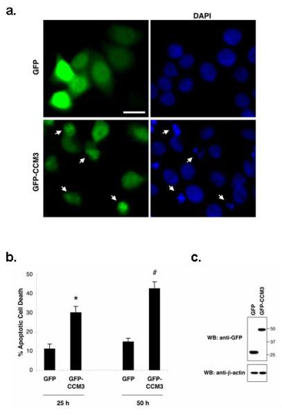
a. HeLa cells were transfected with vector alone or GFP-CCM3 constructs (see Methods). 50 hours (hrs) after transfection, cells were fixed, and nuclei stained with DAPI (blue). Arrows point to the GFP-CCM3-positive cells (green) with condensed or fragmented apoptotic nuclei. Scale bar = 20 μm.
b. GFP-positive cells were quantified for apoptosis by scoring apoptotic nuclear morphology as in (a). The percentage of GFP-positive cells with apoptotic nuclei was represented as mean ± SEM from three and four independent experiments for the 25 and 50 hour (hr) time point, respectively (left panel). *P < 0.002, significant difference from GFP-transfected cells; #P <10-3, significant difference from GFP-transfected cells (by Student’s t-test).
c. Protein expression of GFP and GFP-CCM3 25 hrs after transfection was confirmed by immunoblotting with anti-GFP. Anti-β-actin was used as loading control (bottom panel).